Relationship between aerobic fitness and clinical
indicators of asthma severity in children
*
JOSÉ ALBERTO NEDER1, ANA LUÍZA GODOY FERNANDES2, ANTÔNIO CARLOS SILVA3,
ANNA LÚCIA DE BARROS CABRAL4, LUIZ EDUARDO NERY2
In order to assess the relationship between the physical fitness of asthmatics and the clinical expression of the underlying disease, the authors studied 39 physically active children with moderate to severe but stable asthma. The patients (25 boys and 14 girls, aged between 9 and 16 years) were submitted to clinical evaluation; spirometry before and after bronchodilator (BD); maximal cardiopulmonary exercise test in cycle ergometer with breath-by-breath analysis
of ventilatory and gas exchange variables; and, on a separate day, an exercise challenge test. As expected by the clinical stability, FEV1 post-BD was in the normal range in most of the children (mean ± SD = 93.8 ± 13.7% predicted). Maximal oxygen uptake (VO
2max) was higher than the lower 95% confidence interval in 31/39 children; and in 29/39, the oxygen uptake at the anaerobic threshold (VO2AT) showed values above the lower limit of normality.
Seven patients with low tolerance to exercise (reduced VO
2max) presented suggestions of circulatory limitation (cardiovascular and/or peripheral) and only 1 had ventilatory limitation. There was no association or correlation between the lower ventilatory reserve (VEmax/MVV% ratio > 80%) and the decreased VO2max. Reduction in VO2AT,
but not VO2max, was associated with some clinical indicators of asthma severity, e.g. (i) higher daily inhaled beclomethasone and frequent courses of oral steroids (p < 0.05) and (ii) higher exercise-induced bronchospasm occurrence (p < 0.01). The results show that (i) most patients with moderate to severe asthma, when clinically stable
and physically active, present an adequate level of exercise tolerance; (ii) in estimation of the clinical severity of bronchial asthma in children, VO2AT is a better aerobic index than VO2max. (J Pneumol 1998;24(1):3-10)
Relação entre capacidade aeróbia e indicadores
clínicos da gravidade da asma em crianças
Para avaliar a relação entre o desempenho cardiorrespiratório aeróbio de asmáticos e a expressão clínica da doença, os autores estudaram 39 crianças fisicamente ativas, com asma brônquica estável, de grau moderado a
grave. Os pacientes (25 meninos e 14 meninas, com idade entre 9 e 16 anos) foram submetidos a avaliação clínica, espirometria pré e pós-broncodilatador (BD), teste de exercício cardiopulmonar máximo em cicloergômetro, com análise respiração por respiração da ventilação e das trocas gasosas. Num dia separado, foi realizado um teste de esforço para avaliar broncoespasmo induzido por exercício (BIE). Como esperado pela estabilidade clínica, o VEF1 pós-BD foi normal na maioria das crianças (média ± DP = 93,8 ± 13,7% previsto). O consumo máximo de oxigênio (VO2max) foi maior que o limite de normalidade (95% do intervalo de confiança) em 31 das 39 crianças; e em 29
de 39 o VO
2max e o limiar anaeróbio (VO2LA) mostrou valores acima deste limite. Sete pacientes com baixa
tolerância ao exercício (VO2max reduzido) tiveram sinais de limitação circulatória (cardiovascular e/ou periférica) e somente um teve limitação ventilatória. Não houve associação ou correlação entre a baixa reserva ventilatória (VEmax/VVM% > 80%) e valores reduzidos do VO2max. Redução no VO2LA mas não do VO2max foi associado com: (i)
maior uso diário de beclometasona e freqüentes períodos de uso de corticosteróide oral (p < 0,05); e (ii) maior ocorrência de BIE (p < 0,01). Nossos resultados mostram que a maioria dos pacientes com asma moderada a grave,
quando clinicamente estáveis e ativos, apresentam níveis adequados de tolerância ao exercício. Na avaliação de gravidade clínica da asma brônquica em crianças, VO2LA é um indicador aeróbio melhor que o VO2max.
3 . Professor Adjunto-Doutor do Departamento de Fisiologia da Uni-fesp-EPM.
4 . Médica Pneumologista do Hospital Infantil Darcy Vargas (São Paulo, Brasil).
Address – Prof. Dr. Luiz Eduardo Nery, Disciplina de Pneumologia, Unifesp-EPM, Rua Botucatu, 740, 3º andar – 04023-062 – São Paulo, SP, Brasil. Fax: (011) 549-2127.
Recebido para publicação em 29/8/97. Reapresentado em 12/ 2/98. Aprovado, após revisão, em 18/2/98.
* Partially supported by research grants from CNPq and FAPESP, Brazil.
1 . Doutor em Pneumologia e Pós-doutor em Fisiologia do Exercício pela Universidade Federal de São Paulo, Escola Paulista de Medicina (Unifesp-EPM), São Paulo, Brasil; Research Fellow: Department of Physiology, St. George’s Hospital Medical School University of Lon-don, LonLon-don, United Kingdom.
Key words – Asthma in children. Exercise tolerance. Maximal oxy-gen uptake. Anaerobic threshold. Physical fitness. Exercise-induced bronchospasm.
Descritores – Asma em crianças. Tolerância ao exercício. Consu-mo máxiConsu-mo de oxigênio. Limiar anaeróbico. Condição física. Bron-coespasmos induzidos por exercício.
Abbreviations BD – Bronchodilator
EIB – Exercise-induced bronchospasm f – Respiratory rate
FEV1 – Forced expiratory volume in one second FVC – Forced vital capacity
HR – Heart rate
max – At maximal exercise
MVV – Maximal voluntary ventilation O2 pulse – Oxygen pulse (VO2/HR) PEFR – Peak expiratory flow rate pred – Predicted
R – Respiratory exchange ratio SaO2 – Oxyhemoglobin saturation Ti/Ttot – Inspiratory duty cycle VCO2 – Carbon dioxide production VE – Minute ventilation
VE/VCO2 – Ventilatory equivalent for carbon dioxide VE/VO2 – Ventilatory equivalent for oxygen VO2 – Oxygen uptake
VO2AT – VO2 at the anaerobic threshold VT/Ti – Mean inspiratory flow
WR – Work rate
I
N T R O D U C T I O NCardiorespiratory fitness in normal individuals presents marked differences according to genetic characteristics, sex, age and usual level of physical activity(1). Patients with chronic
respiratory diseases tend to show less tolerance to exercise because of actual pulmonary limitation, self-restriction to activities (fear of dyspnea) or lack of physical activity due to medical orientation or family influence(2). Thus, subjects with
bronchial asthma, primarily those with a clinically more se-vere disease, tend to present a marked sedentary lifestyle(2,3).
This trait is particularly common in patients with exercise-induced bronchospasm who more frequently avoid dyspnea-generating activities(3).
Most studies that analyzed tolerance to dynamic exercise in asthmatic children evaluated patients with mild to moder-ate disease or those taking part in aerobic physical training programs(4-6). Although asthmatics are usually considered to
be less fit for dynamic exercise, the relationship between the clinical aspects of the underlying disease and the objec-tive parameters of exercise performance has been described in a conflicting way(6,7). Thus, Ludwick et al.(6) found that
49% of the sixty-five severe asthmatic children studied had a reduced maximal exercise performance, but no clinical features of disease severity were associated with an abnor-mal exercise response. On the other hand, Strunk et al.(7)
found that an episodic or continuous steroid use in the year before testing was significantly related to a decreased nine-minute running performance (p = 0.0004) in a sample of 76 moderate to severe asthmatic children.
Therefore, the purpose of this study was twofold: (i) to evaluate the overall tolerance to exercise of a group of mod-erate to severe asthmatic children and (ii) to analyze a possi-ble association between aerobic performance and clinical and functional markers of severity of the underlying disease.
M
E T H O D O L O G YSU B J E C T S
Thirty-nine asthmatic children (25 boys and 24 girls), aged between 9 and 16 years (mean ± SD = 12.4 ± 1.8 years), were studied. Means ± SD of body mass and height were 41.4 ± 9.8 kg and 148.8 ± 10.3 cm, respectively (table 1). According to the clinical parameters of the “International Consensus Report on Diagnosis and Management of
Asth-ma”, 1992(8), 24 of 39 patients (61.6%) were classified as
severe asthmatics presenting: a) frequent bronchospasm exa-cerbation and nocturnal asthma symptoms almost daily; b) peak expiratory flow rate (PEFR) values < 60% predicted at baseline despite optimal therapy; c) daily use of inhaled anti-inflammatory agent at high doses (beclomethasone > 800 µg/day) and frequent use of systemic corticosteroids. The remainder 15 children were considered moderate asthmat-ics presenting: a) symptoms requiring inhaled B2-agonist
al-most daily and nocturnal asthma symptoms > 2 times a month but not daily; b) PEFR values between 60-80% pre-dicted at baseline but normal after bronchodilator; c) daily
TABLE 1
Anthropometric and spirometric characteristics of the children with moderate to severe but stable asthma (n = 39; 25 boys and 14 girls)
Variables Values Range
(Mean ± SD)
Age (years) 12.4 ± 1.80 09-16
Weight (kg) 41.4 ± 9.80 27.5-80.0
Height (cm) 148.8 ± 10.30 131-179
FVC pre-BD (% pred) 99.9 ± 13.0 072-130
FEV1 pre-BD (% pred) 82.9 ± 13.8 047-106
FEV1 pre-BD (L) 2.01 ± 0.61 1.01-4.29
FEV1/FVC pre-BD 0.71 ± 0.11 0.59-0.81
FEV1 post-BD (% pred) 93.8 ± 13.7 59.5-112.
FEV1 post-BD (L) 2.40 ± 0.44 1.32-4.56
FEV1/FVC post-BD 0.78 ± 0.07 0.65-0.84
use of anti-inflammatory agent at low or moderate doses (beclomethasone < 800 µg/day). All patients were on in-haled corticosteroid (beclomethasone between 400 and 1500 µg/day) and 24 had a past history of frequent courses of oral corticosteroids; nevertheless, no patient was on system-ic steroids at the time of the study. According to the “Guide-lines for the Evaluation of Impairment/Disability in Patients with Asthma”, 1993(9), 61.6% of the patients (24 out of 39)
presented a medication score > 3, requiring for clinical asth-ma control: bronchodilator on deasth-mand, inhaled beclometha-sone in a daily dose equal to or higher than 800 µg or fre-quent courses of oral steroids.
The patients were referred to the Center of Sport Practic-es of the University of São Paulo (CEPEUSP) where they were undertaking an 8-week multidisciplinary follow-up program for asthmatics, including recreational and sport activities. Although physically active, the patients did not follow an individualized or supervised aerobic training program. All studied children had a regular medical follow-up by a staff physician (A.L.B.C.) and were at a stable phase of the disease, with no symptoms or exacerbation 15 days prior to the tests. None of the tested individuals presented any orthopedic lim-itation to the exercise or contraindication for the exercise tests. Informed consent was obtained from the patients and their parents, after explanation of the procedures to be car-ried out.
ST U D Y DESIGN
The patients were evaluated initially at the Exercise Lab-oratory of the Respiratory Division of the Federal University of São Paulo – Paulista School of Medicine (Unifesp-EPM). They were submitted to a brief clinical history and physical examination, spirometry before and after bronchodilator and then to cardiopulmonary exercise test on a cycle ergometer. Afterwards, on a separate day, the patients were submitted to a protocol for detection of exercise-induced bronchospasm at the Center of Sport Practices of the University of São Paulo (CEPEUSP).
SP I R O M E T R Y
Spirometric tests were performed in all subjects, before and ten minutes after the inhalation of 200 µg of salbuta-mol given by pressurized metered-dose inhaler (MDI) connect-ed to a spacer. The equipment usconnect-ed was a CPF-S (Medical Graphics Corp. – MGC, St. Paul, MN, USA) with flow mea-surement carried out with a pneumotachygraph Fleisch No. 3. Technical procedures, acceptability and reproducibility criteria were those recommended by the American Thorac-ic Society, 1991(10). Predicted normal values for all
spiro-metric variables were those of Knudson et al.(11). Maximal
voluntary ventilation (MVV) was estimated from the product of the forced expiratory volume in one second (FEV
1) x 40
(12).
A positive response to bronchodilator was defined as a 15% and 200 mL increase in FEV
1
(10).
CARDIOPULMONARY EXERCISE TESTING (CPX)
Exercise tests were performed using a digital computer-based exercise system (MGC-CPX System, Medical Graphics Corp. – MGC, St. Paul, MN, USA). The maximal exercise test was carried out on a calibrated electromagnetically braked cycle ergometer (CPE 2000, Medical Graphics Corp. – MGC, St. Paul, MN, USA), modified with children pedal cranks. The selected work rate was continuously increased in a linear ramp pattern (15 watts per minute if height < 150 cm or 20 watts per minute if height > 150 cm)(13) so that the
incre-mental exercise test duration was greater than 8- and lower than 12-minutes(14). During the test, subjects used a
nose-clip and breathed through a mouthpiece connected to a low-resistance, two-way non-rebreathing Hans-Rudolph 2600 valve (60 ml dead space). Expired airflow was measured by a pneumotachygraph Fleisch No. 3 in the flow/volume sys-tem’s module. Expired gases were withdrawn at a flow of 1 ml/sec from a point just proximal to the mouthpiece; sig-nals proportional to the fractional concentrations of carbon dioxide and oxygen were generated by rapidly responding infrared and fuel zirconium cell analyzers, respectively. A microprocessor (“waveform analyzer”) received, through specific channels, the different analogue signals. Since the generation of these signals was not simultaneous, the mi-croprocessor temporally aligned them, promoting a calibrated delay (“phase delay”), allowing breath-by-breath analysis of metabolic, ventilatory and cardiovascular variables(14). The
analogue signals of cardiac electric activity (Funbec 4A-1CN cardioscoper, Funbec ECG-3 electrocardiograph) and oxyhe-moglobin saturation (OxyShuttleTM SensorMedics pulse
The data were calculated automatically using standard formulae(14) and displayed in descriptive numerical (every 20
seconds) and graphical (8 breaths moving average) forms. The following data were obtained breath-by-breath: oxygen uptake (VO
2, ml/min STPD); carbon dioxide production (VCO2, ml/min STPD); respiratory exchange ratio (R); minute venti-lation (VE, l/min BTPS); respiratory rate (f, bpm); ventilatory equivalent for O2 and CO2 (VE/VO2 and VE/VCO2); end-tidal partial pressures of O2 and CO2 (PETO2 and PETCO2, mmHg); inspiratory duty cycle (Ti/Ttot); mean inspiratory flow (VT/Ti, l/s); heart rate (HR, bpm) and oxygen pulse (VO
2/HR, ml/ beat). The predicted VO
2max was calculated from the equa-tions of Cooper et al. for children(16) who consider the VO
2max as the largest breath-by-breath VO
2 value achieved by the subject submitted to a ramp protocol. In order to smooth the high intra- and inter-breath variability of the breath-by-breath data, we took the VO
2max as the highest VO2 value of the last 8 test breaths (“peak VO
2”) and expressed it in abso-lute values (ml/min), corrected for actual weight (ml/min x kg–1) and as percent of the predicted (% pred). Considering
the close demographic and anthropometric similarity of our population and the one studied by Cooper et al.(16) and also
the similarity of the method of VO
2max analysis in both stud-ies, the lower limit of normality for VO
2max was defined ac-cording to the values proposed by these authors (lower 95% confidence limit; mean –1.64 x SD). For girls < 11 years = 26 ml/min x kg–1; girls > 11 years = 27 ml/min x kg–1; boys
< 13 years = 32 ml/min x kg–1; boys > 13 years = 37 ml/
min x kg–1. The VO
2 at the anaerobic threshold (VO2AT) was estimated by the gas exchange method inspecting visually the inflection point of VCO
2 regarding VO2 (modified V-slope)(17) and by the ventilatory method, when V
E/VO 2 and PETO2 increased while VE/VCO
2 and PETCO2 remained stable. The reading was performed independently by two experi-enced observers without knowledge of other results or sub-ject identities. In case of discordance, a third reviewer would be used to reach a consensus or to eliminate the subject. In this study a consensus was always reached and the VO
2AT was identified in all studied patients. The lower limit of nor-mality for VO
2AT was defined at 40% of VO2max predicted, considering that from 109 children evaluated by Cooper et al., only one presented a value below this cutoff(16).
In order to analyze the ventilatory response to VCO 2 dur-ing the incremental test – an useful index of ventilatory stress(18) – a graphical presentation of V
E as a function of
VCO
2 was used to establish the point at which VE increases out of proportion to VCO
2 (respiratory compensation point – RCP). Linear regression techniques were used to fit the best-line from the data starting at 60 seconds after the onset to exercise to the RCP, and the slope (∆VE/∆VCO
2) was calculated for each subject. The obtained values were then compared to weight-based predicted from Cooper et al.(18):
∆VE/∆VCO 2) = –0.099 x weight (kg) + 28.7. The oxygen costs at
maxi-mal exercise for each child were calculated (VO
2max/WRmax, l/min/w) and compared to predicted proposed by Zancona-to et al.(19), as a function of height (–0.095 x height (cm) +
29.11).
Analysis of the factors limiting exercise followed recom-mendations of Wasserman et al.(14), and Nery et al.(20). Briefly,
circulatory limitation (cardiovascular and/or peripheral) was considered in subjects with reduced VO
2max, VO2AT, oxygen pulse and leg pain as the main limiting symptom. Pulmo-nary limitation was suggested in patients with decreased
VO
2max, ventilatory pattern of high frequency and low tidal volume in addition to dyspnea as the main symptom at maximal exercise. The presence of an elevated VEmax/MVV% ratio (> 80%) was suggestive of the ventilatory limitation and a > 4% oxyhemoglobin desaturation showed abnormal-ity in pulmonary gas exchange.
EXERCISE CHALLENGE TEST
The test for exercise-induced bronchospasm (EIB) detec-tion was carried out with a mechanically-braked Monark®.
cycle ergometer. Methylxanthines and B2-adrenoceptor
ag-onist were withheld 12 hours prior to the test. Before the exercise, the patient performed a forced expiratory maneu-ver (Vitatrace® spirometer) with assessment of forced vital
capacity (FVC) and FEV
1 basal values. The test was carried out only in patients with normal spirometry at rest: FEV
1 above 80% of the predicted(11) (n = 32). The tests were
accom-plished on ambient temperature between 18 and 22 C, baro-metric pressure between 680 and 720 mmHg and on air relative humidity between 55 and 80%. After one minute of light exercise on the cycle ergometer, the workload was quick-ly increased until the heart rate corresponding to 80% of predicted (220-age)(14), then being maintained for 6
min-utes(21). This target workload was selected in order to set an
intense exercise, with high ventilatory rates and great heat and water loss via the bronchial mucosa. Spirometric evalu-ations were performed successively at 5, 10 and 20 minutes after the exercise. EIB was defined as present if FEV
1 showed a reduction equal to or greater than 10% of its pre-exercise values(21).
STATISTICAL ANALYSIS
The values are reported as mean ± SD and their ranges. The following statistical tests were used: a) Linear regres-sion technique to determine the slope of the ∆VE/∆VCO
2 rela-tionship during the maximal exercise test and the correla-tion between VEmax/MVV% ratio and VO
2max; b) Paired t test to compare the ∆VE/∆VCO
2 slopes and VO2max/WRmax rela-tionship with their predicted values (Cooper et al.(16)); c)
R
E S U L T SMean ± SD values for spirometry of the clinically stable studied population were within the normal range; FEV
1 post-bronchodilator (BD) being 93.8 ± 13.7% of the predicted (table 1). Only 4 patients presented post-BD FEV
1 below 80% of the predicted. In 17 children (43%) a positive response to BD was obtained.
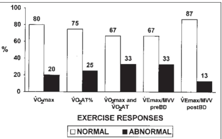
The results of the cardiopulmonary exercise test showed that the patients, as a group, had metabolic and cardiovas-cular variables close to the predicted values for normal chil-dren (table 2)(16). VO
2max was above the lower 95% confi-dence limit (see METHODS) in 31 of the 39 individuals (mean ± SD = 37.8 ± 7.8 ml/min x kg–1). In 29 children (75%), the
VO
2AT was considered normal (> 40% VO2max pred; mean ± SD = 47.4 ± 10.6%) and in 26 patients (67%) both were within the normal range (figure 1). Additionally, the oxygen Figure 1 – Percentage of patients with normal or abnormal metabolic and ventilatory responses at maximal exercise (n = 39)
Normal cutoffs: VO2max (ml/min x kg–1) > lower 95% confidence limit(16); VO2AT% > 40% of VO2max predicted(16); VEmax/MVV% < 80%(14).
Figure 2 – Comparison between observed and predicted ∆VE/∆VCO2 slopes during incremental exercise in asthmatic children with normal (v) or reduced (j) VO2AT
Observed (mean ± SD = 28.5 ± 3.9) higher than predicted values (24.5 ± 0.9) – p < 0.0001.
TABLE 2
Maximal exercise performance of the children with moderate to severe but stable asthma (n = 39; 25 boys and 14 girls)
Variables Values Range
(Mean ± SD)
%VO2 (% pred) 87.3 ± 17.6 057-117
VO2max (ml/min x kg–1) 37.8 ± 7.80 21.6-51.4
VO2max (ml/min) 1,550 ± 5000. 0.895-3,819
WRmax (watts) 110.5 ± 35.20 068-228
VO2AT/VO2max pred (%) 47.4 ± 10.6 33.8-67.3
HRmax (% pred) 90.4 ± 8.10 076.3-110.5
O2 pulse max (% pred) 98.0 ± 19.4 66.8-145.
VEmax/MVV (%) 63.4 ± 13.2 35.1-87.4
SaO2 max (%) 95.2 ± 3.20 92-98
Definition of abbreviations: VO2max: maximum oxygen uptake; WR: work rate; VO2AT: oxygen uptake at the anaerobic threshold; HR: heart rate; VE: minute ventilation; MVV: maximal voluntary ventilation; SaO2: oxygen saturation.
costs of exercise for the whole group (VO
2max/WRmax; mean ± SD = 15.4 ± 3.4) were not significantly different than its predicted values(19) (mean ± SD = 14.9 ± 1.0); on the other hand, as expected, a significantly reduced oxygen costs were
found in patients with reduced VO
2AT (observed = 12.0 ± 0.7; predicted = 14.6 ± 0.6 – p < 0.0001). Analyzing the mechanisms of exercise limitation, we found that from 8 asthmatic children with effective reduction in maximal aero-bic tolerance (reduced VO
2max), 7 presented evidences of cir-culatory (cardiovascular and/or peripheral) and one of venti-latory limitation. None of the patients presented suggestions of pulmonary gas exchange abnormalities.
The VEmax/MVV% ratio was greater than 80% in 13 pa-tients when MVV was estimated from pre-BD FEV
1 and in only 5 when post-BD FEV
1 was used (figure 1). There was no asso-ciation of higher VEmax/MVV% ratio with lower aerobic per-formance, since 4 of these 5 patients had VO
2AT and VO2max above of predicted values. Although ventilatory factors were not related to maximal performance, we found in increased ventilatory response to metabolic load (steeper ∆VE/∆VCO
2 slopes) in 16/39 children, independently of a normal or re-duced VO
2AT (p < 0.0001 – figure 2).
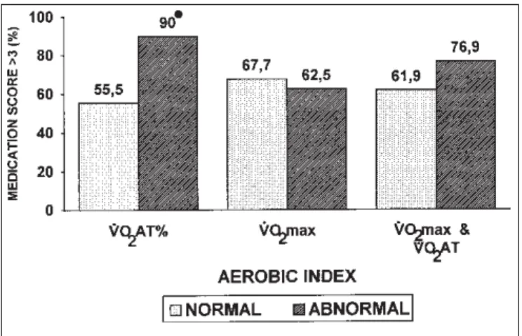
In order to investigate the relationship between aerobic performance at maximal exercise and clinical parameters of the underlying disease, we compared the association of a sensitive indicator of asthma severity (medication score) with the main metabolic variables (VO
2max and VO2AT). There was
a significant association between reduced VO
2AT and high medication score (> 3)(9). Thus, 90% of the patients with reduced VO
For definition of normality, see text and figure 1.
Obs.: Numbers above the blocks are the percentage of patients with the mentioned association.
* p < 0.05 (Fisher’s Exact test).
Figure 3 – Association between high medication score (> 3) and
norma-lity or not of the aerobic indexes at maximal exercise (n = 39)
Figure 4 – Association between a positive exercise challenge test (EIB+)
and normality or not of the aerobic indexes at maximal exercise (n = 32)
For definition of normality, see text and figure 1.
Obs.: Numbers above the blocks are the percentage of patients with the mentioned association.
* p < 0.01 (Fisher’s Exact test).
beclomethasone and frequent courses of oral steroids, con-trasting with 55% of the patients with normal VO2AT and use of high doses of medication (p < 0.05). The reduced VO2max, alone or in addition to reduced VO2AT, was not associated with high medication score (figure 3).
From 39 evaluated patients, 32 performed an exercise challenge test (basal FEV1 > 80% predicted). In 17 of them (53%), a positive response was obtained. Spirometric and clinical indicators of asthma severity did not differ between responders and non-responders, regarding EIB (table 3). How-ever, 88.8% of the patients with reduced VO2AT had EIB and only 30.4% of the individuals with normal VO2AT presented EIB (p < 0.01). Reduced VO2max, alone or in addition to re-duced VO2AT, was not significantly associated with a higher EIB occurrence (figure 4).
TABLE 3
Spirometric and clinical findings of asthmatic children with and without exercise-induced bronchospasm (EIB)
EIB positivea EIB negative (n = 15) (n = 17)
Spirometry (mean ± SD) FEV
1 pre BD (% pred) 79.4 ± 09.1 82.5 ± 17.8
FEV
1 post BD (% pred) 91.8 ± 10.2 93.7 ± 13.8
Clinical (% frequency)
Positive BD responseb 10 (66.6%) 06 (35.2%)
Severe asthma 13 (86.6%) 13 (76.4%)
a Decrease of FEV1 > 10%, determined by 6-minute exercise at 80% of pred HRmax(21); b Positive response to bronchodilator, with increase of FEV1, above 15% and 200 ml in
relation to basal(10).
Obs.: Differences between mean values were not statistically significant (non-paired t test).
D
I S C U S S I O NIn this study we evaluated the aerobic performance of children with moderate to severe bronchial asthma and we found an adequate level of cardiorespiratory fitness, reflect-ed in the VO
2max, VO2max/WRmax relationship, VO2AT, and
maximal O2 pulse values. These findings are at variance with several former studies(3,6,7) and probably influenced by the homogeneity of the group regarding clinical stability and level of physical activity. On the other hand, our data are in accordance with the findings of Fink et al.(23) who showed that moderate to severe trained patients had an adequate overall aerobic performance compared to asthmatic controls. However, a small number of patients presented reduction of exercise tolerance (decreased peak VO
2) primarily due to
“circulatory” factors (7 out of 8). Although Varray et al.(24) have suggested a cardiogenic role for the lower tolerance to exercise in severe asthmatics, our results indicate a periph-eral origin for this limitation, since the clinical evaluation was not able to detect any cardiac abnormality. In agree-ment with most of the previous studies(3,4,6), neither spiro-metric nor clinical severity factors were associated with the lower maximal aerobic power. Although no psychological profile of our patients was obtained, and the effective extent of individual adherence to sport activities was unknown, dif-ferences in attitudes toward the disease and physical exer-cise could, at least partially, explain these findings(25,26). Strunk
et al.(25) evaluated 90 children with moderate and severe
disease adjustment, the medical indicators being of little help. Engström et al.(26) evidenced in a group of 10 severe asth-matic children submitted to physical training that only psy-chological modifications correlated significantly with meta-bolic improvement (p < 0.001). Thus, individual variations regarding acceptance and knowledge of the disease seem to definitely influence the usual level of physical activity of asth-matic children and, therefore, their degree of fitness.
Ventilatory limitation to exercise was evidenced in only 1
child. Although other 4 subjects presented a high VEmax/
MVV% post-BD ratio (> 80%), in these patients the VO
2AT was
normal and the VO
2max was above the lower 95% confidence
interval. Such findings, as a whole, suggest that this low ventilatory reserve did not limit these particularly fit chil-dren. However, considering the low sensitivity and specific-ity of the VEmax/MVV% ratio as isolated indicator of ventilato-ry limitation and the possible different exercise MVV(27), this ratio should be used with caution in the diagnosis of ventila-tory limitation in asthma. Even so, the decrease in the
num-ber of patients with low ventilatory reserve from 13 (MVV
estimated from pre-BD FEV
1) to 5 individuals (MVV estimated
from post-BD FEV
1) illustrates the importance for asthmatics
to perform exercise with the best pre-exercise lung func-tion. A particularly interesting finding was the excessive ven-tilatory response in relation to metabolic load (steeper ∆VE/ ∆VCO
2 slope) in 16 of 39 children. This response could be
linked to a reduced ventilatory efficiency (V/Q inhomogene-ities with larger deadspace ventilation) and/or an increased ventilatory drive (lower CO
2 set-point)
(18).
Analyzing the indicators of aerobic performance to
exer-cise we observed that only the reduced VO
2AT values were
associated with a greater use of daily anti-inflammatory medication, a particularly valuable indicator of the clinical severity of the disease(9). Utilization of the VO
2AT as
effort-independent metabolic index is particularly advantageous in the pediatric group, in which the necessary cooperation and motivation for VO
2max assessment may not be obtained
(26).
The above mentioned association could mean a cause-ef-fect type relationship, assuming the possibility of corticos-teroid-induced myopathy. Chronic steroid use can induce a reduction in proximal muscle strength (reducing endurance capacity), modify lactate metabolism and reduce selectively the type-II anaerobic fibers(29). On the other hand, the asso-ciation may only express the fact the asthmatics with a greater daily need for corticoids to control symptoms present recur-rent periods of physical inactivity, resulting in lower VO
2AT
values to exercise because of more sedentary lifestyle. The association of lower VO
2AT with a higher occurrence
of EIB could be interpreted as secondary to hyperventilation caused by early lactacidosis of the more severe patients(30). Bevegard et al.(31) showed that patients with severe asthma present higher plasma lactate concentration during exercise than those with moderate disease. Henriksen and Nielsen(32)
evidenced a close relationship between plasma lactate dur-ing activity and presence of EIB. So, the sedentary lifestyle of the more severe patients, associated to a greater occurrence of EIB would induce lower VO
2AT values and a greater
ventila-tory response, increasing EIB occurrence(33).
In this study we found a lower occurrence of positive exer-cise challenge test in moderate to severe asthmatic children than previously reported(34); it may be related to the use of a less asthmogenic exercise (cycle ergometer) compared to treadmill running with mouth breathing of dry and cold air. Another possibility is that the ventilatory stress was not suf-ficient for an efsuf-ficient bronchospasm provocation; however, assuming a quasi-linear VO
2-HR relationship in children
(35),
all tests were performed on a supra-AT domain suggesting
an intense ventilatory stimulus.
Generalization of the findings of this study to the usually outpatient population of asthmatic children presents some important limitations. Our patients were clinically stable, received a multidisciplinary follow-up and although they did not participate in a systematic aerobic training, were main-tained physically active. Further, the cardiopulmonary exer-cise testing was performed with the best basal pulmonary function (post-BD). The study of another population with a profile more frequently found in clinical practice could show a higher incidence of ventilatory and peripheral limitation to exercise, with an increase in EIB prevalence. On the other hand, the results obtained with this sample in controlled conditions signal to the effective possibility of fitness main-tenance of asthmatic children, even if they have a clinically more severe disease and are steroid-dependent.
In conclusion, our results show that most children with moderate to severe asthma present adequate tolerance to maximal exercise, when clinically stable and physically ac-tive. The anaerobic threshold (VO
2AT), but not VO2max, was
significantly associated with clinical (higher medication score) and functional (positive exercise challenge test) indices of disease severity. These findings suggest that VO
2AT is a
valu-able effort-independent index to evaluate the clinical severi-ty of bronchial asthma in children.
A
C K N O W L E D G E M E N T SThe authors thank Wandiney A.F. Carvalho, Respiratory Nurse of the Darcy Vargas Children’s Hospital (São Paulo, Brazil), and Sandra R.R. Lucas, Ph.D (Federal University of São Paulo, São Paulo, Brazil), for the technical assistance in performing the exer-cise challenge and cardiopulmonary exerexer-cise tests, respectively. Additionally, the authors are indebted with Mrs. Pat Chapman (Dept. of Physiology, St. George’s Hosp. Med. School, London) by the competent revision of the English language.
R
E F E R E N C E S1 . Ästrand PO, Rodahl K, eds. Textbook of work physiology. 3rd ed. New York: McGraw-Hill, 1986.
2 . McFadden ER. Exercise performance in the asthmatic. Am Rev Respir Dis 1984;129:584-587.
3 . Cochrane LM, Clark CJ. Benefits and problems of a physical training programme for asthmatic patients. Thorax 1990;45:345-351. 4 . Garfinkel SK, Kesten S, Chapman KR, Rebuck AS. Physiologic and
nonphysiologic determinants of aerobic fitness in mild to moderate asth-ma. Am Rev Respir Dis 1992;145:741-745.
5 . Ramonaxto M, Amsalen FA, Merlier JG, Joan R, Prefaut CG. Ventilato-ry control during exercise in children with mild to moderate asthma. Med Sci Sports Exerc 1989;21:11-17.
6 . Ludwick SK, Jones JW, Jones TX, Fukuhara JT, Strunk RC. Normaliza-tion of cardiopulmonary endurance in severely asthmatic children after bicycle ergometry therapy. J Pediatr 1986;109:446-451.
7 . Strunk RC, Rubin D, Kelly L, Sherman B, Fukuhara J. Determination of fitness in children with asthma: use of standardized tests for functional endurance, body fat composition, flexibility, and abdominal strength. Am J Dis Child 1988;142:940-944.
8 . National Heart Lung and Blood Institute. International Consensus Re-port on Diagnosis and Management of Asthma. Publ. No. 92-3091, NHLBI, 1992.
9 . Guidelines for the evaluation of impairment/disability in patients with asthma. Am Rev Respir Dis 1993;147:1056-1061.
1 0 . American Thoracic Society. Lung function testing: selection of refer-ence values and interpretative strategies. Am Rev Respir Dis 1991;144: 1202-1218.
1 1 . Knudson RJ, Lebowitz MD, Holberg CJ, Burrows B. Changes in the normal maximal expiratory flow-volume curve with growth and aging. Am Rev Respir Dis 1983;127:725-734.
1 2 . Campbell SC. A comparison of the maximum voluntary ventilation with forced expiratory volume in one second: an assessment of subject coop-eration. J Occup Med 1982;24:531-533.
1 3 . Godfrey S, Davies CTM, Wozniak E, Barnes CA. Cardiorespiratory re-sponses to exercise in normal children. Clin Sci 1974;40:419-431. 1 4 . Wasserman K, Hansen JE, Sue DY, Whipp BJ, Casaburi R, eds.
Princi-ples of exercise testing and interpretation. 2nd ed. Philadelphia: Lea & Febiger, 1994.
1 5 . Borg G. Psychophysical scaling with applications in physical work and perception of exertion. Scand J Work Environ Health 1990;16:55-58. 1 6 . Cooper DM, Weiler-Ravell D, Whipp BJ, Wasserman K. Aerobic pa-rameters of exercise as a function of body size during growth in children. J Appl Physiol 1984;56:628-634.
1 7 . Beaver WL, Wasserman K, Whipp BJ. A new method for detecting the anaerobic threshold by gas exchange. J Appl Physiol 1986;60:2020-2 0 1986;60:2020-2 7 .
1 8 . Cooper DM, Kaplan MR, Baumgarten L, Weiler-Ravell D, Whipp BJ, Wasserman K. Coupling of ventilation and CO2 production during exer-cise in children. Pediatr Res 1987;21:568-572.
1 9 . Zanconato S, Cooper DM, Armon Y. Oxygen cost and oxygen uptake dynamics and recovery with 1 min of exercise in children and adults. J Appl Physiol 1991;71:993-998.
2 0 . Nery LE, Wasserman K, French W, Oren A, Davis JA. Contrasting car-diovascular and respiratory responses to exercise in mitral valve and chronic obstructive pulmonary disease. Chest 1983;83:446-453.
2 1 . Eggleston PA, Rosenthal RR, Anderson SA, Anderton R, Bierman CW, Bleecker ER, Chai H, Cropp GJA, Johnson JD, Konig P, Morse J, Smith LJ, Summers RJ, Trautlein JJ. Guidelines for the methodology of exer-cise challenge testing of asthmatics. J Allergy Clin Immunol 1979;64: 642-645.
2 2 . Myers J, Walsh D, Sullivan M, Froelicher V. Effect of sampling on vari-ability and plateau in oxygen uptake. J Appl Physiol 1990;68:404-410.
2 3 . Fink G, Kaye C, Blau H, Spitzer SA. Assessment of exercise capacity in asthmatic children with various degrees of activity. Pediatr Pulmonol 1993;15:41-43.
2 4 . Varray A, Mercier J, Savy-Pacaux A-M, Préfaut C. Cardiac role in exer-cise limitation in asthmatic subjects with special reference to disease severity. Eur Respir J 1993;6:1011-1017.
2 5 . Strunk RC, Mrazek DA, Fukuhara JT, Masterson J, Ludwick SK, La-Brecque JF. Cardiovascular fitness in children with asthma correlates with psychologic functioning of the child. Pediatrics 1989;84:460-464.
2 6 . Engström I, Fälström K, Karlborg E, Sten G, Bjure J. Psychological and respiratory physiological effects of a physical exercise programme on boys with severe asthma. Acta Paediatr Scand 1991;80:1058-1062.
2 7 . Haas F, Pineda H, Axen K, Galdino D, Haas A. Effects of physical fit-ness on expiratory airflow in exercising asthmatic people. Med Sci Sports Exerc 1985;17:585-592.
2 8 . Reybrouck TM. The use of the anaerobic threshold in pediatric exercise testing. In: Bar-Or O, ed. Advances in pediatric sport sciences. Vol. III. New York: Human Kinetics Publisher, 1989;131-148.
2 9 . Hollister JR, Bowyer SC. Adverse side effects of corticosteroids. Semin Respir Med 1987;8:400-405.
3 0 . Bungaard A, Ingemann-Hansen T. Schimidt A, Halkjder-Kristensen J. The importance of ventilation in exercise-induced asthma. Allergy 1981; 36:385-389.
3 1 . Bevegärd S, Eriksson BO, Graff-Lonnevig V, Kraepelien S, Saltin B. Respiratory function, cardiovascular dimensions, and work capacity in boys with bronchial asthma. Acta Paediatr Scand 1976;65:289-296.
3 2 . Henriksen JM, Nielsen TT. Effects of physical training on exercise-in-duced broncho-constriction. Acta Paediatr Scand 1983;72:131-136.
3 3 . Arborelius M, Svenonius E. Decrease of exercise-induced asthma after physical training. Eur J Respir Dis 1984;136:25-31.
3 4 . Cropp GJA. The exercise bronchoprovocation test: standardization of procedures and evaluation of response. J Allergy Clin Immunol 1979; 64:627-633.