INTRODUCTION
Address to: Dr. Edgar Leonardo Martínez Salazar. Grupo Malaria/Facultad de Medicina/Universidad de Antioquia. Sede de Investigación Universitaria. Calle 62 #52-59, Laboratorio 610, 050001 Medellín, Colombia.
Phone: 57 4 219-6487 e-mail: [email protected] Received 14 April 2014 Accepted 18 June 2014
Platelet profi le is associated with clinical complications in
patients with vivax and falciparum malaria in Colombia
Edgar Leonardo Martínez-Salazar
[1],[2]and Alberto Tobón-Castaño
[1],[2][1]. Grupo Malaria, Universidad de Antioquia, Medellín, Colombia. [2]. Facultad de Medicina, Universidad de Antioquia, Medellín, Colombia.
ABSTRACT
Introduction: Thrombocytopenia is a common complication in malaria patients. The relationship between abnormal platelet
profi le and clinical status in malaria patients is unclear. In low and unstable endemic regions where vivax malaria predominates, the hematologic profi les of malaria patients and their clinical utility are poorly understood. The aim of this study was to characterize the thrombograms of malaria patients from Colombia, where Plasmodium vivax infection is common, and to
explore the relationship between thrombograms and clinical status. Methods: Eight hundred sixty-two malaria patients were
enrolled, including 533 (61.8%) patients infected with Plasmodium falciparum, 311 (36.1%) patients infected with Plasmodium vivax and 18 (2.1%)patients with mixed infections. Results: The most frequently observed changes were low platelet count
(PC) and high platelet distribution width (PDW), which were observed in 65% of patients; thrombocytopenia with <50,000 platelets/µL was identifi ed in 11% of patients. Patients with complications had lower PC and plateletcrit (PT) and higher PDW values. A higher risk of thrombocytopenia was identifi ed in patients with severe anemia, neurologic complications, pulmonary complications, liver dysfunction, renal impairment and severe hypoglycemia. The presence of thrombocytopenia (<150,000 platelets/µL) was associated with a higher probability of liver dysfunction. Conclusions: Young age, longer duration of illness and higher parasitemia are associated with severe thrombocytopenia. Our study showed that thrombocytopenia is related to
malaria complications, especially liver dysfunction. High PDW in patients with severe malaria may explain the mechanisms of thrombocytopenia that is common in this group of patients.
Keywords: Thrombogram. Thrombocytopenia. Platelet. Plasmodium. Severe malaria. Colombia.
Malaria is a disease caused by infection with Plasmodium parasites1 and is a major health problem worldwide. According
to the World Malaria Report, 219 million malaria cases and 660,000 malaria-related deaths occurred worldwide in 20102.
Plasmodium falciparum is the main cause of malaria-related
death worldwide; however, other species can also cause serious
illness3,4. Clinical complications in malaria patients include
cerebral malaria, severe anemia (SA), acute kidney failure
(AKF), pulmonary edema (PE), severe hypoglycemia, shock, disseminated intravascular coagulation (DIC), acidosis and massive hemolysis. A patient who presents with one or more
of these conditions is diagnosed with severe malaria (SM) and has an increased risk of mortality5.
Different organs can be affected during a malaria episode, which results in localized or systemic injury. Hematological
changes, especially anemia and thrombocytopenia, are common6,7. Thrombocytopenia has been associated with
Plasmodium knowlesi8, Plasmodium falciparum9, Plasmodium
vivax, Plasmodium ovale10 and Plasmodium malariae
infections11. The evidence is mixed regarding the existence of a
correlation between platelet count (PC) and parasitemia8-10,12-14.
In a systematic review, Lacerda et al. found no differences in the
frequency of thrombocytopenia across species of Plasmodium;
the frequency of thrombocytopenia ranged from 24-94% in all
studies6. The frequency of thrombocytopenia ranged from
8.0-64.2% in Colombian patients15-17.
Various mechanisms have been proposed to explain
thrombocytopenia during malaria episodes, including platelet destruction by immune mechanisms14,18-20; low medullar platelet
production14,21,22; low thrombopoietin (TPO) synthesis23; platelet
sequestration in organs, such as the spleen24,25 or brain26-28; and
systemic sequestration29-31. These changes are transient, and in
general, patients recover completely after malaria treatment32.
The 2013 World Health Organization (WHO) criteria
that define complicated malaria do not include severe
thrombocytopenia33. Studies have postulated that malaria
should be suspected in travelers who present with fever and
thrombocytopenia34. In malaria patients, thrombocytopenia
is generally mild or moderate13,17, and bleeding is rare35,36.
METHODS
TABLE 1 - Platelet variables: descriptive statistics and categorical classifi cation.
PCa PTb MPVc PDWd
Number 862 720 720 654
Mean 134,905 0.112 8.3 17.7
Median 121,000 0.102 8.4 17.9
SD 82,249 0.065 1.93 2.2
Minimum 6,000 0.007 2.6 0.6
Maximum 727,000 0.662 14.6 37.9
Classifi cation by category* n % n % n % n %
low 564 65.4 258 35.8 120 16.7 23 3.5
normal 293 34.0 450 62.5 598 83.1 207 31.7
high 5 0.6 12 1.7 2 0.3 424 64.8
PC: platelet count; PT: plateletcrit; MPV: mean platelet volume; PDW: platelet distribution width; SD: standard deviation *Reference values:
aPC (platelets/µL): low <150,000; normal 150,000-450,000; high >450-000. bPT (%): low <0.085; normal 0.085-0.287; high >0.287; cMPV
(fL): low <6.5b; normal 6.5-13.5; high >13.5; dPDW: low <15.4; normal: 15.4-16.8; high >16.8.
was an independent predictor of death (odds ratio (OR)=13.3, 95%CI=3.2-55.1)37, but this finding has been contested9.
Thrombocytopenia is more frequent in pregnant women with
malaria38 and is especially pronounced in primigravidae.
Additionally, there is an inverse relationship between parasite density and PC39.
The relationship between thrombocytopenia and clinical
presentation is not suffi ciently understood, and platelet changes
beyond the PC have been either entirely unstudied or not studied
in relation to other aspects of the clinical presentation. Moreover,
in low and unstable endemic regions such as Colombia, the
hematologic profi les of malaria patients and their clinical utility remain poorly understood. The aim of this descriptive study was to characterize the thrombograms of malaria patients from different regions of Colombia and to explore the relationship
between clinical status and platelet alterations in ambulatory and hospitalized patients.
Patients and sociodemographic characteristics
A retrospective analysis was conducted using clinical and laboratory data obtained from patients from malaria-endemic regions in Colombia. Patients were treated on an outpatient basis for P. falciparum and P. vivax and were enrolled in clinical and epidemiological studies conducted by the Grupo Malaria (Universidad de Antioquia) between 1997 and 200740-42. A
second study group consisted of malaria patients hospitalized between 2005 and 2010 (A. Tobón: unpublished data). The reference population consisted of patients from all age groups who presented at fi rst- or second-tier health-care facilities in the municipalities of Turbo and Necoclí (Urabá Region, Antioquia State), El Bagre (Bajo Cauca Region) and Tumaco, Guapi and Timbiquí (Pacifi c coast), as well as individuals
who were treated on an inpatient basis at third-tier hospitals in Medellin with malaria diagnosed microscopically by thick smear (Table 1). For all patients with a complete blood count
(CBC), a convenience sample was taken. Eight hundred
sixty-two patients were sampled.
Clinical and laboratory procedures
The thick smear was performed according to the WHO
recommendations43. Following a medical assessment, a sample
of blood was collected in ethylenediaminetetraacetic acid (EDTA) for cellular and biochemical studies, which included a CBC within 2 hours after sampling. The analysis was conducted in third- and fourth-generation automated analyzers (Cell-Dyn 3200, from Abbott Diagnostics®, Canada; Coulter HmX from Beckman Coulter®, United States of America; Celltac F MEK 8222, from Nihon Kohden®, United States of America and Sysmex KX-21N®, from Sysmex America, Inc. United States of America). Reference values for the platelet variables for the Colombian population were as follows: PC 150-400 x
103 platelet/µL, mean platelet volume (MPV) 6.5-13.5 fl , platelet
distribution width (PDW) 15.4-16.8% and plateletcrit (PT) 0.085-0.287%44.
The complications were classifi ed according to the major criteria of the WHO5, along with the additional minor criteria
proposed for Colombian endemic regions42. The complications
were classifi ed as follows: 1) Liver failure: severe (total bilirubin >3 mg/dl; AST >120IU/L) or mild (total bilirubin >1.5-3mg/dl; AST >80-120IU/L); 2) Thrombocytopenia: profound (<25,000 platelets/µl) or severe (25-50,000 platelets/µl); 3) Renal impairment: severe (blood urea nitrogen (BUN) >60mg/dl or creatinine >3mg/dl) or mild (BUN 41-60 mg/dl or creatinine 1,5-3mg/dl). 4) Anemia: severe (hemoglobin <5g/dl) or moderate (hemoglobin 5-6.9g/dl); 5) Neurologic complication: cerebral malaria (seizures/coma) or extreme weakness; 6)
RESULTS
pulmonary edema or pleural effusion; 7) Acid-base disturbance: severe acidosis (pH <7.35 and HCO3 <15mEq/L) or acidosis (pH<7.35 and HCO3 15-18mEq/L); and 8) Hypoglycemia:
severe (<40mg/dl) or moderate (40-49mg/dl).
Statistical analysis
The Kolmogorov-Smirnov test was used to test quantitative
variables for normality; the median values were compared using
the Mann-Whitney U test when the variables were not normally distributed. Associations between the qualitative variables were analyzed with the Chi-square test. Spearman’s correlation
coeffi cient was applied to analyze specifi c relationships. The level of statistical significance adopted was 5% (p<0.05). All analyses were conducted using the IBM® SPSS® statistics program, version 19 (licensed for Universidad de Antioquia).
Ethical considerations
All patients provided their informed consent to enroll in this study as required by the ethics committee of the Medical Research Institute of the Medical School of the Universidad de
Antioquia (Acta 31, July 2002 and Acta 05, June 2005). The
review of patient medical records was approved by the hospital ethics committees from Pablo Tobón Uribe Hospital and San Vicente Foundation Hospital (Letter 5282, Oct. 2009).
We reviewed the clinical records of 862 patients with
malaria, including 144 (16.7%) inpatients at the San Vicente
Foundation Hospital and Pablo Tobón Uribe Hospital in Medellin. All patients were classifi ed in 4 age groups: less than 1 year (n=8, 0.9%), 1-5 years (n=49, 5.7%), 5.1-15 years (n=187, 21.7%) and >15 years (n=618; 77.7%); the precise age was known in 610 patients (mean 25.9 years; standard deviation (SD)
16.6; median 22.8). In total, 62.9% of the patients were male. In 592 patients, the mean duration of illness prior to seeking treatment was 6.2 days (SD 6.4; median 5.0; range 1-99). The
Plasmodium species diagnosed were P. falciparum, 533 (61.8%)
patients; P. vivax, 311 (36.1%) patients; and mixed infection,
18 (2.1%) patients. The clinical presentation was classifi ed as
acute malaria in 613 (71.1%) patients and complicated malaria in 248 (28.8%) patients.
Platelet profi le
The values of PC, PT, MPV and PDW were not normally distributed (Kolmogorov-Smirnov, p=<0.001). Categorical sorting indicated that the most signifi cant alterations were the low PC and high PDW, which were present in approximately 65% of patients (Table 1).
In the 564 (65.4%) patients with PC values below 150,000
platelets/µL, thrombocytopenia was classifi ed in 4 categories
(Table 2). Signifi cant thrombocytopenia (<50,000 platelets/µL) was observed in 93 (11%) patients, which included 2% with
<25,000 platelets/µL. Other frequent abnormalities included low values for PT and MPV.
The thrombogram and other variables
We did not identify differences by gender in the median values of PC (p=0.299), PT (p=0.833), MPV (p=0.660) or PDW (p=0.115) or by age (children-adults) in the median values of PC (p=0.914), PT (p=0.972), MPV (p=0.328) or PDW (p=0.640) (Mann-Whitney U test).
The analysis by species is presented in Table 2. The PT, MPV and PDW were signifi cantly lower in the P. vivax cases compared with the P. falciparum cases (Mann-Whitney U test, p<0.05). PC was lower in the P. vivax cases, but this result was not signifi cantly different from that in the P. falciparum cases (p=0.832). The frequency of thrombocytopenia (<150,000 platelets) in the patients with P. falciparum was 66.6% compared with 63.7% in the P. vivax cases. Severe or profound thrombocytopenia (<50,000 platelets) was observed in 55 (10.3%) of the 533 P. falciparum cases, 34 (10.9%) of the 311 P. vivax cases and 4 (22.2%) of the 18 mixed infections (Chi2, p>0.05).
The plateletcrit was higher in mixed infections compared with P. vivax infections (p=0.041), and the PDW was lower in mixed infections compared with P. falciparum infections (p=0.011); additional comparisons by species were not signifi cantly different.
Thrombocytopenia severity and others variables
To explore the relationships between the severity of thrombocytopenia and age, duration of illness and hematological variables, the variables were compared among the patients with no alteration in PC (group A) and the patients with mild (B), severe (C) or profound (D) thrombocytopenia (Table 3). Patient age was not different between the patients with normal or low PCs (p>0.05) but was signifi cantly different between the patients with mild and severe thrombocytopenia (p<0.05), which indicates that younger age could be a risk factor for severe platelet reduction. The median duration of illness was similar between the non-thrombocytopenic patients and the patients with mild changes; however, the duration was signifi cantly higher in the patients with PCs below 50,000/µL (p<0.05). Parasitemia was signifi cantly higher and the erythrocyte count was signifi cantly lower in the patients with PCs below 50,000/µL compared to the other patients groups (Table 3).
The comparison between the patients with normal PC and the patients with any degree of thrombocytopenia showed significant differences in the leukocyte count, with the latter group characterized by signifi cantly lower counts of lymphocytes (p<0.001), monocytes (p=0.003), neutrophils (p=0.010) and eosinophils (p=0.029).
In the cases of severe or profound thrombocytopenia, all variables with the exception of age were signifi cantly different compared with the patients with normal counts. Lower leukocyte and erythrocyte counts, higher coagulation times [prothrombin time (PTi) and partial thromboplastin time (PTT)] and higher parasitemia were observed in the patients with severe or profound thrombocytopenia.
TABLE 2 -Thrombocytopenia level and platelet profi le by species of Plasmodium.
Thrombocytopenia level P. falciparum P. vivax Mixed infection Total Cumulative
Thrombocytopenia level Platelets/µl n % n % n % n % %
Profound < 25,000 15 2.2 5 1.3 ― ― 20 2.3 2.3
Severe 25,000-49,999 40 5.8 29 7.6 4 21.1 73 8.5 10.8 Moderate 50,000-74,999 59 8.6 48 12.6 3 15.8 110 12.8 23.5
Mild 75,000-149,999 241 34.9 116 30.5 4 21.1 361 41.9 65.4
Normal count 150,000-449,000 175 25.4 111 29.2 7 36.8 293 34.0 99.4
High count > 450,000 3 0.4 2 0.5 ― ― 5 0.6 100.0
Platelet count n=533 n=311 n=18
mean 136,710 132,002 131,611
median 119,000 124,000 99,000
SD 84,909 75,938 107,626
Plateletcrit n=467 n=241 n=12
mean 0.118 0.102 0.109
median 0.110a 0.100a 0.129d
SD 0.068 0.057 0.041
Mean platelet volume n=467 n=241 n=12
mean 8.5 7.8 8.7
median 8.5b 8.3b 8.8
SD 1.7 2.2 1.9
Platelet distribution width n=407 n=237 n=10
mean 18.2 17.5 17.2
median 18.5c 17.2c 16.7e
SD 2.2 2.2 0.8
P.: Plasmodium; SD: standard deviation. Mann-Whitney U test, ap=0.007; bp=0.003; cp=0.000; dp=0.041; ep=0.011.
TABLE 3 - Thrombocytopenia level stratifi ed by quantitative variables (median values).
Group A B C D C+Db A vs B A vs C+D
Thrombocytopenia No Mild- Severe Profound Severe or p= (Mann-Whitney U test) (platelet count) (>150, 000) moderate (25,000-49,999) (<25,000) profound (<50,000)
(50,000-150,000)
Age 23.3 23.0 19.0 17.0 19.0 0.112 0.129
Duration of disease (days) 4.0 4.0 7.0 7.0 7.0 0.683 0.009
Parasitemia (parasites/µL) 3,720 5,000 8,960 30,556 10,800 0.115 0.000
Hemoglobin (g/dL) 12.2 11.9 11.4 11.5 11.4 0.155 0.029
Erythrocyte count (#/µL) 4.46 4.4 4.2 4.2 4.2 0.461 0.002
Leucocytes (#/µL) 7,000 5,700 5,800 5,450 5,700 0.217 0.000
Lymphocytes (#/µL) 1,955 1,421 1,425 1,073 1,372 0.187 0.000
Monocytes (#/µL) 400 360 318 216 273 0.086 0.012 Neutrophils (#/µL) 4,379 3,732 3,949 3,397 3,700 0.483 0.001
PTia 12.1 11.9 14.1 13.9 14.1 0.195 0.001
PTTa 33.0 33.0 35.5 39.0 36.7 0.871 0.002
TABLE 4 - Median contrast of platelet features by clinical complications (major or minor).
Platelet Mean platelet Platelet
count Plateletcrit volume distribution width
Group n median p n median p n median p n median p
CM
yes 248 72,000
0.000 183 0.070 0.000 149 8.6 0.537 154 18.9 0.000
no 614 137,000 537 0.111 571 8.4 500 17.5
CM except thrombocytopenia
yes 155 105,000
0.000 110 0.086 0.000 110 8.2 0.466 83 19.1 0.000
no 614 137,000 537 0.111 537 8.4 500 17.5
Liver complication
yes 170 90,500
0.000 123 0.073 0.000 123 8.4 0.126 99 19.1 0.289
no 264 129,000 184 0.100 184 8.0 144 18.7
Renal complication
yes 31 80,000
0.033 25 0.120 0.850 25 8.8 0.212 22 16.7 0.007
no 401 114,000 280 0.090 280 8.1 219 18.9
Neurologic complication
yes 15 41,000
0.000 11 0.13 0.876 11 9.7 0.024 11 16.7 0.003
no 847 122,000 709 0.10 709 8.4 643 17.7
Pulmonary complication
yes 17 55,000
0.001 11 0.13 0.649 11 9.3 0.278 10 16.7 0.525
no 845 122,000 709 0.10 709 8.4 644 17.7
Hypoglycemia (<49g/dl)
yes 4 40,500
0.004 3 0.04 0.010 3 9.8 0.649 2 18.3 0.751
no 333 119,000 242 0.09 242 8.0 182 19.2
Severe anemia
yes 26 105,500
0.307 20 0.11 0.956 20 8.8 0.776 18 17.6 0.336
no 836 121,500 700 0.10 700 8.4 636 17.7
Acidosis (severe or moderate)
yes 11 32,000
0.276 11 0.13 0.134 11 9.8 0.119 10 16.7 0.392
no 18 56,500 13 0.13 13 8.3 14 16.7
CM: complicated malaria.
of illness and higher parasitemia were related to severe thrombocytopenia, a lower erythrocyte count and a longer coagulation time.
Clinical aspects
Clinical complications were diagnosed in 248 (28.8%)
patients, and 206 (23.9%) patients exhibited complications
other than severe thrombocytopenia. The complications were
classifi ed using major, 105 (12.2%) and minor, 143 (16.6%)
criteria in accordance with the guidelines established by the
WHO5 and the Colombian endemic regions, respectively42.
The complications were distributed as follows: 1) Liver
failure: mild in 96 (11.1%) patients or severe in 74 (8.6%) patients; 2) Thrombocytopenia: severe in 73 (8.5%) patients or profound in 20 (2.3%) patients; 3) Renal impairment: mild in 21 (2.4%) patients or severe in 10 (1.2%) patients; 4) Anemia: moderate in 22 (2.6%) patients or severe in 4 (0.5%) patients; 5) Neurological complications: extreme weakness in 4 (0.5%) patients or seizures/coma in 11 (1.3%) patients; 6) Pulmonary injury: pleural effusion in 5 (0.6%) patients or ARDS/pulmonary edema in 12 (1.4%) patients; 7) Acid-base disturbances: acidosis in 5 (0.6%) patients or severe acidosis in 4 (0.5%) patients; and 8) Hypoglycemia: moderate in 3 (0.4%) patients or severe
Patients with complicated malaria (with or without severe
thrombocytopenia) exhibited signifi cantly lower median values
for PC and PT and higher median values for PDW (Table 4).
Median PC values were signifi cantly lower in the patients with
complications other than severe anemia or acidosis. In the
patients who experienced complications, the median PT was
lower and the median PDW was higher compared with the
patients who did not experience complications. Furthermore, the PDW was signifi cantly lower in the patients with renal or
neurologic complications. The MPV was similar across patients.
Severe and profound thrombocytopenia (<50,000 platelets/µL) were related to clinical complications (O.R. 1.6; 95% 95%CI=1.5-1.8; p<0.001). A higher probability of thrombocytopenia was observed in patients with severe anemia (OR=3.2; 95% CI=1.3-7.9; p=0.007), neurologic complications (OR=12.0;
95%CI=4.0-35.0; p<0.001), pulmonary complications
(OR=8.0; 95%CI=3.0-21.0; p<0.001), liver dysfunction
(OR=3.9; 95%CI=2.2-6.9; p<0.001), renal impairment
(OR=3.7; 95%CI=1.7-8.1; p=0.001) and severe hypoglycemia (OR=38.6; 95%CI 3.9-385; Fisher, p=0.002). The presence of thrombocytopenia (<150,000 platelets/µL) was associated with a higher probability of liver dysfunction (OR=2.4; 95%CI=1.5-3.8; p<0.001).
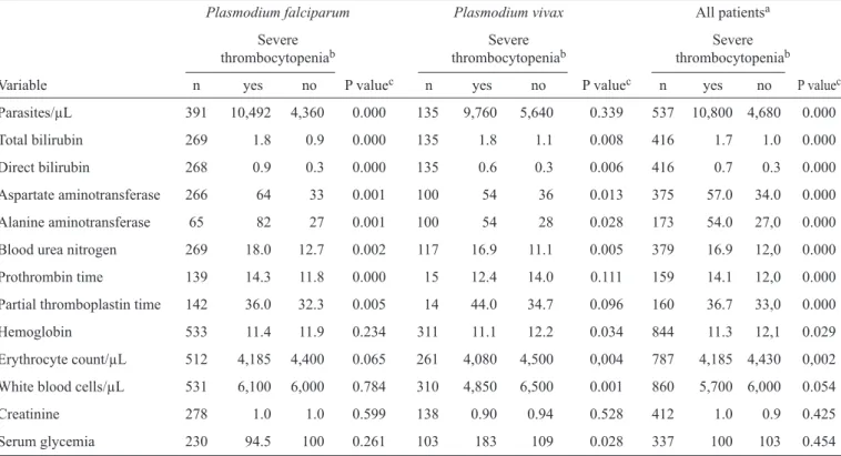
Finally, we compared the laboratory data among malaria
patients with and without severe thrombocytopenia and stratifi ed
by Plasmodium specie (Table 5). The laboratory data were
signifi cantly different across categories, with the exception of the white blood cell count, serum creatinine and serum
glycemia. In the patients infected with P. falciparum, severe thrombocytopenia was related to abnormal values on all
liver-function tests, BUN, PTi and PTT; in the patients with P. vivax,
a relationship was identifi ed between severe thrombocytopenia and abnormal values on liver-function tests, BUN, serum
glucose, erythrocyte and white cell counts. TABLE 5 - Median values of hematological laboratory tests in relation to severe thrombocytopenia.
Plasmodium falciparum Plasmodium vivax All patientsa
Severe Severe Severe thrombocytopeniab thrombocytopeniab thrombocytopeniab
Variable n yes no P valuec n yes no P valuec n yes no P valuec
Parasites/µL 391 10,492 4,360 0.000 135 9,760 5,640 0.339 537 10,800 4,680 0.000
Total bilirubin 269 1.8 0.9 0.000 135 1.8 1.1 0.008 416 1.7 1.0 0.000
Direct bilirubin 268 0.9 0.3 0.000 135 0.6 0.3 0.006 416 0.7 0.3 0.000
Aspartate aminotransferase 266 64 33 0.001 100 54 36 0.013 375 57.0 34.0 0.000 Alanine aminotransferase 65 82 27 0.001 100 54 28 0.028 173 54.0 27,0 0.000
Blood urea nitrogen 269 18.0 12.7 0.002 117 16.9 11.1 0.005 379 16.9 12,0 0.000
Prothrombin time 139 14.3 11.8 0.000 15 12.4 14.0 0.111 159 14.1 12,0 0.000
Partial thromboplastin time 142 36.0 32.3 0.005 14 44.0 34.7 0.096 160 36.7 33,0 0.000
Hemoglobin 533 11.4 11.9 0.234 311 11.1 12.2 0.034 844 11.3 12,1 0.029
Erythrocyte count/µL 512 4,185 4,400 0.065 261 4,080 4,500 0,004 787 4,185 4,430 0,002
White blood cells/µL 531 6,100 6,000 0.784 310 4,850 6,500 0.001 860 5,700 6,000 0.054
Creatinine 278 1.0 1.0 0.599 138 0.90 0.94 0.528 412 1.0 0.9 0.425
Serum glycemia 230 94.5 100 0.261 103 183 109 0.028 337 100 103 0.454
aIncluding mixed infections; b<50,000 platelets/µL; cMann-Whitney U test.
DISCUSSION
In addition to anemia, a reduction in the number of platelets is one of the more well-known hematologic changes observed
in patients with malaria. While platelet-count changes are particularly associated with P. falciparum malaria9,12,45, this
pathology has also been identified in malaria caused by
other species, such as P. vivax36,46. Changes in other platelet
parameters, such as PT, MPV or PDW, have not been well described in malaria patients.
Some studies have found that patients infected with P. vivax or P. falciparum develop anemia, monocytosis, eosinopenia and lymphopenia45. This study found a progressive reduction
in hemoglobin levels and white blood cell counts with reduced PCs, which resulted in anemia, monocytopenia, neutropenia
and lymphopenia. Thrombocytopenia presented frequently in the patients infected with P. falciparum (67%) and P. vivax
(64%). The PC did not differ by species, which suggests that
within the normal range. The PDW was signifi cantly higher in the patients infected with P. falciparum compared with
those infected with P. vivax; however, both median values
were above the normal range. These fi ndings and the results of various studies that have demonstrated a rapid recovery of PCs following the initiation of antimalarial treatment suggest
that thrombocytopenia is related to a systemic endothelial and platelet activation rather than medullary suppression. Systemic endothelial activation likely mediates platelet sequestration
and does not depend exclusively on the formation of platelet
aggregates and parasitized erythrocytes, as observed in P. falciparum infections47.
Interestingly, we found that the patients with P. vivax
infection and severe thrombocytopenia had more signifi cant
changes in leukocyte and erythrocyte counts compared with the patients without thrombocytopenia. These changes, which were
not identifi ed in the patients infected with P. falciparum, favor
a stronger infl ammatory response in severe P. vivax infections
that can affect bone marrow via the production of tumor necrosis factor alpha (TNF-α). This hypothesis is supported by a study that identifi ed increased production of TNF-α in
P. vivax malaria and a stronger host immune response in P. vivax malaria compared with P. falciparum malaria48.
Various mechanisms have been proposed to explain thrombocytopenia in malaria; however, its clinical signifi cance
remains uncertain. Thrombocytopenia can be interpreted as
evidence of damage and has been associated with clinical
complications and high parasitemia9,12,37. However, some studies
have suggested that thrombocytopenia has a protective effect, which reduces the probability of red blood cell aggregation28
or mediates parasite destruction. The latter effect has been
observed in vitro in the early stages of erythrocytic infection with P. falciparum and in a mouse model with Plasmodium chabaudi49.
In this study, we identifi ed a signifi cant relationship between
severe thrombocytopenia and other clinical complications, but we were not able to establish causality.
Thrombocytopenia has been associated with disturbances in
coagulation and has been implicated in cases of disseminated
intravascular coagulation (DIC)50. In children with severe
malaria, prolonged PTi, PTT and thrombin time (in 47.5,
35.0 and 62.5% of patients, respectively) may lead to platelet
hypoaggregation46. Activation of coagulation may also be
responsible, in part, for thrombocytopenia6. Our study showed
that the patients infected with P. falciparum, but not P. vivax, had more prolonged PTi and PTT when the thrombocytopenia
was more severe. This difference may be the result of increased
procoagulant activity in P. vivax compared with P. falciparum
infections, which develops in response to increased endothelial activity induced by TNF-α levels and the consequent formation of thrombin-antithrombin III48.
In this study, patients with severe or profound
thrombocytopenia showed higher parasitemia compared with patients without thrombocytopenia, which is in agreement with
previous fi ndings. Low PCs have been associated with increased parasite load. This association may result from sensitization
induced by the parasitized red blood cells in the platelet,
with consequent increased platelet sensitivity to adenosine diphosphate (ADP) and higher dense-granule secretion45,46.
These changes could ultimately promote platelet aggregation on the endothelium, which has been demonstrated in cerebral malaria27.
Plateletcrit is a measure of platelet mass, the clinical signifi cance of which should be interpreted in terms of the number and size of the platelets. Whitfi eld et al.51 demonstrated
that in healthy subjects, there was an inverse relationship between the platelet size (MPV) and the platelet number (PC) and proposed that the total platelet mass is closely regulated
by changes on MPV or PC. We found that the PT was low in 36% of patients with malaria. This fi nding may be explained by a combination of low PC with normal MPV (63%), low
PC and MPV (35%) and normal PC with low MPV (2%). The
reduction in circulating platelet mass is best explained by a
reduction in platelet number rather than a reduction in platelet size. It is suspected that in malaria, sequestration leads to pseudothrombocytopenia52.
Some studies have identifi ed a relationship between the decrease in PC and the appearance of clinical complications,
such as anemia, respiratory distress syndrome and cerebral malaria37. We found that the patients with severe
(25,000-49,999 platelets/µL) or profound thrombocytopenia (<25,000
platelets/µL) had more pathological changes in their clinical status compared with the patients without thrombocytopenia.
Furthermore, parasitemia, the duration of disease, and TP and TPP increased with an increasing severity of thrombocytopenia
(mildmoderatesevereprofound), whereas erythrocyte and
leukocyte counts decreased.
In this study, the patients who experienced complications had lower PC and PT values and higher levels of PDW compared with the patients who did not experience such complications. Specifi cally, the median PCs were signifi cantly lower in the patients with hepatic complications (90,500 vs. 129,000; p≤0.001), renal complications (80,000 vs. 114,000; p=0.017), neurologic complications (42,000 vs. 122,000; p≤0.001), pulmonary complications (55,000 vs. 122,000; p=0.001) and severe hypoglycemia (40,500 vs. 119,000; p=0.004). The low PC may result from the adhesion of platelets to the sites of vascular injuries and the mediation of malaria-parasitized red
blood cells (pRBC) sequestration to the endothelium through
von Willebrand-factor strings53.
When platelets decrease in number, bone marrow megakaryocytes are stimulated by thrombopoietin, and their nucleus becomes hyperlobulated with a higher deoxyribonucleic acid (DNA) content. These stimulated megakaryocytes produce larger platelets. Thus, platelets with a higher MPV are
expected to be present in autoimmune thrombocytopenia when
megakaryocytic stimulation is present. Conversely, platelets with
a lower MPV are expected in thrombocytopenic states associated
with marrow hypoplasia or aplasia54. Interestingly, 83% of the
patients studied had normal MPV, and it appears that neither of
these two mechanisms associated with thrombocytopenia was
The examination of PDW with MPV provides information
regarding thrombocyte volume disturbance. Thrombocyte
volume heterogeneity occurs because of the production factors in the bone marrow. PDW is an index of thrombocyte
volume heterogeneity similar to erythrocyte distribution, and
both MPV and PDW are markers of platelet immaturity and
platelet activation55. A high PDW predominated in the patients
studied (65%), and we identifi ed signifi cantly higher PDW in the total sample of severe patients; however, the analysis for each complication indicated confl icting results, which could be a result of the differential patterns of infl ammation in the spectrum of clinical complications or statistical effects based on the low number of cases when the complications were
individually analyzed.
In healthy human subjects, no correlation was identifi ed
between PCT, MPV or PDW values and platelet aggregation results54; however, these relationships could be modifi ed in the
presence of infl ammation.
It has been reported that PDW increases over storage
time because of the formation of abnormally small and large
platelets54, and it is well known that pseudo-thrombocytopenia
occurs as a result of exposure to EDTA when used in blood
sampling56. There is a need for multi-center and prospective
studies with large sample sizes to elucidate the utility and clinical
importance of these parameters in malaria.
Our results indicate that thrombocytopenia accompanies
malaria complications, especially liver dysfunction.
Major changes in total bilirubin, direct bilirubin, aspartate
aminotransferase, alanine aminotransferase, prothrombin
time and partial thromboplastin time were observed in severe
thrombocytopenia (<50,000 platelets/µL). The PCs were significantly lower in the patients who experienced liver,
renal, neurologic, and pulmonary complications and severe hypoglycemia.
The liver is a key regulator of platelet production through the synthesis of TPO. Thus, hepatic dysfunction may explain, in part, the thrombocytopenia observed in these patients;
however, more data are required because TPO appears to rise
during infection by P. falciparum, even when liver function is compromised23.
Differences between P. falciparum and P. vivax infections
may arise from the different mechanisms by which the physiopathology of thrombocytopenia is mediated, i.e., via
clumping in P. falciparum and via medullar suppression in P. vivax. A better understanding of these mechanisms is still needed.
Despite the inherent limitations of retrospective analyses, these results may contribute to the understanding of severe
thrombocytopenia as a negative prognostic marker in patients
with malaria. Our fi ndings suggest a need to study hepatic function and to establish close clinical monitoring of malaria patients. Furthermore, there is a substantial need for prospective studies to elucidate the utility and clinical importance of these
parameters in malaria.
ACKNOWLEDGMENTS
REFERENCES
The authors declare that there is no confl ict of interest.
CONFLICT OF INTEREST
Our sincere thanks to Grupo Malaria, Universidad de Antioquia, who provided partial data analyzed in this study, and to Felipe Miranda, M.D., Ana del Mar Cortina, M.D., Ana Maria Cadavid, Lia Palacio and Shirley Jolianiz, who are
medical students at the School of Medicine at the University of Antioquia, for their help in acquiring hospital medical records. This project was funded by the Universidad de Antioquia,
Colombia (Grupo Malaria de la Facultad de Medicina, Programa Jóvenes Investigadores de Vicerrectoría de Investigación y CODI- Estrategia de Sostenibilidad 2014-2015).
1. World Health Organization. The Roll Back Malaria partnership. Geneva: WHO; 2008.
2. World Health Organization. World malaria report 2012. Geneva:WHO; 2012.
3. Price RN, Douglas NM, Anstey NM. New developments in Plasmodium vivax malaria: severe disease and the rise of chloroquine resistance.
Curr Opin Infect Dis 2009; 22:430-435.
4. Picot S, Bienvenu AL. Plasmodium vivax infection: not so benign.
Med Sci (Paris) 2009; 25:622-626.
5. World Health Organization. Severe falciparum malaria. World Health Organization, Communicable Diseases Cluster. Trans R Soc Trop Med Hyg 2000; 94 (suppl I):1-90.
6. Lacerda MV, Mourão MP, Coelho HC, Santos JB. Thrombocytopenia in malaria: who cares? Mem Inst Oswaldo Cruz 2011; 106:52-63.
7. Quintero JP, Siqueira AM, Tobón A, Blair S, Moreno A, Arévalo-Herrera M, et al. Malaria-related anaemia: a Latin American perspective. Mem Inst Oswaldo Cruz 2011; 106:91-104.
8. Daneshvar C, Davis T, Cox-Singh J, Rafa’ee MZ, Zakaria SK, Divis PCS, et al. Clinical and laboratory features of human Plasmodium knowlesi
infection. Clin Infect Dis 2009; 49:852-860.
9. Ladhani S, Lowe B, Cole AO, Kowuondo K, Newton CR. Changes in white blood cells and platelets in children with falciparum malaria: relationship to disease outcome. Br J Haematol 2002; 119:839-847. 10. Horstmann R, Dietrich M. Haemostatic alterations in malaria correlate to
parasitaemia. Ann Hematol 1985; 51:329-335.
11. Hong YJ, Yang SY, Lee K, Kim TS, Kim HB, Park KU, et al. A case of imported Plasmodium malariae malaria. Ann Lab Med 2012; 32:229-233.
12. Richards MW, Behrens RH, Doherty JF. Short report: hematologic changes in acute, imported Plasmodium falciparum malaria. Am J Trop Med Hyg 1998; 59:859.
13. Kochar DK, Das A, Kochar A, Middha S, Acharya J, Tanwar GS, et al. Thrombocytopenia in Plasmodium falciparum, Plasmodium vivax and mixed infection malaria: a study from Bikaner (Northwestern India). Platelets 2010; 21:623-627.
14. Touze JE, Mercier P, Rogier C, Hovette P, Schmoor P, Dabanian C, et al. Platelet antibody activity in malaria thrombocytopenia. Pathol Biol (Paris) 1990; 38:678-681.
16. Echeverri M, Tobon A, Alvarez G, Carmona J, Blair S. Clinical and laboratory fi ndings of Plasmodium vivax malaria in Colombia, 2001. Rev Inst Med Trop Sao Paulo 2003; 45:29-34.
17. Tobon A. Danger signs in the malaria patient. Biomedica 2009; 29: 320-329.
18. Ohtaka M, Ohyashiki K, Iwabuchi H, Iwabuchi A, Lin KY, Toyama K. A case of vivax malaria with thrombocytopenia suggesting immunological mechanisms. Rinsho Ketsueki 1993; 34:490-492.
19. Kelton JG, Keystone J, Moore J, Denomme G, Tozman E, Glynn M, et al. Immune-mediated thrombocytopenia of malaria. J Clin Invest 1983; 71:832-836.
20. Rios A, Alvarez T, Carmona J, Blair S. Temporal evolution of platelets and anti-platelet antibodies in patients of endemic area with non complicated malaria. An Med Interna 2005; 22:561-568.
21. Abdalla SH. Hematopoiesis in human malaria. Blood Cells 1990; 16: 401-416.
22. Beale PJ, Cormack JD, Oldrey TB. Thrombocytopenia in malaria with immunoglobulin (IgM) changes. Br Med J 1972; 1:345-349.
23. Kreil A, Wenisch C, Brittenham G, Looareesuwan S, Peck-Radosavljevic M. Thrombopoietin in Plasmodium falciparum malaria. Br J Haematol 2000;
109:534-536.
24. Skudowitz RB, Katz J, Lurie A, Levin J, Metz J. Mechanisms of thrombocytopenia in malignant tertian malaria. Br Med J 1973; 2:515-518. 25. Watier H, Verwaerde C, Landau I, Werner E, Fontaine J, Capron A, et al.
T-cell-dependent immunity and thrombocytopenia in rats infected with
Plasmodium chabaudi. Infect Immun 1992; 60:136-142.
26. Gall C, Spuler A, Fraunberger P. Subarachnoid hemorrhage in a patient with cerebral malaria. N Engl J Med 1999; 341:611-613.
27. Grau GE, Mackenzie CD, Carr RA, Redard M, Pizzolato G, Allasia C, et al. Platelet accumulation in brain microvessels in fatal pediatric cerebral malaria. J Infect Dis 2003; 187:461-466.
28. Wassmer SC, Taylor T, Maclennan CA, Kanjala M, Mukaka M. Platelet-induced clumping of Plasmodium falciparum-infected erythrocytes from
Malawian patients with cerebral malaria-possible modulation in vivo by thrombocytopenia. J Infect Dis 2008; 197:72-78.
29. de Mast Q, Groot E, Lenting PJ, de Groot PG, McCall M, Sauerwein RW, et al. Thrombocytopenia and release of active von Willebrand factor during early Plasmodium falciparum malaria. J Infect Dis 2007; 196:622-628.
30. Renard N, Nooijen PT, Schalkwijk L, De Waal RM, Eggermont AM, Lienard D, et al. VWF release and platelet aggregation in human melanoma after perfusion with TNF alpha. J Pathol 1995; 176:279-287. 31. Tacchini-Cottier F, Vesin C, Redard M, Buurman W, Piguet PF. Role
of TNFR1 and TNFR2 in TNF-induced platelet consumption in mice. J Immunol 1998; 160:6182-6186.
32. Taylor WR, Widjaja H, Basri H, Ohrt C, Taufi k T, Tjitra E, et al. Changes in the total leukocyte and platelet counts in Papuan and non Papuan adults from northeast Papua infected with acute Plasmodium vivax or uncomplicated Plasmodium falciparum malaria. Malar J 2008; 7:259.
33. World Health Organization. Management of severe malaria - A practical handbook. 3rd edition. Geneva: 2013.
34. Eriksson B, Hellgren U, Rombo L. Changes in erythrocyte sedimentation rate, C-reactive protein and hematological parameters in patients with acute malaria. Scand J Infect Dis 1989; 21:434-441.
35. Moulin F, Lesage F, Legros A-H, Maroga C, Moussavou A, Guyon P, et al. Thrombocytopenia and Plasmodium falciparum malaria in children with different exposures. Arch Dis Child 2003; 88:540-541.
36. Kumar A, Shashirekha. Thrombocytopenia--an indicator of acute vivax malaria. Indian J Pathol Microbiol 2006; 49:505-508.
37. Gerardin P, Rogier C, Ka AS, Jouvencel P, Brousse V, Imbert P. Prognostic value of thrombocytopenia in African children with falciparum malaria. Am J Trop Med Hyg 2002; 66:686-691.
38. Tan SO, McGready R, Zwang J, Pimanpanarak M, Sriprawat K, Thwai KL, et al. Thrombocytopaenia in pregnant women with malaria on the Thai-Burmese border. Malar J England 2008; 7:209.
39. Erhabor O, Jeremiah Z, Adias T. Thrombocytopenia in plasmodium parasitized pregnant women in the Niger Delta of Nigeria. Pathology 2010; 2:1-5.
40. Blair S, Álvarez G, Campuzano G. Relación entre anemia y malaria en una población rural de Colombia. Bol Dir Malariol Saneam Amb (Venezuela) 1997; 37:7-13.
41. Tobón AC, Giraldo S, Pineros JG, Arboleda N, Blair S, Carmona-Fonseca J. The epidemiology of complicated falciparum malaria: case and controls study in Tumaco and Turbo, Colombia, 2003. Rev Bras Epidemiol 2006; 9:283-296.
42. Tobón A, Giraldo C, Blair S. Utilidad de los signos clínicos y parasitológicos en el pronóstico de la malaria grave en Colombia. Biomedica. 2012; 32:79-94.
43. World Health Organization. New perspectives: malaria diagnosis. Geneva: 2000.
44. Campuzano G. Del hemograma manual al hemograma de cuarta generación. Medicina & Laboratorio. 2007; 13:511-550.
45. Wickramasinghe S, Abdalla S. Blood and bone marrow changes in malaria. Best Pract Res ClinHaematol 2000; 13:277-299.
46. Prasad R, Das BK, Pengoria R, Mishra OP, Shukla J, Singh TB. Coagulation status and platelet functions in children with severe falciparum malaria and their correlation of outcome. J Trop Pediatr England; 2009; 55:374-378.
47. Chotivanich K, Sritabal J, Udomsangpetch R, et al. Platelet-induced autoagglutination of Plasmodium falciparum-infected red blood cells
and disease severity in Thailand. J Infect Dis 2004; 189:1052-1055. 48. Hemmer CJ, Holst FG, Kern P, Chiwakata CB, Dietrich M, Reisinger EC.
Stronger host response per parasitized erythrocyte in Plasmodium vivax
or ovale than in Plasmodium falciparum malaria. Trop Med Int Health
2006; 11:817-823.
49. McMorran BJ, Marshall VM, de Graaf C, Drysdale KE, Shabbar M, Smyth GK, et al. Platelets kill intraerythrocytic malarial parasites and mediate survival to infection. Science 2009; 323:797-800.
50. Dennis LH, Eichelberger JW, Inman MM, Conrad ME. Depletion of coagulation factors in drug-resistant Plasmodium falciparum malaria. Blood 1967; 29:713-721.
51. Whitfi eld J, Martin N, Rao D. Genetic and environmental infl uences on the size and number of cells in the blood. Genetic epidemiology 1985; 2:133-144.
52. Pain A, Ferguson DJ, Kai O, Urban BC, Lowe B, Marsh K, et al. Platelet-mediated clumping of Plasmodium falciparum-infected erythrocytes is
a common adhesive phenotype and is associated with severe malaria. Proc Natl Acad Sci USA 2001; 98:1805-1810.
53. Bridges DJ, Bunn J, van Mourik JA, Georges Grau G, Roger JS. Preston RJS, et al. Rapid activation of endothelial cells enables
Plasmodium falciparum adhesion to platelet-decorated von Willebrand factor strings. Blood 2010; 115:1472-1474.
54. Beyan C, Kaptan K, Ifran A. Platelet count, mean platelet volume, platelet distribution width, and plateletcrit do not correlate with optical platelet aggegation responses in healthy volunteers. J Thromb Thrombolysis 2006; 22:161-164.
55. Aydogan A, Akkucuk S, Arica S, Motor S, Karakus A, Ozkan OV, et al. The analysis of mean platelet volume and platelet distribution width levels in appendicitis. Indian J Surg 2013:1-6. DOI:10.1007/s12262-013-0891-7.