R E V B R A S R E U M A T O L . 2 0 1 4 ;5 4 ( 4 ): 3 1 8 – 3 2 1
www.reumatologia.com.br
REVISTA BRASILEIRA DE
REUMATOLOGIA
Case report
Rhabdomyolysis associated with dengue fever in a
lupic patient
Louise D. Verdolin*, Alice R. Borner, Henrique Mussi, Ronaldo A. Gismondi, Bruno Schau,
Ricardo C. Ramos
Department of Internal Medicine, Hospital Universitário Antônio Pedro, Universidade Federal Fluminense, Niterói, RJ, Brazil
a r t i c l e i n f o
Article history:
Received on 3 April 2012 Accepted on 19 February 2013
Keywords: Rhabdomyolysis Dengue fever
Systemic lupus erythematous
a b s t r a c t
This report describes the case of a woman with systemic lupus erythematous (SLE) that developed rhabdomyolysis after being infected by dengue virus. There are only a few cases of SLE accompanied by rhabdomyolysis, none of them associated with dengue fever. Initially, the woman presented high fever, myalgia, muscular weakness, mild headache, polyarthralgia and thrombocytopenia reminding a lupus lare, but since the number of people infected by dengue at that time was high and the symptoms from both conditions are similar, a dengue serology was requested. After a few days, the patient developed rhabdomyolysis. She was then submitted to immunosuppressive drugs, urinary alkalization and vigorous hydration, which improved her muscle damage and inlammatory condition. The positive dengue serology was only available after the therapy above had been established. She was discharged in an asymptomatic state.
This case demonstrates how alike dengue fever and a lupus lare are, warning clinicians that, especially during an epidemic, both diseases should be carefully differentiated in order to establish a correct and eficient therapy.
© 2014 Sociedade Brasileira de Reumatologia. Published by Elsevier Editora Ltda. All rights reserved.
* Corresponding author.
E-mail: [email protected] (L.D. Verdolin).
0482-5004/$ - see front matter. © 2014 Sociedade Brasileira de Reumatologia. Published by Elsevier Editora Ltda. All rights reserved. http://dx.doi.org/10.1016/j.rbr.2013.02.003
A rabdomiólise está associada à febre dengue em um paciente lúpico
Palavras-chave: Rabdomiólise Febre dengue
Lúpus eritematoso sistêmico
r e s u m o
Esse relato descreve o caso de uma mulher com lúpus eritematoso sistêmico (LES) que sofreu rabdomiólise em seguida à sua infecção pelo vírus da dengue. Foram relatados ape-nas alguns casos de LES com manifestação de rabdomiólise, nenhum deles associados à febre dengue.
319
R E V B R A S R E U M A T O L . 2 0 1 4 ;5 4 ( 4 ): 3 1 8 – 3 2 1
medicamentos imunossupressivos, alcalinização urinária e hidratação vigorosa, medidas que melhoraram seus danos musculares e a condição inlamatória. A sorologia positiva para dengue nos foi disponibilizada apenas depois da instauração do tratamento descrito acima. A paciente recebeu alta em estado assintomático.
Esse caso demonstra a grande semelhança entre a febre dengue e uma exacerbação lúpi-ca; isso deve alertar o clínico para que, especialmente durante uma epidemia, faça uma cuidadosa diferenciação entre essas doenças, de forma a estabelecer uma terapia correta e eiciente.
© 2014 Sociedade Brasileira de Reumatologia. Publicado por Elsevier Editora Ltda. Todos os direitos reservados.
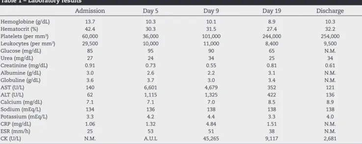
Table 1 – Laboratory results
Admission Day 5 Day 9 Day 19 Discharge
Hemoglobine (g/dL) 13.7 10.3 10.1 8.9 10.3
Hematocrit (%) 42.4 30.3 31.5 27.4 32.2
Platelets (per mm3) 60,000 36,000 101,000 244,000 254,000
Leukocytes (per mm3) 29,500 10,000 11,000 8,400 9,500
Glucose (mg/dL) 85 95 90 65 N.M.
Urea (mg/dL) 27 24 34 25 34
Creatinine (mg/dL) 0.91 0.73 0.55 0.81 0.61
Albumine (g/dL) 3.0 2.6 2.2 3.1 N.M.
Globuline (g/dL) 3.6 3.7 3.0 3.4 N.M.
AST (U/L) 140 6,601 4,679 352 121
ALT (U/L) 62 1,115 1,325 422 136
Calcium (mg/dL) 7.1 7.1 7.0 8.5 8.9
Sodium (mEq/L) 134 136 138 138 138
Potassium (mEq/L) 3.3 4.2 4.4 3.3 4.0
CRP (mg/dL) 1.06 1.32 4.84 1.51 N.M.
ESR (mm/h) 25 53 51 38 N.M.
CK (U/L) N.M. A.U.L 45,265 9,117 2,681
AST, aspartate aminotransferase; ALT, alanine aminotransferase; CRP, C-reactive protein; ESR, erythrocyte sedimentation rate; CK, creatine kinase; N.M., not measured; A.U.L., above upper limit.
Introduction
From the irst semester of 2011 to the irst week of October 2011, there were 721,546 reported cases of dengue in Brazil.1
Dengue fever is an arbovirus that typically manifests high fever, severe myalgia, arthralgia, headache, retro-orbital pain, and rash. Rare clinical manifestations include hepati-tis, rhabdomyolysis and neurological presentations, such as encephalopathy, peripheral neuropathy, and Guillain-Barré syndrome.2 Rhabdomyolysis is a disorder characterized by
skeletal muscle injury, associated with the extravasation of intracellular constituents into the plasma, resulting in elec-trolyte disorders and even acute kidney injury.3
Symptoms such as fever, thrombocytopenia, or arthral-gia are found in many diseases, including SLE, which is an inlammatory autoimmune disorder characterized by peri-ods of remissions and relapses. Epidemiological studies of incidence and prevalence of SLE are scarce in Brazil, where high racial miscegenation and different weather condi-tions can inluence the disease’s complicacondi-tions. In Brazil, lupus is more common in the black population than any other. Within the entire Brazilian population, lupus affects women of reproductive age more than men, with a ratio of 9-10:1.4
A new infection by dengue virus in patients with previ-ous diagnosis of lupus may mimic its reactivation.5 There
have been a few reports illustrated in the literature of rhab-domyolysis in lupic patients and in other patients infected by dengue fever,2,6 but there are no cases combining all of these
conditions: lupus, dengue and rhabdomyolysis together. Our objective is to describe the case of a patient previously diag-nosed with lupus that manifested rhabdomyolysis secondary to dengue fever.
Case report
In 2010, a thirty-nine year-old black woman developed malar rash, photosensitivity, oral ulcers and polyarthritis; she was diagnosed with SLE. At that time, her antinuclear antibody was 1/160 with a ibrillar cytoplasmic pattern, and she was treated with prednisone, azathioprine and hydroxicloroquine. Her symptoms resolved. However, in the irst semester of 2011, she was admitted to the emergency room of a university hos-pital with high fever (39 degrees Celsius), chills, myalgia, mus-cular weakness, mild headache and polyarthralgia (shoul-ders, elbow, wrists, hips and knees). Her admission exams (Table 1) showed thrombocytopenia (60,000 per mm3), high
320
R E V B R A S R E U M A T O L . 2 0 1 4 ;5 4 ( 4 ): 3 1 8 – 3 2 1hypoalbuminemia, erythrocyte sedimentation rate (ESR) dis-cretely elevated, high liver enzymes [aspartate transaminase (AST) higher than alanine transaminase (ALT)]. Complement levels were in the inferior normal range. Antibodies to Deoxy-ribonucleic Acid (anti-DNA), anti-SSB/La, anti-Smith and an-tibodies to nuclear ribonucleoproteins (anti-RNP) were in the normal range. Urine analysis revealed proteinuria (30mg/dL). Dengue serology was requested upon admission, due to an epidemiologic suspicion: it was summer time in Rio de Janei-ro, and there were a lot of patients affected by this arbovirus. She was medicated with symptomatic medications.
Five days after admission her symptoms worsened, and she manifested muscular weakness, red urine (Urine analy-sis: pH 8,5, density 1,015, false positive for hemoglobinuria (+++) without erythrocytes, no evidence of erythrocyte di-morphism; 24-hour urine collection revealed 4,681.46mg of proteinuria) and increased edema, more pronounced in the arms. Creatine kinase (CK) was much higher than the upper limit detection rate of our test (dimension RXLMAX clinical chemistry system from Siemens, USA). The muscle injury was so elevated that four days passed before this enzyme could be detected, at which time it had reached a level of 45,265 (U/L). She also presented high AST (6,601U/L) and ALT (1,115U/L), hypocalcemia (7.1mg/dL), and metabolic acidosis, but electro-lytes and renal function were still normal. Rhabdomyolysis was diagnosed and urinary alkalization plus vigorous hydra-tion were started.
After 48 hours, the patient evolved with clinical and labo-ratory deterioration. She manifested generalized edema, now much worse on her lower limbs, making it dificult to palpate the leg’s arterial pulses. Compartmental syndrome was sus-pected, and it was decided to submit her to pulse therapy with methylprednisolone (1g per day for three days). At this point, lupus lare was the main hypothesis for rhabdomy-olysis. Only after administration of the pulse therapy was the dengue serology result available. IgM antibody was 20.38 Panbio (ELISA). Levels above 11 Panbio suggest recent dengue infection. Hepatitis virus and HIV serology were all negative. Urinary alkalization and vigorous hydration were maintained and symptoms improved. After 34 days in hospital, the patient was discharged and sent home with a supply of chloroquine, prednisone and azathioprine. At this moment, her laboratory exams showed improvement: liver enzymes were dropping (AST: 121U/L, ALT 136U/L), and her CK levels (2,681U/L) de-creased 94.07% from the highest measurable value. She had no clinical symptoms.
Discussion
Several conditions may lead to rhabdomyolysis, such as trau-ma, drugs, genetic disorders, endocrine, metabolic and infec-tious diseases.3 This condition is rarely described in
rheuma-tologic diseases. Six cases of rhabdomyolysis in lupic patients have been reported in the literature: two of them had discoid lupus, and four had SLE. The precipitating factors responsible for the induction of rhabdomyolysis in three of these patients were: the use of myotoxic drugs in one case and bacterial in-fection in two cases. In the remaining cases, no precipitating factors were identiied.6
When the patient arrived at the hospital, the irst hypoth-esis was lupus reactivation, since her main complaint was ar-thralgia and her laboratory exams showed thrombocytopenia, with high CRP. After the positive serology for dengue was re-ceived, it was clearer the infection was the precipitating factor for the rhabdomyolysis. The clinical manifestations of dengue and lupus lare may be similar, and this may confound physi-cians. However, in a review of Medline’s database, only a few reports of dengue mimicking a lupus lare were found.5
It is important that, in countries where dengue is a com-mon disease, clinicians carefully distinguish a lupus lare from a dengue infection. In this case, the epidemiology of dengue (summer time and the high number of infection cases at the time) was the leading clue to request the serology.
Evidence suggests that bacterial and viral infection could become a trigger for a new or relapsing lupus lare in geneti-cally predetermined individuals. This has been proven, for example, in Cytomegalovirus infection. Studies show that Ep-stein-Barr virus infection presents immunologic aberrations of B cells and apoptosis and molecular mimicry that perpetu-ate autoimmunity in SLE.7 There are no studies proving a
re-lationship between dengue and SLE, but recently there has been a report which considers the possibility of dengue virus triggering a dysfunctional immune response resulting in the development of autoimmunity and SLE with lupus nephritis.8
In the literature, there are no reports of rhabdomyolysis as-sociated with dengue fever in a lupic patient.
Viruses that commonly cause rhabdomyolysis are inluen-za A and B, HIV, Coxsackie and Epstein-Barr.3 Dengue shares
several features with these viruses, and there have been few cases of dengue with rhabdomyolysis.9 There is no proved
cause for this association, but some hypotheses have been proposed. Direct viral invasion of the muscle had not been demonstrated, but some muscle biopsies showed marked in-lammation, varying from mild lymphocytic iniltrate to foci of severe myonecrosis. Myotoxic cytokines, especially TNF released in response to virus infection, may have been the cause of the muscle injury.10-12
In the case reports describing rhabdomyolysis due to dengue, patients were treated with urinary alkalization and vigorous hydration, with favorable outcome in one case and death in the other.9 The patient described in our case was
in-fected with a viral disease, but initially she was treated with pulse therapy with methylprednisolone because her clinical deterioration was attributed to lupus exacerbation. The den-gue serology was only released after this treatment was es-tablished. Her clinical outcome was favorable.
No speciic treatment for dengue exists, apart from analge-sia and medications to reduce fever.2 In this case,
methylpred-nisolone was used because of the suspicion that the patient was clinically deteriorating due to a lupus lare. Some authors argue there is no evidence in vivo to support the use of an-tiviral agents, drugs that reduce vascular permeability, nor corticosteroid, and that high-dose methylprednisolone fails to reduce mortality in severe dengue shock syndrome (DSS).13
Others suggest that a single dose of intravenous methyl pred-nisolone, 1g intravenous over 20 min as rescue medication to highly selected patients who develop hypotensive DSS, reduc-es mortality (p=0.01).14 Nevertheless, the author of this article
321
R E V B R A S R E U M A T O L . 2 0 1 4 ;5 4 ( 4 ): 3 1 8 – 3 2 1
conirm this fact. Regarding the rhabdomyolysis treatment, there is only one case reporting that steroid therapy should be considered for the treatment of rhabdomyolysis or myopathy associated with Cytomegalovirus infection in order to prevent renal failure or fatal progression of the disease.15 Therefore it
is impossible to know if the steroids used in the patient had any inluence in her outcome, since there are no clinical trials available addressing this speciic case.
Conclusion
There are few cases of SLE manifesting rhabdomyolysis, none of them previously associated with dengue fever as described in our case report. Although rhabdomyolysis is an uncommon presentation of this viral infection, dengue fever clinical mani-festations may mimic lupus lare. Rheumatologists, clinicians and infectologists should be aware of common manifestations of these diseases in order to identify more severe cases.
Acknowledgments
Michele Veldhoen.
Conlicts of interest
The authors declare no conlict of interest.
R E F E R E N C E S
1. Coordenação Geral do Programa Nacional de Controle da Dengue. Balanço Dengue: Semana Epidemiológica 1 a 39 de 2011 [Internet]. 2011 [accessed in 11 November 2011]. Available at: http://portal.saude.gov.br/portal/arquivos/pdf/ informe_dengue_2011_37_39.pdf.
2. Karakus A, Banga N, Voorn GP, Meinders AJ. Dengue shock syndrome and rhabdomyolysis. Neth J Med. 2007;65:78-81. 3. Bosch X, Poch E, Grau JM. Rhabdomyolysis and acute kidney
injury. New Engl J Med. 2009;361:62-72.
4. Nakashima CAK, Galhardo AP, Silva JFPM, Fiorenzano GR, Santos ABS, Leite MFS, Nogueira MA et al. Incidence and clinical-laboratory aspects of systemic lupus erythematosus in a southern Brazilian city. Rev Bras Reumatol. 2011;51:231-39.
5. Souza SP, Moura CGG. Dengue mimicking a lupus lare. J Clin Rheumatol. 2010;16:47-78.
6. Carvalho JF, Mota LM, Bonfa E. Fatal rhabdomyolysis in systemic lupus erythematosus. Rheumatol Int. 2010;31:1243-5.
7. Zandman-Goddard G, Shoenfeld Y. Infections and SLE. Autoimmunity. 2005;38:473-85.
8. Rajadhyaksha A, Mehra S. Dengue fever evolving into systemic lupus erythematosus and lupus nephritis: a case report. Lupus [Internet]. 2012 [accessed in 8 March 2012]; 0:1-4. Available at: http://lup.sagepub.com/content/ early/2012/02/21/0961203312437807.
9. Davis SJ, Bourke P. Rhabdomyolysis associated with dengue virus infection. Clin Infect Dis. 2004;38:109-11.
10. Halstead SB. Dengue. Curr Opin Infect Dis. 2002;15:471-6. 11. Konrad RJ, Goodman DB, Davis WL. Tumor necrosis factor and
coxsackie B4 rhabdomyolysis. Ann Intern Med. 1993;119:861. 12. Gagnon S, Mori M, Kurane I. Cytokine gene expression and
protein production in peripheral blood mononuclear cells of children with acute dengue virus infections. J Med Virol. 2002;67:41-6.
13. Rigau-Perez JG, Clark GC, Gubler DJ, Reiter P, Sanders EJ, Vorndam AV. Dengue and dengue haemorrhagic fever. Lancet. 1998;352:971-7.
14. Premaratna R, Jayasinghe KG, Liyanaarachchi EW, Weerasinghe OM, Pathmeswaran A, Silva HJ. Effect of a single dose of methyl prednisolone as rescue medication for patients who develop hypotensive dengue shock syndrome during the febrile phase: a retrospective observational study. Int J Infect Dis. 2011;15:433-4.
15. Sato K, Yoneda M, Hayashi K, Nakagawa H, Higuchi I, Kuriyama M. A steroid-responsive case of severe