Letters to the Editor
Radiol Bras. 2017 Jan/Fev;50(1):62–68
64
http://dx.doi.org/10.1590/0100-3984.2015.0159 The symptoms of SMD vary depending on the age of the
pa-tients(1), the principal symptoms being as follows: limited postna-tal growth; rhizomelic shortening of the limbs in early childhood evolving to shortening of the trunk by the age of 10 years; tho-racic hypoplasia, which causes respiratory problems in the neona-tal period and increases susceptibility to respiratory tract infec-tion(4); scoliosis with dorsal kyphosis; abnormalities of the meta-physes and pelvis(5); odontoid hypoplasia; and valgus of the knees and claudication(6), the latter typically being the first sign of the disease(2). There might be little or no ossification of the cervical vertebrae, leading to cervical instability and swan neck deformity(7). A review of the literature revealed that there are currently 10 recognized subtypes of SMD. However, there in no consensus in the medical literature regarding those subtypes, because they are based characteristics that are minimally different. Some subtypes are based on reports of only one case, and others can be diagnosed only after years of follow-up, which is difficult. For example, the longest follow-up period in a report of Sedaghatian-type SMD was 161 days. Therefore, there is no acceptable standard for subclas-sifying the disease.
REFERENCES
1. Nural MS, Diren HB, Sakarya O, et al. Kozlowski type spondylometaphyseal
Márcio Luís Duarte1, Élcio Roberto Duarte2, Daniela Brasil Solorzano2, Edgar Brasil Solorzano2, Jael Brasil de Alcântara Ferreira2
1. WebImagem, São Paulo, SP, Brazil. 2. Brasil Imagem Medicina Diagnóstica, Santos, SP, Brazil. Mailing Address: Dr. Márcio Luís Duarte. Avenida Ana Costa, 259, Gonzaga. Santos, SP, Brazil, 11060-001. E-mail: [email protected].
dysplasia: a case report with literature review. Diagn Interv Radiol. 2006;12:70–3.
2. Jones KL. Smith’s Recognizable patterns of human malformation. 6th ed. Philadelphia: Elsevier Saunders; 2006.
3. Nemec SF, Cohn DH, Krakow D, et al. The importance of conventional radiography in the mutational analysis of skeletal dysplasias (the TRPV4 mutational family). Pediatr Radiol. 2012;42:15–23.
4. Suzuki S, Kim OH, Makita Y, et al. Axial spondylometaphyseal dysplasia: additional reports. Am J Med Genet A. 2011;155A:2521–8.
5. Krakow D, Vriens J, Camacho N, et al. Mutations in the gene encoding the calcium-permeable ion channel TRPV4 produce spondylometaphyseal dysplasia, Kozlowski type and metatropic dysplasia. Am J Hum Genet. 2009;84:307–15.
6. Ferrari D, Sudanese A. Spondylometaphyseal dysplasia: description of a case. Chir Organi Mov. 1994;79:325–9.
7. Simon M, Campos-Xavier AB, Mittaz-Crettol L, et al. Severe neurologic manifestations from cervical spine instability in spondylo-megaepiphyseal-metaphyseal dysplasia. Am J Med Genet C Semin Med Genet. 2012;160C: 230–7.
Multiple primary malignancies: synchronous urothelial carcinoma of the bladder and adenocarcinoma of the colon
Dear Editor,
A 75-year-old White male presented with a three-month his-tory of pain in the left hypochondrium. The patient also reported experiencing an episode of gross hematuria six months prior. He had quit smoking 20 years prior, having previously smoked 30 ciga-rettes/day for 30 years. He had also undergone surgery for a gas-tric ulcer 20 years prior. He reported no other comorbidities.
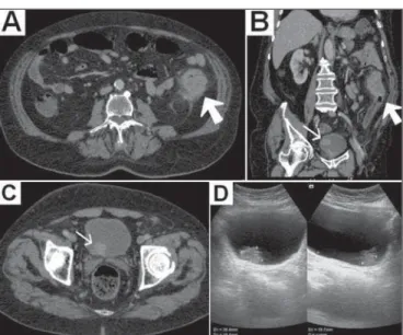
Computed tomography of the abdomen showed a solid, ir-regular, concentric mass, which was expansive and stenotic, in the middle third of the descending colon (Figures 1A and 1B). The mass showed heterogeneous uptake of the intravenous iodi-nated contrast medium and increased density of adjacent fat tis-sue, suggesting that it had expanded through the serosa. In addi-tion, a vegetative lesion, with irregular borders and showing con-trast enhancement, was observed in the right posterolateral wall of the bladder (Figures 1B and 1C).
Colonoscopy with biopsy of the intestinal mass led to a histo-logical diagnosis of moderately differentiated adenocarcinoma of the colon, and the patient was therefore submitted to segmental colectomy with colostomy. The anatomopathological study re-vealed a hard, annular tumor, which was ulcerative and vegeta-tive, infiltrating the intestinal wall and surrounding fat, thus con-firming the result of the microscopy study of the biopsy. Subse-quently, ultrasound of the urinary tract confirmed bladder nodula-tion (Figure 1D), with no perceptible flow on color Doppler. Com-plete transurethral resection of the nodulation was performed, and histopathological analysis of the resected specimen led to a diagnosis of superficial low-grade papillary urothelial carcinoma (World Health Organization grade I). A subsequent computed tomography scan of the abdomen and pelvis, for staging, showed no suspicious lesions. The final diagnosis was multiple, synchro-nous primary malignancies, probably secondary to smoking.
Colon cancer is the fourth most common malignancy in men, accounting for 90% of the cases that occur after the fifth decade of life, adenocarcinoma being the most common type(1).
Figure 1. A,B: Computed tomography scan of the abdomen, obtained in the portal phase after intravenous administration of contrast medium, in an axial view (A) and oblique coronal reconstruction (B), showing a solid, irregular, concentric mass, which was expansive and stenotic, in the descending colon, presenting heterogeneous enhancement, together with increased density of the adjacent fat tissue (large arrow). Note also the vegetative lesion, with irregular borders and showing contrast enhancement (small arrow in B). C: Axial computed tomography slice, obtained in the portal phase after intravenous administration of iodinated contrast medium, showing the vegetative lesion, with irregular borders, located in the right posterolateral wall of the bladder (arrow). D: Abdominal ultrasound, con-firming the lesion in the bladder wall.
In 5–10% of cases, adenocarcinoma is associated with he-reditary syndromes (e.g., familial adenomatous polyposis, heredi-tary non-polypoid colorectal cancer, etc.), especially in young adults(1). It is related to obesity, a sedentary lifestyle, a diet low in fiber, and inflammatory bowel diseases(1–4). Smoking and alco-holism can also play roles(2–4).
Letters to the Editor
Radiol Bras. 2017 Jan/Fev;50(1):62–68
65
http://dx.doi.org/10.1590/0100-3984.2015.0114 of age, 75–80% of whom are men, urothelial carcinoma being
the predominant form(5,6). Urothelial carcinoma can be multifo-cal/multicentric, can occur in the upper or lower urinary tract, and is often recurrent(5). Smoking is implicated in 50–65% of all cases in men and in 20–30% of all cases in women(4). Other, less com-mon causes include chemotherapy, exposure to aromatic or het-erocyclic amines, radiotherapy, and chronic infection(2,4–6).
Multiple primary malignancies are defined as those that are confirmed, independent, and of non-metastatic origin(7). They are classified as synchronous if they are identified within the first six months after the appearance of the first lesion or as metachronous if they are identified thereafter(7).
The overall prevalence of multiple primary malignancies is 0.7–11.7%, increasing proportionally with patient age(2,3,7,8). It is estimated that 75% of cases occur in individuals over 50 years of age(7). These values are on the rise due to the effectiveness of treatments, the variety of therapeutic techniques now available, the improvement of diagnostic methods, the increased longevity of the population, and contemporary lifestyles(3,7). Hayat et al.(2) reported a probability of developing a second malignancy, depend-ing on the primary tumors diagnosed, rangdepend-ing from 1% (history of hepatic neoplasia) to 16% (previous bladder tumors)(2). Braisch et al.(4) observed that 1.2–2.5% of cancer patients who were smok-ers developed another distinct malignant lesion within the first year of follow-up.
In smokers, multiple primary malignancies can affect sev-eral organs, notably the lungs, upper aerodigestive tract, and kid-neys, as well as the upper and lower urinary tract. Other potential sites include the thyroid gland, stomach, colon, rectum, and pan-creas(4,6,8).
Rodolfo Mendes Queiroz1, Daniel Roque1, Eduardo Miguel Febronio1
1. Documenta – Hospital São Francisco, Ribeirão Preto, SP, Brazil. Mailing address: Dr. Rodolfo Mendes Queiroz. Documenta – Centro Avançado de Diagnóstico por Imagem. Rua Bernardino de Campos, 980, Centro. Ribei-rão Preto, SP, Brazil, 14015-130. E-mail. [email protected].
REFERENCES
1. Tiferes DA, Jayanthi SK, Liguori AAL. Cólon, reto e apêndice. In: D’Ippolito G, Caldana RP, editores. Gastrointestinal – Série CBR. São Paulo: Elsevier; 2011. p. 203–51.
2. Hayat MJ, Howlader N, Reichman ME, et al. Cancer statistics, trends, and multiple primary cancer analyses from the Surveillance, Epide-miology, and End Results (SEER) Program. Oncologist. 2007;12:20– 37.
3. VanderWalde AM, Hurria A. Second malignancies among elderly survivors of cancer. Oncologist. 2011;16:1572–81.
4. Braisch U, Meyer M, Radespiel-Tröger M. Risk of tobacco-related multiple primary cancers in Bavaria, Germany. BMC Cancer. 2012; 12:250.
5. Prando A. Tumores uroteliais. In: Prando A, Baroni RH, editores. Urinário – Série CBR. São Paulo: Elsevier; 2013. p. 321–58. 6. Bermejo JL, Sundquist J, Hemminki K. Bladder cancer in cancer
patients: population-based estimates from a large Swedish study. Br J Cancer. 2009;101:1091–9.
7. Demandante CGN, Troyer DA, Miles TP. Multiple primary malig-nant neoplasms: case report and a comprehensive review of the lit-erature. Am J Clin Oncol. 2003;26:79–83.
8. Tabuchi T, Ito Y, Ioka A, et al. Tobacco smoking and the risk of subsequent primary cancer among cancer survivors: a retrospective cohort study. Ann Oncol. 2013;24:2699–704.
Dural fistula with bilateral arterial supply, mimicking a brainstem tumor
Dear Editor,
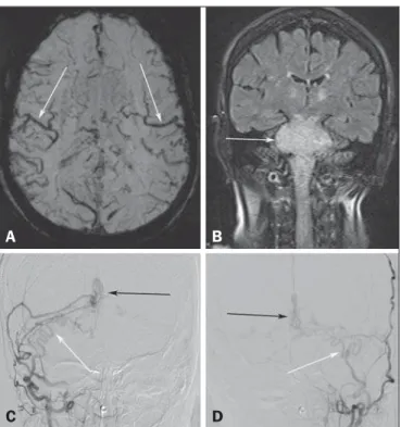
A 73-year-old woman presented with a history of at least four episodes of deep vein thrombosis. In the last five months, she had experienced severe ataxia, difficulty in swallowing, bilateral tinni-tus, and symptoms related to intracranial hypertension, such as nausea and vomiting. Magnetic resonance imaging (MRI) re-vealed a hyperintense signal on T2-weighted images and an en-larged brainstem, the swelling extending to the thalamus, cerebel-lar peduncles, and to the cervical portion of the spinal cord (Fig-ures 1A and 1B). The images could erroneously indicate a diag-nosis of brainstem tumor, glioma in particular, due to the infiltra-tive pattern of the lesion and the increased organ volume. How-ever, thorough evaluation with advanced imaging techniques, such as magnetic susceptibility-weighted sequences, demonstrated an extensive network of dilated peripheral veins, together with pro-nounced collateral circulation. Cerebral angiography showed a dural arteriovenous fistula (DAVF) with bilateral arterial supply via branches of the maxillary arteries. Venous drainage was mostly through the rectum and galenic system (Figures 1C and 1D). Involvement of the brainstem and cervical spinal cord was due to venous congestive injury. The classical surgical approach was precluded by the deep, inaccessible location, whereas endovascular therapy was precluded by the extensive involvement and bilateral nature of the fistula-sustaining arterial supply. The patient un-derwent gastrostomy and was discharged to palliative home care. Vascular lesions are often difficult to diagnose(1–8). DAVFs, which are characterized by abnormal communication between
Figure 1. A: Axial slice in a susceptibility-weighted sequence showing numerous large-caliber superficial veins, representing venous congestion. B: Coronal slice in a fluid-attenuated inversion recovery sequence showing a hyperintense signal and increased brainstem volume, mimicking a brain tumor. C,D: Digital angiography with subtraction technique, revealing the bilateral nature of the arterial supply (white arrows) and the nidus (black arrows) formed by the fistula.