256
DOI:
10.1590/0004-282X20150214
IMAGES IN NEUROLOGY
Neurophysiological and neuroimaging
changes (crossed cerebrocerebellar
atrophy) after prolonged non-convulsive
status epilepticus
Alterações de neuroimagem e de neurofisiologia após estado de mal epiléptico
não-convulsivo
Jivago S. Sabatini
1, Fabrício Stewan Feltrin
2, Márcio Andriani Rahal
1, Luiz César Lopes
1, Marcelo Rezende
Young Blood
1, Leilane Hoffmann Nogueira
1, Diogo Lago Pinheiro
2, Carlos Henrique Ferreira Camargo
1A 33-year-old female patient sufering from epilepsy since the
age of seven years (Figure 1) presented with left-sided hemipare
-sis following non-convulsive status epilepticus (NCSE) (Figure 2).
Neuroimaging showed a reduction in volume of the right cere
-bral hemisphere and left cerebellar hemisphere (Figure 3). EEG
showed asymmetry with right hemispheric slowing (Figure 4).
NCSE can have deleterious efects on the CNS because
of neuronal death after 30 to 60 minutes of continuous con
-vulsive activity
1,2. Following an episode of NCSE, 10 to 50%
of patients present with incapacitating neurological deicit
3.
Cranial MRI may show a persistent hyperintense lesion in
T2- or difusion-weighted images
4,5.
1Universidade Estadual de Ponta Grossa, Hospital Universitário, Serviço de Neurologia, Ponta Grossa PR, Brazil; 2Universidade Estadual de Ponta Grossa, Hospital Universitário, Unidade de Radiologia, Ponta Grossa PR, Brazil.
Correspondence: Carlos Henrique Ferreira Camargo; Departamento de Medicina, UEPG; Rua Carlos Cavalcanti, 4748; 84030-900 Ponta Grossa PR, Brasil; E-mail: [email protected]
Conflict of interest: There is no conflict of interest to declare.
Received 13 July 2015; Received in final form 14 August 2015; Accepted 04 September 2015.
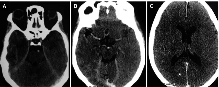
Figure 1.
Cranial computed tomography before the patient was admitted to hospital due to non-convulsive status epilepticus
(January 2015).
257
Jivago S. Sabatin et al. Crossed cerebrocerebellar atrophy after status epilepticus (A) Computed tomography performed without intravenous contrast on the day of the episode showing swelling and cortical edema in the right brain hemisphere and left cerebellar hemisphere as well as calcification of the parietal region of the right cerebral hemisphere. (B) Coronal FLAIR MRI image showing cortical edema in the right cerebral hemisphere. Left hippocampal formation with reduced volume and hyperintense signal. (C) and (D) Diffusion-weighted axial MRI images showing diffusion restriction in the regions mentioned above, indicating cytotoxic edema.
Figure 2.
Brain images (April 2015) after the patient was admitted to hospital.
A
B
C
D
(A) Axial FLAIR MRI image showing marked atrophy of the right brain hemisphere and discrete signal hyperintensity in the white matter caused by Wallerian degeneration. T2-weighted (B) Axial T1-weighted MRI MRI image showing marked atrophy of the right brain hemisphere and left cerebellum (C) MRI images showing cortical thinning in the right cerebral hemisphere more clearly. (D) Coronal FLAIR-weighted MRI images showing a marked reduction in volume and an abnormal signal in both hippocampal formations.
Figure 3.
Brain MRI after the patient was admitted to hospital for non-convulsive status epilepticus (June 2015)
Figure 4.
EEG performed in May 2015 showing asymmetry due to slowing of the right brain hemisphere (a sequela of prolonged
non-convulsive status epilepticus).
A
B
C
D
References
1. Fountain NB, Lothman EW. Pathophysiology of status epilepticus. J Clin Neurophysiol. 1995;12(4):326-42.
2. Chapman MG, Smith M, Hirsch NP. Status epilepticus. Anaesthesia. 2001;56(7):648-59. doi:10.1046/j.1365-2044.2001.02115.x 3. Cooper AD, Britton JW, Rabinstein AA. Functional and cognitive
outcome in prolonged refractory status epilepticus. Arch Neurol. 2009;66(12):1505-9. doi:10.1001/archneurol.2009.273
4. Donaire A, Carreno M, Gómez B, Fossas P, Bargalló N, Agudo R et al. Cortical laminar necrosis related to prolonged focal status epilepticus. J Neurol Neurosurg Psychiatry. 2006;77(1):104-6. doi:10.1136/jnnp.2004.058701