Abstract
Objective: Speciic phobia (SP) is characterized by irrational fear associated with avoidance of
speciic stimuli. In recent years, neuroimaging techniques have been used in an attempt to better understand the neurobiology of anxiety disorders. The objective of this study was to perform a systematic review of articles that used neuroimaging techniques to study SP. Method: A literature search was conducted through electronic databases, using the keywords: imaging, neuroimaging, PET, spectroscopy, functional magnetic resonance, structural magnetic resonance, SPECT, MRI, DTI, and tractography, combined with simple phobia and speciic phobia. One-hundred ifteen articles were found, of which 38 were selected for the present review. From these, 24 used fMRI, 11 used PET, 1 used SPECT, 2 used structural MRI, and none used spectroscopy. Result: The search showed that studies in this area were published recently and that the neuroanatomic substrate of SP has not yet been consolidated. Conclusion: In spite of methodological differences among studies, results converge to a greater activation in the insula, anterior cingulate cortex, amygdala, and prefrontal and orbitofrontal cortex of patients exposed to phobia-related situations compared to controls. These indings support the hypotheses of the hyperactivation of a neuroanatomic structural network involved in SP.
©2012 Elsevier Editora Ltda. All rights reserved.
Neuroimaging in speciic phobia disorder: a
systematic review of the literature
Ila M.P. Linares,
1Clarissa Trzesniak,
1Marcos Hortes N. Chagas,
1Jaime E. C. Hallak,
1Antonio E. Nardi,
2José Alexandre S. Crippa
1¹ Department of Neuroscience and Behavior of the Ribeirão Preto Medical School, Universidade de São Paulo (FMRP-USP). INCT Translational Medicine (CNPq). São Paulo, Brazil
2 Panic & Respiration Laboratory. Institute of Psychiatry, Universidade Federal do Rio de Janeiro (UFRJ). INCT Translational Medicine (CNPq). Rio de Janeiro, Brazil
Received on August 03, 2011; accepted on October 12, 2011
DESCRIPTORS Neuroimaging; Speciic Phobia; Review;
Anxiety Disorder; Phobia.
REVIEW ARTICLE
Corresponding author: Ila M. P. Linares; Hospital das Clínicas, 3º andar; Av. Bandeirantes, 3900; 14048-900, Ribeirão Preto, SP, Brazil; Phone: (+55 16) 36022703, Fax: (+55 16) 36022544; E-mail: [email protected]
The authors have no conlict of interest related to the topic of this article. The funding agency had no further role in study design; and in the decision to submit the paper for publication.
1516-4446 - ©2012 Elsevier Editora Ltda. All rights reserved.
Official Journal of the Brazilian Psychiatric Association Volume 34 • Number 1 • March/2012
Psychiatry
Neuroimagem do transtorno de fobia especíica: uma revisão sistemática da
literatura
Resumo
A Fobia Especíica (SP do inglês) é caracterizada por medos irracionais associados à evitação de estímulos especíicos. Nos últimos anos, técnicas de neuroimagem vêm sendo empregadas na tentativa de melhor compreender a neurobiologia dos transtornos de ansiedade. O objetivo do presente estudo é realizar uma revisão sistemática dos artigos que utilizaram neuroimagem para estudar a SP. A busca na literatura foi realizada por intermédio de indexadores eletrônicos,
utilizando-se as palavras-chave: imaging, neuroimaging, PET, spectroscopy, functional magnetic
ressonance, structural magnetic ressonance, SPECT, MRI, DTI e tractography, cruzadas
individualmente com os termos simple phobia e speciic phobia. Foram encontrados 115 artigos, sendo 38 deles selecionados para a presente revisão. Desses, 24 usaram fMRI, 11 usaram PET, 1 usou SPECT, 2 usaram MRI estrutural e nenhum artigo de espectroscopia. Veriica-se que os estudos na área foram publicados recentemente e que, até o momento, o substrato neuroanatômico deste transtorno não está consolidado. Apesar das diferenças metodológicas entre os estudos, os resultados convergem para maior ativação na ínsula, cíngulo anterior, amídala e córtex pré-frontal e orbitopré-frontal dos pacientes expostos a situações phobia related quando comparados aos controles. Esses achados reforçam hipóteses a respeito da hiperativação de uma determinada rede de estruturas neuroanatômicas envolvidas no transtorno de SP.
©2012 Elsevier Editora Ltda. Todos os direitos reservados. DESCRITORES:
Neuroimagem; Fobia especíica; Revisão; Transtorno de ansiedade; Fobia.
Introduction
Speciic phobia (SP), or simple phobia, is an anxiety disor-der characterized by increased and persistent excessive or irrational fear in the presence or anticipation of an object or phobic situation causing, almost invariably, an immedi-ate anxiety response (DSM-IVR – APA, 2000).1 DSM-IV deined ive subtypes of SP: animal, natural environment, blood-injection-injury (BII), situational, and others. Speciic phobias are considered the most frequent anxiety disorders and are among the most common psychiatric disorders in general population, with a prevalence of about 12.5%.2
Although environmental, constitutional, and genetic fac-tors are believed to contribute to the pathogenesis of the disorder,3-5 little attention had been dedicated to the study of SP neurobiology and etiology. The neuroimaging techniques, in particular, have helped to deepen the understanding of the neural circuitry underlying SP. Since its advent, neuroimaging has enabled the in vivo analysis of anatomical and functional structures, as well as analyses of regional metabolism in different psychiatric disorders. Methods like magnetic reso-nance imaging (MRI), functional magnetic resoreso-nance imag-ing (fMRI), positron emission tomography (PET), and simag-ingle photon emission computed tomography (SPECT) have been used in the investigation of biological processes involved in the neurocircuitry of SP.
In order to expose a panoramic view to the readers, this article presents a systematic qualitative review of published studies on neuroimaging of patients with SP. The qualitative approach was chosen because quantita-tive methods, such as meta-analysis, show that: (a) the information necessary to calculate result size is not always available, and may limit this analysis to a small subset of studies;6 (b) methods and extent of detailed information to deine regions of interest vary greatly among studies,
hindering accurate comparisons; (c) there are great dif-ferences regarding secondary variables in studies (for instance, gender, medication, co-morbidity, SP subsets); (d) studies used different functional (PET, SPECT, fMRI, relaxometry) and structural (MRI) neuroimaging methods, which precludes comparative statistical analyses; (e) dif-ferent methods are used to analyze images (automatic vs. ROI) in different forms of investigation, and (f) meta-analyses have intrinsic limitations in estimating negative results which are not published (the ile drawer problem;7). Thus, a quantitative analysis is not adequate for a review with large amplitude (with no time limit set). Finally, it is relevant to point out that although animal research is valu-able to understanding anxiety disorders, the current study will focus only on research performed with humans, as the analysis of animal research is beyond the scope proposed.
Method
Searches were performed in LILACS, SciElo and Web of Science databases, using the following key words: imag-ing, neuroimagimag-ing, PET, spectroscopy, functional magnetic resonance, structural magnetic resonance’, SPECT, MRI, DTI e tractography, individually crossed with the terms simple phobia and speciic phobia. Additional articles were sought manually in references from previously selected material.
Results
From the 38 studies included in this review, 24 used fMRI, 11 used PET, 1 used SPECT, and 2 used MRI (cortical thickness). None of them used MRS, VBM techniques, or Graph analysis. For heuristic reasons, the articles will be grouped according to the neuroimaging techniques applied.
Structural magnetic resonance: Cortical Thickness
To the best of our knowledge, only two articles used MRI. One of the studies, while investigating cortical thickness of subjects with animal phobia and healthy volunteers, found that when compared to control, the SP group presented increased thickness in the insular bilateral cortex, bilateral pregenual anterior cingulum, bilateral posterior cingulate cortex, and left visual cortex.8
The other study investigated the existence of correla-tion between the degree of anxiety sensitivity and cortical thickness of the right anterior insula in patients with animal phobia and healthy individuals. The authors observed that patients presented high anxiety scores compared to controls, however, there was no difference in cortical thickness or insula volume in both groups9 (Table 1).
The reduced number of participants in the studies, the need to assess volumetric differences in structures knowingly implicated in SP, such as the hippocampus and subcortical structures, and the absence of other articles using VBM in SP make the continuation of complementary studies in the area an issue of paramount importance.
Positron emission tomography (PET)
The irst studies that sought to investigate the neuroanatomic substrate of SP made use of PET. By using tracers marked with a radioactive isotope, the technique provides brain activity assessment. To date, eleven articles that made use of PET in SP were found. The irst study using PET in SP did not ind signiicant differences between patients with animal phobia and healthy volunteers.10 However, subsequent studies have shown changes in rCBF (regional cerebral blood low) in paralimbic and sensory regions. Female patients with snake phobia11-13 and spider phobia12,14 were shown phobia-related images, neutral images, and aversive images unrelated to phobia. Signiicant increase was found in rCBF in the visual cortex,13,14 amygdala, thalamus, and striatum12 and reduced rCBF in the orbitofrontal cortex, temporopolar cortex, pos-terior cingulate cortex, and hippocampus.11,14
With the same experimental group of sixteen phobic subjects, a study tried to replicate some of the results previously described,15 and another study investigated the activity of the NK1 receptor system (neuropeptide substance P binds to the neurokinin receptor SP-NK1) of the amygdale.16 Viewing phobia-related images was correlated with increased activation of the right amygdala, cerebellum, left visual cortex, and circumscribed frontal areas. Decreased activity of prefrontal, orbitofrontal, ventromedial, somatosensso-rial, and hearing cortices was observed. Additionally, during non-aversive stimulation, prefrontal activity was negatively correlated with rCBF of the amygdala. These results suggest that phobic reactions activate areas of object recognition and deactivate prefrontal areas involved in cognitive control of emotions, triggering areas like the amygdala, related to motor readiness for ight or light.15 The second study observed that the antagonist reuptake in SP-NK1 receptors was signiicantly reduced in the right amygdala during pho-bic stimulation, suggesting a decrease in the availability of NK1 receptors, which may relect the increased release of endogenous substance P.16
Table 1 Structural Magnetic Ressonance studies in SP
Name Subjects (n) F/M Age (m ± SD ) Handedness Sample Procedure T Results (SP > controls)
Rauch et al. 8 10 SP- animal
20 controls 4-6 8-12
32.1 ±1 1.6 28.6 ± 6.5
Right-handed Right-handed
Comorbidity: no. Medication: free
Regional cortical thickness to investigate structural
abnormalities
1.5 SP: ↑ cortical thickness of paralimbic cortex, sensory cortex (bilateral insular, bilateral
pregenual anterior, cingulated, posterior cingulated, visual
cortical regions)
Rosso et al. 9 19 SP - animal
20 SP - controls 14-5 11-9
28.68 ± 6.36 29.70 ± 6.22
Right-handed Comorbidity: no. Medication: free
Regression and correlation analyses examined anxiety sensitivity scores in relation to anterior and posterior insular cortex volume and
thickness
3.0 SP: ↑ anxiety scores than controls but did not differ volumes or thickness of insula Anxiety scores predicted ↑ thickness of the right anterior insular cortex in SP group
↓, decrease; ↑, increase; F, female; M, male; SD, standard deviation; m, mean; SP, speciic phobia
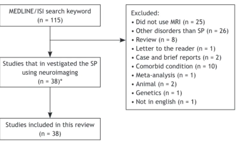
Figure 1 Flow chart showing study selection for the review.
MEDLINE/ISI search keyword (n = 115)
Studies included in this review (n = 38)
Studies that in vestigated the SP using neuroimaging
(n = 38)*
Excluded:
• Did not use MRI (n = 25)
• Other disorders than SP (n = 26)
• Review (n = 8)
• Letter to the reader (n = 1)
• Case and brief reports (n = 2)
• Comorbid condition (n = 10)
• Meta-analysis (n = 1)
• Animal (n = 2)
• Genetics (n = 1)
• Not in english (n = 1)
In tasks related to the imagination17 and anticipatory anxiety18 participants with small animal phobia showed in-creased rCBF in the anterior cingulate cortex, insular cortex, anterior temporal cortex, and somatosensory cortex17 and decreased rCBF in the primary visual cortex.18 The authors suggest the existence of a neurophysiologic correlate of avoidant anticipatory coping.
Brain activation during aversive modulation in phobics through pairing phobia-related and neutral images to an aversive noise was investigated.19 There was an increase in ACC and in the left hippocampal-amygdaloid area activa-tion during exposure to phobia-related images associated with aversive noise. This raised the hypothesis that these two regions could be part of a neural system connected to attention and danger orientation.
Habituation to feared stimuli has also been studied.20 The referred protocol caused a decrease in anxiety measured by SUD-S and regression analyses showed that anxiety was correlated to increased activity in left amygdala, perirhinal bilateral cortex, right FG, and periaquedutal gray matter. In its turn, phobic fear seems to be associated with increased right hippocampus activity. While habituation to stimuli promoted a decrease of physiologic and subjective anxiety, administration of diazepam did not affect the rCBF of sub-jects with phobia.14 Table 2 provides detailed information on methodologic studies with PET included in this review.
Single photon emission tomography (SPECT)
This technique is used in the investigation of brain activity through tracers marked with a single photon emitter. SPECT has been little used in the study of SP, the only study that applied the SPECT technique to SP (Table 3) investigated whether brain changes associated with increases in anxiety states are different from those observed during a cognitive activation experiment.21 Participants underwent two tests: one in which they would hear audio with relaxing content, and another in which they would hear phobia-related audio content. Psychophysiologic and subjective measures showed substantial changes in the induction of fear. Additionally, a decrease in the radiotracer reuptake in regions of bilateral occipital cortex, right posterior temporal cortex, and right anterior cingulate cortex could be observed.
Functional magnetic resonance imaging (fMRI)
Currently, fMRI is the most widely used neuroimaging meth-odology in the study of SP. So far, twenty four studies were conducted in order to better understand the neurobiology of SP, ive of these related to psychotherapeutic treatment of the disorder. In the study of SP, the predominance of this technique over the others can be attributed to the fact that it provides data on brain functioning pattern with high temporal solution and greater patient safety, as it does not make use of radioisotopes.
One of the irst studies to use fMRI to investigate SP was conducted by Wright et al. (2003).22 At that moment, studies pointed at an increase in brain activity in sensory areas and increase or decrease in anterior and hippocampal paralimbic regions.11,13,14,17 However, studies on SP did not show amygdala hyperresponsivity against a phobic stimulus. Similarly, Wright et al. (2003)22 assessed the amygdala response to emotional faces versus neutral faces in subjects with animal phobia and
in healthy individuals. Authors did not ind signiicant differ-ences regarding amygdala activation; however, consistently with other studies,17,23,24 they observed an increase in the response of right insular cortex in phobic individuals exposed to aversive images. This insular activation occurred after the introduction of stimuli that represented threat but were not particular to the disorder.
Contrary to these indings, several subsequent studies consistently observed changes in amygdala activation of individuals with arachnophobia exposed to phobia-related images.24-28 In general, studies have observed increase in amygdala activation,24,26-29 and in one study this response was also conirmed upon exposure of images related to the feeling of disgust.27 Additionally, increase has been conirmed in insula activation,22,24,26-28orbitofrontal cortex and uncus,24 associative visual cortex, DLPFC, hippocampus,25 bilateral ACC,26,27 pre-frontal medial cortex, thalamus,26 thalamus pulvinar nucleus, and supplementary motor area.27These indings suggest that the amygdala has a relevant role in fear processing, and they also suggest brain activation patterns when phobic individuals are confronted with phobia-related stimuli.
Still trying to clarify the role of the amygdala in SP, Alpers et al. (2009)30 investigated if the attribution of attention to overlap images modulates the activation of this structure. Authors observed increase in amygdala activation of subjects with phobia in response to mixed images (spider and bird), static or in movement. It is also relevant to highlight that the amygdala activation happened in a dose-response rela-tion, which means the higher the exposure to the feared stimulus the greater the structure activation. Larson et al. (2006),31 while investigating the time of amygdala activation of individuals with fear of spiders, observed shorter starting time for the activation peak in this brain region. That means phobic individuals presented intense and short responses in the amygdala, while the control group presented weaker and sustained responses. These indings conirm the involvement of the amygdala in the processing of phobia-relevant stimuli.
Although a central role in the pathophysiology of SP has been attributed to the amygdala, some studies have inves-tigated the relationship among other brain regions in SP. Martis et al. (2004)32 exposed phobic individuals and healthy participants to a “serial reaction time task paradigm” in or-der to investigate an implicit learning sequence. During the implicit learning sequence, signiicant differences between patients and control group concerning striatum activation were not found. The existence of different pathophysiological mechanisms when comparing SP to other disorders was sug-gested; in obsessive compulsive disorder (OCD), for instance, striatum activation was decreased in patients.33
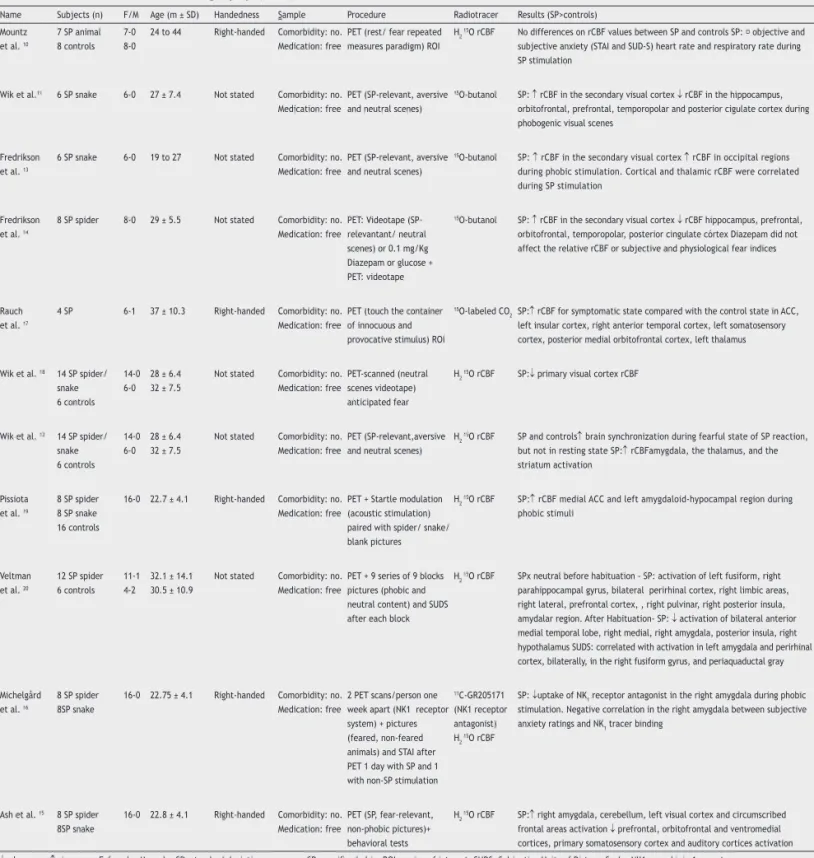
Table 2 Positron Emission Tomography (PET) studies in SP
Name Subjects (n) F/M Age (m ± SD) Handedness Sample Procedure Radiotracer Results (SP>controls) Mountz
et al. 10
7 SP animal 8 controls
7-0 8-0
24 to 44 Right-handed Comorbidity: no. Medication: free
PET (rest/ fear repeated measures paradigm) ROI
H2
15O rCBF No differences on rCBF values between SP and controls SP: objective and
subjective anxiety (STAI and SUD-S) heart rate and respiratory rate during SP stimulation
Wik et al.11 6 SP snake 6-0 27 ± 7.4 Not stated Comorbidity: no.
Medication: free
PET (SP-relevant, aversive and neutral scenes)
15O-butanol SP: ↑ rCBF in the secondary visual cortex ↓ rCBF in the hippocampus,
orbitofrontal, prefrontal, temporopolar and posterior cigulate cortex during phobogenic visual scenes
Fredrikson et al. 13
6 SP snake 6-0 19 to 27 Not stated Comorbidity: no. Medication: free
PET (SP-relevant, aversive and neutral scenes)
15O-butanol SP: ↑ rCBF in the secondary visual cortex ↑ rCBF in occipital regions
during phobic stimulation. Cortical and thalamic rCBF were correlated during SP stimulation
Fredrikson et al. 14
8 SP spider 8-0 29 ± 5.5 Not stated Comorbidity: no. Medication: free
PET: Videotape (SP-relevantant/ neutral scenes) or 0.1 mg/Kg Diazepam or glucose + PET: videotape
15O-butanol SP: ↑ rCBF in the secondary visual cortex ↓ rCBF hippocampus, prefrontal,
orbitofrontal, temporopolar, posterior cingulate córtex Diazepam did not affect the relative rCBF or subjective and physiological fear indices
Rauch et al. 17
4 SP 6-1 37 ± 10.3 Right-handed Comorbidity: no. Medication: free
PET (touch the container of innocuous and provocative stimulus) ROI
15O-labeled CO
2 SP:↑ rCBF for symptomatic state compared with the control state in ACC,
left insular cortex, right anterior temporal cortex, left somatosensory cortex, posterior medial orbitofrontal cortex, left thalamus
Wik et al. 18 14 SP spider/
snake 6 controls
14-0 6-0
28 ± 6.4 32 ± 7.5
Not stated Comorbidity: no. Medication: free
PET-scanned (neutral scenes videotape) anticipated fear
H2
15O rCBF SP:↓ primary visual cortex rCBF
Wik et al. 12 14 SP spider/
snake 6 controls
14-0 6-0
28 ± 6.4 32 ± 7.5
Not stated Comorbidity: no. Medication: free
PET (SP-relevant,aversive and neutral scenes)
H2 15O rCBF SP and controls↑ brain synchronization during fearful state of SP reaction,
but not in resting state SP:↑ rCBFamygdala, the thalamus, and the striatum activation
Pissiota et al. 19
8 SP spider 8 SP snake 16 controls
16-0 22.7 ± 4.1 Right-handed Comorbidity: no. Medication: free
PET + Startle modulation (acoustic stimulation) paired with spider/ snake/ blank pictures
H2 15O rCBF SP:↑ rCBF medial ACC and left amygdaloid-hypocampal region during
phobic stimuli
Veltman et al. 20
12 SP spider 6 controls
11-1 4-2
32.1 ± 14.1 30.5 ± 10.9
Not stated Comorbidity: no. Medication: free
PET + 9 series of 9 blocks pictures (phobic and neutral content) and SUDS after each block
H2 15O rCBF SPx neutral before habituation - SP: activation of left fusiform, right
parahippocampal gyrus, bilateral perirhinal cortex, right limbic areas, right lateral, prefrontal cortex, , right pulvinar, right posterior insula, amydalar region. After Habituation- SP: ↓ activation of bilateral anterior medial temporal lobe, right medial, right amygdala, posterior insula, right hypothalamus SUDS: correlated with activation in left amygdala and perirhinal cortex, bilaterally, in the right fusiform gyrus, and periaquaductal gray
Michelgård et al. 16
8 SP spider 8SP snake
16-0 22.75 ± 4.1 Right-handed Comorbidity: no. Medication: free
2 PET scans/person one week apart (NK1 receptor system) + pictures (feared, non-feared animals) and STAI after PET 1 day with SP and 1 with non-SP stimulation
11C-GR205171
(NK1 receptor antagonist) H2
15O rCBF
SP: ↓uptake of NK1 receptor antagonist in the right amygdala during phobic
stimulation. Negative correlation in the right amygdala between subjective anxiety ratings and NK1 tracer binding
Ash et al. 15 8 SP spider
8SP snake
16-0 22.8 ± 4.1 Right-handed Comorbidity: no. Medication: free
PET (SP, fear-relevant, non-phobic pictures)+ behavioral tests
H2
15O rCBF SP:↑ right amygdala, cerebellum, left visual cortex and circumscribed
frontal areas activation ↓ prefrontal, orbitofrontal and ventromedial cortices, primary somatosensory cortex and auditory cortices activation
↓, decrease; ↑, increase; F, female; M, male; SD, standard deviation; m, mean; SP, speciic phobia; ROI, region of interest; SUDS, Subjective Units of Distress Scale; NK1,neurokinin 1 receptor ; rCBF, regional cerebral blood low; STAI, Spielberger Trait.
with SP. Another study showed an increase in the activation of the amygdala, insula, ACC, and DMPFC (dorsomedial pre-frontal cortex) during the identiication task, and bilateral activation in the amygdala of phobics during the “distrac-tion task”.35Notably, in the face of phobia-related stimuli, amygdala activation was observed independently of attention resources, unlike the activation of ACC, DMPFC, and insula that were dependent on the resources of attention.35,36This
inding was reproduced in a later study,23 which investigated the defensive response mobilization. These data suggest that the activation of the amygdala in fMRI studies primar-ily indexes the detection of motivationally relevant stimuli, whereas the insula might be more speciically connected to defensive response mobilization.
of phobia-related words was related to increased activation in prefrontal cortex, insula, and posterior cingulate cortex of subjects with phobia compared to healthy subjects. A neural network for the processing of these threatening stimuli was suggested. Conirming some of the results already described, in 2007 Straube reported an increase in activation of the dorsal ACC, insula, thalamus, and visual areas in phobic in-dividuals during an anticipatory stimulus of phobia relevant images compared to the anticipatory stimulus of neutral pictures. This study provided evidence for the hypothesis that there is increased activation in speciic brain regions during anticipatory anxiety in individuals with SP.
Assuming that SP is characterized by a deicit in the automatic regulation of emotions, fourteen subjects with spider phobia were assessed while visualizing phobia-related and aversive images; then, individuals were asked to up- and down-regulate the emotions elicited by the images. An in-crease was observed in the activation of the insula, as well as a decrease in activation of the ventromedial prefrontal cortex of patients, suggesting they have deicits in both automatic and effortful regulation of emotions elicited by phobic compared with aversive stimuli.39
Most studies on SP and neuroimaging concern the animal subtype;therefore, little is known about the neurocircuitry related to other subtypes of phobia. Herman et al. (2007)40 examined the effects of symptom induction in the neural activation of individuals with BII phobia. When compared to the control group, there was decreased activity in the medial prefrontal cortex and increased activation of the supplemen-tary motor area in the BII group exposed to phobia-related images. The results might relect reduced cognitive control of emotions in BII phobic individuals during the experience of phobic symptoms as well as during states of disgust.
Some studies have compared the neurocircuitry of healthy individuals to that of patients with animal and BII phobia subtypes. Lueken et al. (2011)41 observed greater activation of structures such as ACC, insula, and thalamus in individuals with the animal phobia subtype, unlike indi-viduals with BII phobia that had circumscribed activation in the prefrontal and orbitofrontal cortices. Caseras et al. (2010)42 observed similar activation in brain structures, such as the occipitotemporal region and thalamus, in both groups with SP. Individuals with spider phobia showed activation of bilateral dorsal ACC, anterior insula, inferior frontal gyrus, and visual cortex; however, compared with BII phobics, there was reduced activation of the medial frontal cortex extend-ing later to the rostral ACC and greater activation in dorsal anterior cingulus and anterior insula. When viewing images of blood-injection-injury, the BII phobics showed increased activation in the thalamus and occipitotemporoparietal cortex compared to the other two groups.
The results described suggest partially distinct neurobio-logical substrates between animal and BII phobic subtypes; thus, Caseras et al. (2010b)43 sought to evaluate if by the display of phobia-related images, the BII group would present different responses in the insula and ACC compared to those with spider phobia. The BII group showed higher activity of ventral prefrontal cortex compared to control, and reduced peak activity in left amygdala compared to those with spider phobia. Similarly, both groups took long time to reach the peak of activation in the right amygdala, but only the spider phobia group showed signiicant changes for this parameter in the left amygdala compared to control group. In contrast to previous indings, no differences were found in the insula and ACC activity in patients with BII phobia and spider phobia. Considering these and other previous results, the authors suggest that the two phobia subtypes (spider and BII) showed similar responses when exposed to phobia-related pictures; however, there are signiicant differences in their sustained responses to these stimuli.
As for the pharmacotherapy of the disorder, there is a limited number of studies that investigated the neuronal ef-fect of drugs in people with SP. The efef-fects of D-cycloserine (DCS-a partial agonist to NMDA receptors) were assessed in patients with spider phobia.44 The authors found that patients with phobia who received DCS showed increased activation of the PFC, dorsal ACC, and insula during exposure to phobia-related images. Still, the placebo group showed a positive correlation between lateral PFC and amygdala activation. Results suggest that during the initial phobic symptom in-ducement, DCS increases activation in regions involved in cognitive control and interoceptive integration.
Some studies have used fMRI in order to investigate the main effects of psychotherapy on SP. Spider phobia patients underwent fMRI before and after four45 and two psycho-therapy (CBT - Cognitive Behavioral Therapy) sessions.28Upon exposure to phobia-related images in the irst examination, there was increased activity in the parahippocampal gyrus, associative visual cortex, and right dorsolateral prefrontal cortex of subjects with phobia45 and increased amygdala activity compared to controls.28 After CBT phobic subjects showed decreased symptoms and increased activity in areas, such as associative visual cortex, superior parietal lobule, bilateral inferior frontal gyrus, absence of parahippocampal gyrus, and right dorsolateral prefrontal cortex activation.45 Additionally, there is a decrease in amygdala, insula, and ACC hyperactivity.28 These indings suggest that the CBT psychotherapeutic treatment has the potential to modify the dysfunctional neural circuitry associated with SP.
Other studies related to psychotherapy and SP were per-formed in which participants were divided into controls, sub-jects with arachnophobia on the waiting list, and individuals
Table 3 Single Positron Emission Tomography (SPECT) study in SP
Name Subjects (n) F/M Age (m ± SD) Handedness Sample Radiotracer Drug Procedure Results (SP>controls)
O’Carrol et al. 21
10 SP 9-1 51 ± 15.1 Right-handed Comorbidity: no. Medication: free
99mTc-HMPAO/rCBF Volunteers
listened relaxing tape/ phobic tape + SPECT
↓ tracer uptake in posterior regions ↓ right temporal/ occipital regions and left ACC
with spider phobia who underwent CBT – psychotherapy.29,46 Upon exposure to neutral, disgust, fear and phobia-related images, brain activation in the irst fMRI session did not dif-fer between the two groups with phobia in which there was increased activation in the insula, ACC29,46 parahippocampal gyrus, amygdala, and lateral orbitofrontal cortex29 compared to control group. In the second fMRI scan, after one and two psychotherapy sessions,29 there was decreased activity of the insula,29,46 amygdala,29,46 and ACC46 and increased activa-tion in the medial orbitofrontal cortex29 in the group that underwent psychotherapy compared to waiting list group. The authors concluded that successful treatment of SP is primarily accompanied by functional changes of the medial OFC (orbitofrontal cortex), a key brain region for the self regulation of emotions and the relearning of stimulus rein-forcement associations. Straube claims that reducing the activity of the insula and ACC may relect the attenuation of the phobic response after treatment.
In an attempt to prove the eficacy of CBT after six months of treatment, Schienle et al. (2009)47 once again exposed ten participants from their previous study29 to the same sequence of photos and performed fMRI scans. It was observed that the psychotherapy group showed a decrease of clinical symptoms. Moreover, when comparing results of the irst and second fMRI sessions, there is increased activity of the medial orbitofrontal cortex, and decreased activity in the insula and lateralorbitofrontal cortex. The authors high-lighted that the results of the medial OFC would be related to the neural basis of the lasting positive outcome of CBT.
Consistent evidence resulting from the change of brain functioning in SP has been observed by fMRI studies with predominance of limbic structures, especially the amygdala, insula, and ACC. It is likely that the correlation between the indings is largely due to the experimental procedures used in examining the emotional processing in SP, despite having different goals and speciic methodology.
Discussion
Neuroimaging may be considered an important technique to help understand the anatomical, functional and meta-bolic neurocircuitry underlying the pathophysiology of SP. Most of the studies analyzed in this review used functional neuroimaging techniques to assess brain response against exposure to phobia-related images, and results consistently point to activation mainly in the amygdala, ACC, and insular cortex (Figure 2). Despite the high prevalence of SP in the general population,2 only two MRI studies on the disorder were found: cortical thickness8 and SPECT.21 Also, in contrast to the numerous studies of other anxiety disorders, such as obsessive compulsive disorder (OCD) and post traumatic stress disorder (PTSD), no studies on SP have been performed with techniques such as MRI/VBM, MRS, and graph analysis.48
Contrary to panic disorder or OCD, there is still no de-initive model for the neuroanatomical substrate of SP. One possibility is that they are learned, characterized by fear conditioned to a stimulus or situation. Moreover, the phobias may be due to dysregulation of systems that are speciic for the detection of stimuli or threatening situations.49
Experimental evidence indicates the existence of speciic neural circuits that modulate emotional behavior, which responds according to the nature of the stimulus to which
the individual is exposed. The Brain Aversive System (BAS), represented by the medial hypothalamus, the dorsal periaq-ueductal gray matter; and the amygdala, would be associated with the emission of unconditioned responses (innate and not learned); for example, elevated blood pressure, increased heart rate and breathing, piloerection, urination, defeca-tion, and exophthalmos.50,51 There are hints that anatomic and functional abnormalities connected to the Behavioral Inhibition System (BIS) could cause abnormal defense reac-tions, such as panic attacks and phobic symptoms develop-ment.52 It is believed that the organization of conditioned responses is carried out by the BIS, represented by the septo-hippocampal system, the median raphe nucleus, and ventral periaqueductal gray matter.53 It is known that the amygdala is involved in the activation of both BAS and BIS, acting as the primary interface between both.54,55
The amygdala seems to play a key role in the neurobiol-ogy of anxiety disorders in general and particularly in SP. The irst neuroimaging studies on SP explored patterns of brain activity in phobic symptoms caused by visual stimuli,11,13,14,56 tactile stimuli,10,17 or auditory cues.21 The initial hypothesis was that the amygdala, with its role in surveillance and negative affection, would show increased activity during fear inducement.10,13,17 In fact, several studies have demon-strated greater activation of this structure in patients with SP.20,23-25,27-29,34,35,44However, some studies failed to show this increased activity of the amygdale.12,18,22,31,37,40,45,57 It is pos-sible that these negative results are due to methodological differences between studies, such as time of exposure to the phobia-related stimulus and the cognitive/emotional nature of the task applied.24
Other structures that showed hyperactivity in phobic individuals exposed to phobia-related stimuli were the anterior insula, hippocampus, dorsal and rostral ACC, and somatosensory and occipitoparietal cortex.6,24,25,27,29,35,46 In particular, the ACC (anterior cingulate córtex) has numer-ous subdivisions that perform cognitive, emotional, motor, nociceptive, and visual-spatial functions58 and is connected to the amygdala, periaqueductal gray matter, accumbens nucleus, hypothalamus, anterior insula, hippocampus, and orbitofrontal cortex.59 The dorsal ACC is mainly involved in assessing information of emotional and motivational nature,
Figure 2 Figurative summary of the major indings of the reviewed studies (number of studies/total number of studies) ACC - Anterior Cingulate Cortex, PFC - Prefrontal Cortex.
PFC (11/38)
ACC (13/38)
Amygdala (16/38)
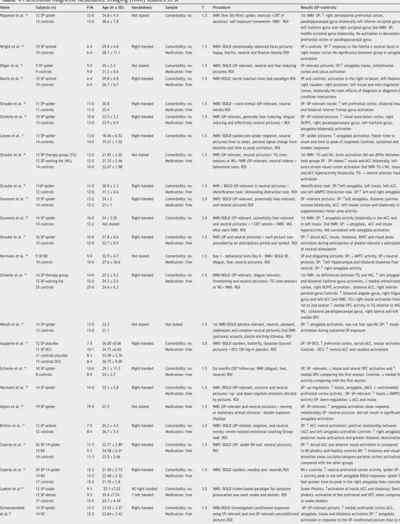
Table 4 Functional Magnetic Resonance Imaging (fMRI) studies in SP
Name Subjects (n) F/M Age (m ± SD) Handedness Sample T Procedure Results (SP>controls)
Paquette et al. 45 12 SP spider 13 controls
12-0 13-0
24.8 ± 4.5 28.6 ± 7.8
Not stated Comorbidity: no. 1.5 fMRI (ive 30s ilms: spider, neutral) +CBT (4 sessions)/ self-exposure homework+ fMRI - ROI
1st fMRI- SP: ↑ right dorsolateral prefrontal cortex, parahippocampal gyrus bilaterally left inferior occipital gyrus, left fusiform gyrus and right occipital gyrus 2nd fMRI- SP: middle occipital gyrus bilaterally. No activation in dorsolateral prefrontal cortex or parahippocampal gyrus
Wright et al. 22 10 SP animal 10 controls
6-4 6-4
29.8 ± 6.8 28.7 ± 11.1
Right-handed Comorbidity: no. Medication: free
1.5 fMRI/ BOLD (emotionally-valenced faces pictures - happy, fearful, neutral and ixation blocks) ROI
SP x controls- SP:↑ response to the fearful x neutral faces in right insular cortex No signiicance between-group in amygdala activation
Dilger et al. 24 9 SP spider 9 controls
9-0 9-0
25 ± 2.3 21.3 ± 0.6
Not stated Comorbidity: no. Medication: free
1.5 fMRI/ BOLD (SP-relevant, neutral and fear-inducing pictures) ROI
SP-relevant pictures: SP:↑ amygdala insula, orbitofrontal cortex and uncus activation
Martis et al. 32 10 SP animal 10 controls
6-4 6-4
29.8 ± 6.8 26.7 ± 6.7
Right-handed Comorbidity: no. Medication: free
1.5 fMRI/BOLD +serial reaction time task paradigm ROI SP and controls: activation in the right striatum, left thalamus, right caudate, right putamen, left insula and mid-cingulated cortex, bilaterally No main effects of diagnosis or diagnosis-by-condition interactions
Straube et al. 38 11 SP spider 11 controls
11-0 11-0
20.8 22.4
Right-handed Comorbidity: no. Medication: free
1.5 fMRI/ BOLD + word stimuli (SP-relevant, neutral words) ROI
SP- SP-relevant words: ↑ left prefrontal cortex, bilateral insula and bilateral inferior frontal gyrus activation
Schienle et al. 25 10 SP spider 13 controls
10-0 13-0
22.5 ± 2.2 23.9 ± 6.9
Right-handed Comorbidity: no. Medication: free
1.5 fMRI (SP-relevant, generally fear-inducing, disgust-inducing and affectively neutral pictures) / ROI
SP- SP-related pictures: ↑ visual association cortex, right DLPFC, right parahippocampal gyrus, left fusiform gyrus, amygdala bilaterally activation
Larson et al. 31 13 SP spider 14 controls
13-0 14-0
18.46 ± 0.52 19.21 ± 1.02
Right-handed Comorbidity: no. 1.5 fMRI/ BOLD (spider,non-spider negative, neutral pictures) time to onset, percent signal change from baseline and time to peak activation. ROI
SP- spider pictures: ↑ amygdala activation /faster time to onset and time to peak of responses Controls: sustained and weaker responses
Straube et al. 46 13 SP therapy group (TG) 12 SP waiting list (WL) 14 controls
13-0 12-0 14-0
21.92 ± 2.02 21.33 ± 2.46 22.07 ± 1.98
Not stated Comorbidity: no. Medication: free
1.5 fMRI (SP-relevant, neutral pictures)+ TG (two-session) or WL+ fMRI (SP-relevant, neutral videos) + behavioral tests. ROI
1st fMRI- TG and WL: brain activation did not differ between both groups SP - SP-videos:↑ insula and ACC bilaterally, left extra striate visual cortex activation 2nd fMRI TG x WL: insula and ACC hyperactivity bilaterally. TG: ↓ ventral anterior insula activation
Straube et al. 35 11SP spider 12 controls
11-0 12-0
20.9 ± 2.3 21.3 ± 0.6
Right-handed Comorbidity: no. Medication: free
1.5 fMRI / BOLD (SP-relevant in neutral pictures) + identiication task/ demanding distraction task. ROI
Identiication task- SP:↑left amygdala, left insula, left ACC, and left DMPFC Distraction task -SP:↑ left and right amygdala Goossens et al. 27 15 SP spider
14 controls
13-2 12-2
24 ± 2 23 ± 1
Right-handed Comorbidity: no. Medication: free
3.0 fMRI/ BOLD (SP-relevant, potentially fear-relevant, and neutral pictures) ROI
SP -relevant pictures: SP: ↑left amygdala, thalamic pulvinar nucleus bilaterally, ACC, left insular cortex and bilaterally in supplementary motor area activity
Goossens et al. 28 16 SP spider 14 controls
16-0 12-2
24 ± 3.02 Not stated
Right-handed Comorbidity: no. 3.0 fMRI/BOLD (SP-relevant, potentially fear-relevant and neutral pictures) + 1 CBT session + fMRI. VAS after each fMRI. ROI
1st fMRI: SP: ↑ amygdala activity bilaterally in the ACC and in left insula. 2nd fMRI: SP: ↓ amygdala, ACC and insula hyperactivity. VAS correlated with amygdala activation Straube et al. 37 16 SP spider
15 controls
16-0 15-0
21.8 ± 0.6 22.7 ± 0.9
Right-handed Comorbidity: no. Medication: free
1.5 fMRI (SP and neutral pictures) + each picture was preceded by an anticipatory period and symbol. ROI
SP: ↑ dorsal ACC, insula, thalamus, BNST and visual areas activation during anticipation of phobia-relevant x anticipation of neutral stimulation
Hermann et al. 40 9 SP BII 10 control
9-0 10-0
22.9 ± 4.7 27.6 ± 10.6
Not stated Comorbidity: no. Medication: free
1.5 Day 1 - behavioral tests Day II - fMRI/ BOLD SP, disgust, fear, neutral pictures). ROI
SP and disgusting pictures: SP: ↓ MPFC activity. SP x neutral pictures: SP: ↑left hippocampus and bilateral thalamus Fear x neutral: SP: ↑ right amygdala activity
Schienle et al. 29 14 SP therapy group 12 SP waiting list 25 controls
14-0 12-0 25-0
27.2 ± 9.2 24.3 ± 2.0 24.6 ± 6.3
Right-handed Comorbidity: no. Medication: free
1.5 fMRI/BOLD (SP-relevant, disgust-relevant, threatening and neutral pictures)+ TG (one-session) or WL+ fMRI. ROI
1st fMRI: no differences between TG and WG. ↑ left amygdala and bilateral fusiform gyrus activation, ↓ medial orbitofrontal cortex, right DLPFC activation , bilateral ACC, right inferior parietal gyrus Controls: ↑ bilateral angular gyrus, right lingual gyrus and left ACC 2nd fMRI: TG↓ right insula activation from 1st to 2nd session ↑ medial OFC activity in TG relative to WG WL: ↓bilateral parahippocampal gyrus, right lateral and left medial OFC
Wendt et al. 23 13 SP spider 13 controls
13-0 13-0
23.2 21.1
Not stated Not stated 1.5 1st fMRI/BOLD (phobia relevant, neutral, pleasant, unpleasant and complex neutral pictures) 2nd fMRI (pictures) acoustic startle eliciting stimulus. ROI
SP: ↑ amygdala activation, was not fear speciic SP: ↑ insula activation during sustained SP-exposure
Aupperle et al. 47 12 SP placebo 11 SP DCS 11 controls placebo 12 controls DCS
7-5 10-1 8-3 8-4 26.00 ±8.06 24.73 ±6.65 23.09 ± 3.70 26.75 ± 9.09
Right-handed Comorbidity: no. Medication: free
3.0 fMRI/ BOLD (spiders, butterly, Gaussian-blurred pictures) + DCS 100 mg or placebo. ROI
SP: SP-DCS: ↑ prefrontal cortex, dorsal ACC, insular activations Controls - DCS: ↑ ventral ACC and caudate activations
Schienle et al. 47 10 SP spider 8 controls
10-0 8-0
29.1 ± 11.5 24 ± 3.7
Right-handed Comorbidity: no. Medication: free
1.5 Six months CBT follow-up: fMRI (disgust, fear, neutral) ROI
SP: SP -relevant: ↓ insula and lateral OFC activation and ↑
medial OFC comparing the irst session. Controls: ↓ medial OFC activity comparing with the irst session.
Hermann et al. 39 14 SP spider 14-0 22.1 ± 2.8 Right-handed Comorbidity: no. Medication: free
1.5 fMRI /BOLD (SP-relevant, aversive and neutral pictures) /up- and down-regulate emotions elicited by pictures. ROI
SP: up-regulation: ↑ insula, amygdala, dACC ↓ ventromedial prefrontal cortex activity , SP: SP-relevant: ↑ insula ↓ DMPFC activity SP: down-regulation: ↓ ACC and insula Alpers et al. 30 19 SP spider 19-0 22.5 Not stated Medication: free 1.5 fMRI (SP-relevant and neutral pictures) + moving
or stationary animal stimulus - double exposure displays
SP: SP-relevant: ↑ amygdala activation (dose–response relationship) SP: neutral pictures: did not result in signiicant amygdala activation
Britton et al. 34 12 SP animal 12 controls
7-5 8-4
25.2 ± 4.5 26.7 ± 5.5
Right-handed Comorbidity: no. Medication: free
3.0 fMRI/ BOLD (SP-related, negative, and neutral words) +event-related emotional counting Stroop task. ROI
SP: ↑ ACC rostral activation/ positive relationship between rACC and left amygdala activation Controls: ↑ right amygdala, posterior insula activations and greater thalamic deactivation Caseras et al. 42 26 SP 14 spider
12 BII 14 controls
11-3 9-3 11-3
22.71 ± 2.89 24.58 ± 4.31 23.0 ± 2.66
Right-handed Comorbidity: no. Medication: free
1.5 fMRI/ BOLD (SP: spider BII and neutral pictures). ROI
SP: ↑ dorsal ACC and anterior insula activation in compared to BII phobics and healthy controls BII: ↑ thalamus and visual/ attention areas (occipito-temporo-parietal cortex) activation compared with the other groups
Caseras et al. 43 29 SP 14 spider 15 BII 17 controls
12-2 13-2 15-2
21.50 ± 2.73 22.40 ± 2.32 21.76 ± 2.8
Right-handed Comorbidity: no. Medication: free
1.5 fMRI/ BOLD (spiders, needles and neutral) ROI BII x controls: ↑ ventral prefrontal cortex activity. spider SP: ↓ activity peak in the left amygdale BOLD responses -spider SP had quicker time-to-peak in the right amygdala than controls Lueken et al.41 12 SP snake
12 SP dental 17 controls 9-3 9-3 12-5 25.1 ±7.03 25.6 ±7.54 23.7 ± 4.44
40 right handed 1 left handed
Comorbidity: no. Medication: free
3.0 fMRI/ BOLD (video-based paradigm for symptom provocation was used -snake and dental). ROI
Snake Phobics: ↑activation of insula,ACC and thalamus; Dental phobics: activation of the prefrontal and OFC when compared to snake phobics
Schweckendiek et al.26
15 SP spider 14 HC
13-2 12-2
23.53 ± 3.27 23.64 ± 3.43
Right-handed Comorbidity: no. Medication: free
1.5 fMRI/BOLD (investigated conditioned responses using SP-relevant and non-SP-relevant unconditioned picture).ROI
SP: SP-relevant picture: ↑ medial prefrontal cortex,ACC, amygdala, insula and thalamus activation SP: ↑ amygdala activation in response to the SP conditioned picture than to the non-SP conditioned
besides the regulation of emotional responses.60 Studies show that lesions in this structure have produced various symptoms, including apathy, inattention, dysregulation of autonomic functions, and emotional instability. In individuals with animal phobia, activation of the ACC has been consis-tently observed when subjects were shown a phobia-related picture.6,24,37
The medial prefrontal cortex (MPFC) is understood as having speciic roles in risk assessment and cognitive pro-cessing during defensive reactions.61 Thus, it is likely that the abnormal activity in the MPFC might be relevant in anxiety disorders, including SP.62 Some studies showed de-creased responses in areas of the medial prefrontal cortex (MPFC)29,36,40,56,63 in people with SP who were shown phobia-related pictures, which was associated with reduced cogni-tive control of aversive emotions in these patients.
The increased activation in the anterior insula observed in SP studies17,24 is consistent with the role attributed to the structure, which may be related to the representation of internal bodily states.64 The insular cortex exerts control over visceral and autonomic functions via connection to brain stem, diencephalon and amygdale.65 In addition, it has an important role in the mediation among the sensory input, autonomic systems and brain regions involved in higher-order processing.22
It is important to emphasize that results related to the activation of paralimbic and limbic structures as the amyg-dala, hippocampus, insula and medial prefrontal cortex are not exclusive to SP. Similar observations were conirmed in PTSD,66,67 OCD,68,69 social phobia,70 and panic disorder (PD).71 Together, these results suggest that the increased activity of limbic structures is associated with fear and anxiety, which are present in SP but are not speciic to the disorder.
The few studies investigating the neural substrate of drug treatment in SP were inconclusive or contradictory. Traditional drugs such as benzodiazepines have been inves-tigated, but there was no change in levels of subjective fear and physiological studies on patients with SP.14 However, after conirming that D-cycloserine (partial agonist of NMDA neuronal receptors) caused activation in structures such as prefrontal DCS, dorsal ACC, and insula of phobic individuals,44 it was suggested that this substance might contribute to a possible clinical improvement of patients and as a possible therapeutic option.
As for the psychotherapeutic treatment of the disorder, it is known that behavioral therapy with in vivo exposure is currently the most effective method of intervention.72 Patients are advised to approach the feared stimulus in a gradual manner until the anxiety experienced decreases substantially. The immediate, positive, and long-term ef-fects of CBT are documented,73-78 and it was reported that beneits from immediate treatment were maintained or even improved over a 12 months follow-up period.29
It is relevant to mention the methodological charac-teristics of the studies described in this review, because methodological differences hamper the comparability and generalization of the results. For example, most samples were small (typically N < 20), composed of young adults with low rates of comorbidity, which reinforces the importance of assessing the consistency of results found. Similarly, methodological limitations of previous studies included the
absence of control groups, no sample matching procedures (prevalence of women), the absence of data on laterality, and software and MRI equipment differences (Tables 1-4). Although authors claim that these peculiarities do not affect the results, the generalization and reproducibility of indings may be impaired.
Conclusion
The different techniques used in neuroimaging studies showed consistent results regarding the role of the amygdala, insula, cingulate cortex, prefrontal and orbitofrontal corti-ces, thus, reinforcing the hypothesis that there is a speciic brain circuit, consisting of cortical and limbic areas, that is involved in the disorder due to a dysfunction. Differences in the role of certain brain regions in patients with different phobia subtypes were found; however, further investigation is required. Regarding the treatment of the disorder, psy-chotherapy (CBT) has proven more eficient, contributing positively to the reduction of activation in regions that were previously hyperactive. Still, it is of critical importance that further studies should be conducted in order to expand and consolidate literature indings.
Regarding methodological aspects, continuous ad-vances have been made in the use of functional imaging techniques, with more homogeneous samples, with the combination of cognitive and behavioral tests and, more recently, pursuing the association of neuroimaging and psychotherapeutic treatment. Furthermore, fMRI can raise hypotheses regarding functional mechanisms, which, in the future, may lead to new insights for the diagnosis and treatment of SP.
Yet, despite research advances in this area, only one study used SPECT, only two studies have investigated corti-cal thickness in brains of individuals with SP and no study has investigated possible metabolite changes through MRS, or structure changes by graph analysis, or volume changes through VBM. Therefore, it is of fundamental importance to carry out further complementary and longitudinal studies with larger and more homogeneous samples, and with the use of the same sample in different neuroimaging techniques in order to contribute to a better understanding of the un-derlying pathogenesis of SP.
Disclosures
Ila M. P. Linares
Employment: Ribeirão Preto Medical School, Universidade de São Paulo (FMRP- USP), São Paulo, Brazil. Research grant: Fundação de Amparo a Pesquisado Estado de São Paulo (FAPESP), São Paulo, Brazil.
Clarissa Trzesniak
Employment: Ribeirão Preto Medical School, Universidade de São Paulo (FMRP- USP), São Paulo, Brazil. Research grant: Fundação de Amparo a Pesquisado Estado de São Paulo (FAPESP), São Paulo, Brazil.
Marcos H.N. Chagas
Employment: Ribeirão Preto Medical School, Universidade de São Paulo (FMRP- USP), São Paulo, Brazil.
Jaime E. Hallak
Employment: Ribeirão Preto Medical School, Universidade de São Paulo (FMRP- USP), São Paulo, Brazil. Research grant: Fundação de Amparo a Pesquisado Estado de São Paulo (FAPESP), São Paulo, Brazil. Other re-search grant or medical continuous education: Astra zenica; Janssen; Lundbeck; Novartis; Eli Lilly.
Antonio E. Nardi
Employment: Universidade Federal do Rio de Janeiro (UFRJ) Rio de Janeiro, Brazil. Research grant: Conselho Nacional de Desenvolvimento
José Alexandre S. Crippa
Employment: Universidade Federal do Rio de Janeiro (UFRJ) Rio de Janeiro, Brazil. Research grant: Fundação de Amparo a Pesquisado Estado
de São Paulo (FAPESP); Conselho Nacional de Desenvolvimento Cientíico e
Tecnológico (CNPq); Coordenação de Aperfeiçoamento de Pessoal de Nível Superior (CAPES); Fundação de Apoio ao Ensino, Pesquisa e Assistência (FAEPA), Brazil. Other research grant or medical continuous education: Moksha8; Janssen; Servier.
* Modest
** Signiicant
*** Signiicant: Amounts given to the author’s institution or to a colleague for
research in which the author has participation, not directly to the author.
References
1. American Psychiatry Association. Diagnostic and statistical manual of mental disorders (Text revision) Washington DC: American Psychiatric Press; 2000.
2. Kessler RC, Berglund P, Demler O, Jin R, Merikangas KR, Walters EE. Lifetime prevalence and age-of-onset distributions of DSM-IV disorders in the National Comorbidity Survey Replication. Arch Gen Psychiatry. 2005;62(6):593-602.
3. Fyer AJ. Current approaches to etiology and pathophysiology of
speciic phobia. Biol Psychiatry. 1998;44(12):1295-304.
4. Kendler KS, Karkowski LM, Prescott CA. Fears and phobias: reliability and heritability. Psychol Med. 1999;29(3):539-53. 5. Kendler KS, Jacobson KC, Myers J, Prescott CA. Sex differences
in genetic and environmental risk factors for irrational fears and phobias. Psychol Med. 2002;32(2):209-17.
6. Etkin A, Wager TD. Functional neuroimaging of anxiety: a meta-analysis of emotional processing in PTSD, social anxiety disorder,
and speciic phobia. Am J Psychiatry. 2007;164(10):1476-88.
7. Rosenthal R, editor. Judgement studies. Design, analysis, and metaanalysis. Cambridge: Cambridge University Press; 1987. 8. Rauch SL, Wright CI, Martis B, Busa E, McMullin KG, Shin LM, Dale
AM, Fischl B. A magnetic resonance imaging study of cortical thickness in animal phobia. Biol Psychiatry. 2004;55(9):946-52. 9. Rosso IM, Makris N, Britton JC, Price LM, Gold AL, Zai D, Bruyere
J, Deckersbach T, Killgore WD, Rauch SL. Anxiety sensitivity correlates with two indices of right anterior insula structure in
speciic animal phobia. Depress Anxiety. 2010;27(12):1104-10.
10. Mountz JM, Modell JG, Wilson MW, Curtis GC, Lee MA, Schmaltz S, Kuhl DE. Positron emission tomographic evaluation of cerebral
blood low during state anxiety in simple phobia. Arch Gen
Psychiatry. 1989;46(6):501-4.
11. Wik G, Fredrikson M, Ericson K, Eriksson L, Stone-Elander S, Greitz T. A functional cerebral response to frightening visual stimulation. Psychiatry Res. 1993;50(1):15-24.
12. Wik G, Fredrikson M, Fischer H. Evidence of altered cerebral blood-flow relationships in acute phobia. Int J Neurosci. 1997;91(3-4):253-63.
13. Fredrikson M, Wik G, Greitz T, Eriksson L, Stone-Elander S, Ericson
K, Sedvall G. Regional cerebral blood low during experimental
phobic fear. Psychophysiology. 1993;30(1):126-30.
14. Fredrikson M, Wik G, Annas P, Ericson K, Stone-Elander S. Functional neuroanatomy of visually elicited simple phobic fear: additional data and theoretical analysis. Psychophysiology. 1995;32(1):43-8.
15. Ahs F, Pissiota A, Michelgard A, Frans O, Furmark T, Appel L, Fredrikson M. Disentangling the web of fear: amygdala reactivity and functional connectivity in spider and snake phobia. Psychiatry Res. 2009;172(2):103-8.
16. Michelgard A, Appel L, Pissiota A, Frans O, Langstrom B,
Bergstrom M, Fredrikson M. Symptom provocation in speciic
phobia affects the substance P neurokinin-1 receptor system. Biol Psychiatry. 2007;61(8):1002-6.
17. Rauch SL, Savage CR, Alpert NM, Miguel EC, Baer L, Breiter HC, Fischman AJ, Manzo PA, Moretti C, Jenike MA. A positron emission tomographic study of simple phobic symptom provocation. Arch Gen Psychiatry. 1995;52(1):20-8.
18. Wik G, Fredrikson M, Fischer H. Cerebral correlates of
anticipated fear: a PET study of speciic phobia. Int J Neurosci.
1996;87(3-4):267-76.
19. Pissiota A, Frans O, Michelgard A, Appel L, Langstrom B, Flaten MA, Fredrikson M. Amygdala and anterior cingulate cortex activation during affective startle modulation: a PET study of fear. Eur J Neurosci. 2003;18(5):1325-31.
20. Veltman DJ, Tuinebreijer WE, Winkelman D, Lammertsma
AA, Witter MP, Dolan RJ, Emmelkamp PM. Neurophysiological correlates of habituation during exposure in spider phobia. Psychiatry Res. 2004;132(2):149-58.
21. O’Carroll RE, Moffoot AP, Van Beck M, Dougall N, Murray C, Ebmeier KP, Goodwin GM. The effect of anxiety induction on the regional uptake of 99mTc-exametazime in simple phobia as shown by single photon emission tomography (SPET). J Affect Disord. 1993;28(3):203-10.
22. Wright CI, Martis B, Schwartz CE, Shin LM, Fischer HH, McMullin K, Rauch SL. Novelty responses and differential effects of order in the amygdala, substantia innominata, and inferior temporal cortex. Neuroimage. 2003;18(3):660-9.
23. Wendt J, Lotze M, Weike AI, Hosten N, Hamm AO. Brain activation and defensive response mobilization during sustained exposure to phobia-related and other affective pictures in spider phobia. Psychophysiology. 2008;45(2):205-15.
24. Dilger S, Straube T, Mentzel HJ, Fitzek C, Reichenbach JR, Hecht H, Krieschel S, Gutberlet I, Miltner WH. Brain activation to phobia-related pictures in spider phobic humans: an event-related functional magnetic resonance imaging study. Neurosci Lett. 2003;348(1):29-32.
25. Schienle A, Schafer A, Walter B, Stark R, Vaitl D. Brain activation of spider phobics towards disorder-relevant, generally disgust- and fear-inducing pictures. Neurosci Lett. 2005;388(1):1-6. 26. Schweckendiek J, Klucken T, Merz CJ, Tabbert K, Walter B,
Ambach W, Vaitl D, Stark R. Weaving the (neuronal) web: fear learning in spider phobia. Neuroimage. 2011;54(1):681-8. 27. Goossens L, Schruers K, Peeters R, Griez E, Sunaert S. Visual
presentation of phobic stimuli: amygdala activation via an extrageniculostriate pathway? Psychiatry Res. 2007;155(2):113-20.
28. Goossens L, Sunaert S, Peeters R, Griez EJ, Schruers KR. Amygdala hyperfunction in phobic fear normalizes after exposure. Biol Psychiatry. 2007;62(10):1119-25.
29. Schienle A, Schafer A, Hermann A, Rohrmann S, Vaitl D. Symptom provocation and reduction in patients suffering from spider phobia: an fMRI study on exposure therapy. Eur Arch Psychiatry Clin Neurosci. 2007;257(8):486-93.
30. Alpers GW, Gerdes AB, Lagarie B, Tabbert K, Vaitl D, Stark R. Attention and amygdala activity: an fMRI study with spider pictures in spider phobia. J Neural Transm. 2009;116(6):747-57. 31. Larson CL, Schaefer HS, Siegle GJ, Jackson CA, Anderle MJ,
Davidson RJ. Fear is fast in phobic individuals: amygdala activation in response to fear-relevant stimuli. Biol Psychiatry. 2006;60(4):410-7.
32. Martis B, Wright CI, McMullin KG, Shin LM, Rauch SL. Functional magnetic resonance imaging evidence for a lack of striatal dysfunction during implicit sequence learning in individuals with animal phobia. Am J Psychiatry. 2004;161(1):67-71.
33. Rauch SL, Savage CR, Alpert NM, Dougherty D, Kendrick A, Curran T, Brown HD, Manzo P, Fischman AJ, Jenike MA. Probing striatal function in obsessive-compulsive disorder: a PET study of implicit sequence learning. J Neuropsychiatry Clin Neurosci. 1997;9(4):568-73.
34. Britton JC, Gold AL, Deckersbach T, Rauch SL. Functional MRI
study of speciic animal phobia using an event-related emotional
counting stroop paradigm. Depress Anxiety. 2009;26(9):796-805. 35. Straube T, Mentzel HJ, Miltner WH. Neural mechanisms of
automatic and direct processing of phobogenic stimuli in speciic
36. Carlsson K, Petersson KM, Lundqvist D, Karlsson A, Ingvar M, Ohman A. Fear and the amygdala: manipulation of awareness generates differential cerebral responses to phobic and fear-relevant (but nonfeared) stimuli. Emotion. 2004;4(4):340-53. 37. Straube T, Mentzel HJ, Miltner WH. Waiting for spiders: brain
activation during anticipatory anxiety in spider phobics. Neuroimage. 2007;37(4):1427-36.
38. Straube T, Kolassa IT, Glauer M, Mentzel HJ, Miltner WH. Effect of task conditions on brain responses to threatening faces in social phobics: an event-related functional magnetic resonance imaging study. Biol Psychiatry. 2004;56(12):921-30.
39. Hermann A, Schafer A, Walter B, Stark R, Vaitl D, Schienle A. Emotion regulation in spider phobia: role of the medial prefrontal cortex. Soc Cogn Affect Neurosci. 2009;4(3):257-67.
40. Hermann A, Schafer A, Walter B, Stark R, Vaitl D, Schienle A.
Diminished medial prefrontal cortex activity in blood-injection-injury phobia. Biol Psychol. 2007;75(2):124-30.
41. Lueken U, Kruschwitz JD, Muehlhan M, Siegert J, Hoyer
J, Wittchen HU. How speciic is speciic phobia? Different neural response patterns in two subtypes of speciic phobia.
Neuroimage. 2011;56(1):363-72.
42. Caseras X, Giampietro V, Lamas A, Brammer M, Vilarroya O, Carmona S, Rovira M, Torrubia R, Mataix-Cols D. The functional
neuroanatomy of blood-injection-injury phobia: a comparison
with spider phobics and healthy controls. Psychol Med. 2010;40(1):125-34.
43. Caseras X, Mataix-Cols D, Trasovares MV, Lopez-Sola M, Ortriz
H, Pujol J, Soriano-Mas C, Giampietro V, Brammer MJ, Torrubia
R. Dynamics of brain responses to phobic-related stimulation in
speciic phobia subtypes. Eur J Neurosci. 2010;32(8):1414-22.
44. Aupperle RL, Hale LR, Chambers RJ, Cain SE, Barth FX, Sharp SC, Denney DR, Savage CR. An fMRI study examining effects of acute D-cycloserine during symptom provocation in spider phobia. CNS Spectr. 2009;14(10):556-71.
45. Paquette V, Levesque J, Mensour B, Leroux JM, Beaudoin G, Bourgouin P, Beauregard M. “Change the mind and you change the brain”: effects of cognitive-behavioral therapy on the neural correlates of spider phobia. Neuroimage. 2003;18(2):401-9. 46. Straube T, Glauer M, Dilger S, Mentzel HJ, Miltner WH. Effects
of cognitive-behavioral therapy on brain activation in speciic
phobia. Neuroimage. 2006;29(1):125-35.
47. Schienle A, Schafer A, Stark R, Vaitl D. Long-term effects of cognitive behavior therapy on brain activation in spider phobia. Psychiatry Res. 2009;172(2):99-102.
48. Trzesniak C, Araújo D, Crippa JAS. Magnetic resonance
spectroscopy in anxiety disorders. Acta Neuropsychiatrica. 2008;20(2):56-71.
49. Rauch SL, Shin LM. Structural and Functional Imaging of Anxiety and Stress Disorders. In: Davis KL, Dennis Charney D, Coyle JT, editors. Neuropsychopharmacology The Fifth Generation of Progress: Lippincott Williams & Wilkins; 2002. p. 953-66. 50. Brandao ML, Cardoso SH, Melo LL, Motta V, Coimbra NC. Neural
substrate of defensive behavior in the midbrain tectum. Neurosci Biobehav Rev. 1994;18(3):339-46.
51. Brandao ML, Anseloni VZ, Pandossio JE, De Araujo JE, Castilho
VM. Neurochemical mechanisms of the defensive behavior in the dorsal midbrain. Neurosci Biobehav Rev. 1999;23(6):863-75. 52. Dantendorfer K, Prayer D, Kramer J, Amering M, Baischer W,
Berger P, Schoder M, Steinberger K, Windhaber J, Imhof H, Katschnig H. High frequency of EEG and MRI brain abnormalities in panic disorder. Psychiatry Res. 1996;68(1):41-53.
53. Gray JA, McNaughton N. The neuropsychology of anxiety: reprise. Nebr Symp Motiv. 1996;43:61-134.
54. McNaughton N, Corr PJ. A two-dimensional neuropsychology of defense: fear/anxiety and defensive distance. Neurosci Biobehav Rev. 2004;28(3):285-305.
55. Brandao ML, Vianna DM, Masson S, Santos J. [Neural organization of different types of fear: implications for the understanding of anxiety]. Rev Bras Psiquiatr. 2003;25 Suppl 2:36-41.
56. Johanson A, Gustafson L, Passant U, Risberg J, Smith G, Warkentin S, Tucker D. Brain function in spider phobia. Psychiatry Res. 1998;84(2-3):101-11.
57. Straube T, Mentzel HJ, Glauer M, Miltner WH. Brain activation
to phobia-related words in phobic subjects. Neurosci Lett.
2004;372(3):204-8.
58. Vogt BA, Finch DM, Olson CR. Functional heterogeneity in cingulate cortex: the anterior executive and posterior evaluative regions. Cereb Cortex. 1992;2(6):435-43.
59. Devinsky O, Morrell MJ, Vogt BA. Contributions of anterior cingulate cortex to behaviour. Brain. 1995;118 ( Pt 1):279-306.
60. Bush G, Luu P, Posner MI. Cognitive and emotional inluences in
anterior cingulate cortex. Trends Cogn Sci. 2000;4(6):215-22. 61. Phan KL, Taylor SF, Welsh RC, Ho SH, Britton JC, Liberzon I.
Neural correlates of individual ratings of emotional salience: a trial-related fMRI study. Neuroimage. 2004;21(2):768-80. 62. LeDoux JE. Emotion circuits in the brain. Annu Rev Neurosci.
2000;23:155-84.
63. Johanson A, Risberg J, Tucker DM, Gustafson L. Changes in frontal lobe activity with cognitive therapy for spider phobia. Appl Neuropsychol. 2006;13(1):34-41.
64. Guenot M, Isnard J, Sindou M. Surgical anatomy of the insula. Adv Tech Stand Neurosurg. 2004;29:265-88.
65. Mufson EJ, Mesulam MM, Pandy DN. Insular connections with the amygdala in the rhesus monkey. Neuroscience. 1981;6:1231-48. 66. Woodward SH, Kaloupek DG, Streeter CC, Martinez C, Schaer M,
Eliez S. Decreased anterior cingulate volume in combat-related PTSD. Biol Psychiatry. 2006;59(7):582-7.
67. Armony JL, Corbo V, Clement MH, Brunet A. Amygdala response in patients with acute PTSD to masked and unmasked emotional facial expressions. Am J Psychiatry. 2005;162(10):1961-3. 68. Mataix-Cols D, Cullen S, Lange K, Zelaya F, Andrew C, Amaro
E, Brammer MJ, Williams SC, Speckens A, Phillips ML. Neural correlates of anxiety associated with obsessive-compulsive symptom dimensions in normal volunteers. Biol Psychiatry. 2003;53(6):482-93.
69. van den Heuvel OA, Veltman DJ, Groenewegen HJ, Dolan RJ, Cath DC, Boellaard R, Mesina CT, van Balkom AJ, van Oppen P, Witter MP, Lammertsma AA, van Dyck R. Amygdala activity in obsessive-compulsive disorder with contamination fear: a study with oxygen-15 water positron emission tomography. Psychiatry Res. 2004;132(3):225-37.
70. Straube T, Mentzel HJ, Miltner WH. Common and distinct brain activation to threat and safety signals in social phobia. Neuropsychobiology. 2005;52(3):163-8.
71. Sakai Y, Kumano H, Nishikawa M, Sakano Y, Kaiya H, Imabayashi E, Ohnishi T, Matsuda H, Yasuda A, Sato A, Diksic M, Kuboki T. Cerebral glucose metabolism associated with a fear network in panic disorder. Neuroreport. 2005;16(9):927-31.
72. Choy Y, Fyer AJ, Lipsitz JD. Treatment of speciic phobia in adults.
Clin Psychol Rev. 2007;27(3):266-86.
73. Ost LG, Ferebee I, Furmark T. One-session group therapy of spider phobia: direct versus indirect treatments. Behav Res Ther. 1997;35(8):721-32.
74. Arntz A, Lavy E. Does stimulus elaborationpotentiate exposure invivo treatment? Two forms of one-session treatment of spider phobia. Behavioural Psychotherapy. 1993;21:1-12.
75. Hellstrom K, Ost LG. One-session therapist directed exposure vs two forms of manual directed self-exposure in the treatment of spider phobia. Behav Res Ther. 1995;33(8):959-65.
76. Ost LG. One-session group treatment of spider phobia. Behav Res Ther. 1996;34(9):707-15.
77. Gotestam KG, Hokstad A. One session treatment of spider phobia in a group setting with rotating active exposure. European Journal of Psychiatry. 2002;16:129-34.