CHANGES IN MANDIBULAR AND CERVICAL MOTOR
CONTROL OF CHILDREN WITH CEREBRAL PALSY
Alterações no controle motor mandibular e cervical
de crianças com paralisia cerebral
Kelly Cristine Schmidt (1), Marianne Briesemeister (2), Lilian Gerdi Kittel Ries (3)
(1) Fundação Catarinense de Educação Especial – FCEE, Flo-rianópolis, SC, Brazil.
(2) UDESC, Florianópolis, SC, Brazil.
(3) Department of Physical Therapy – UDESC, Florianópolis, SC, Brazil.
Conlict of interest: non-existent
between occlusal, anatomical, psychological, neuro-muscular and postural factors4. Its main features are
pain, joint sounds, abnormal mandibular function, including disorders related to articulation and the
masticatory and cervical muscle complex.
Several studies have been published on the effects of the association of cervical and mandibular movements during mastication5,6. It is believed that
during mastication, the mutual inluence between the
trigeminal and cervical system can allow the system trigeminal to modulate the cervical movements6.
In children with CP, the righting and balance reactions, necessary to maintain posture and head control are incomplete. In contrast, many times
the pathological relexes are intense stopping this
cervical control and which can lead to changes in the stomatognathic system7. The functional
performance of the CP is connected to the motor impairment and there may be involvement of the orofacial muscles8.
There are few studies that aim to study the motor control of chewing activity of children with CP. Most studies on posture and movement disorders in these
INTRODUCTION
Children with Cerebral Palsy (CP) have devel-opmental disorders of movement and posture causing activity limitation. Abnormalities of muscle tone, muscle weakness, muscle synergisms limited, awkwardness, contractures and altered biome-chanics are common1. Such disturbances result in a
developmental delay and can also affect the devel-opment of orofacial organs providing inadequate performance the functions of speech, sucking, chewing, swallowing and respiratory changes2,3.
It is considered that the temporomandibular disorder (TMD) is triggered by multifactorial processes related to the combination of imbalances
ABSTRACT
Purpose: to study was to analyze the electrical activity of Masseter and Temporalis muscles and the pattern of posture and movement of the head and jaws of children with cerebral palsy (CP). Methods:
the sample comprised 32 volunteers with spastic CP and with normal development, with ages ranging
from 7 to 13 years of age, characterized based on the Classiication of Angle and Research Diagnostic
Criteria for Temporomandibular Disorders (RDC/TMD). Simultaneously, we evaluated the position and movement of the head and jaw and electrical activity of Temporalis and Masseter muscles by means of kinematic and electromyography. Results: the CP was not associated with the presence of TMD or with the alteration of dental occlusion. In the CP group, there was greater asymmetry of the
temporalis muscle (p<0.05), more head extension at maximum mouth opening (p<0.05), greater range of head extension (p<0.01) and greater range of anterior projection of the head (p<0.05). Conclusion:
the greater asymmetry in muscle activity, the greater extension and projection of the head during the
chewing cycle can be causes of disorders of the oral motor function of children with CP.
evaluation (simultaneous), chosen at random, after wearing appropriate clothing.
Clinical Evaluation
For both groups, the clinical evaluation of the morphological aspects of dental occlusion was
based on Angle’s classiication of malocclusion, with
visual inspection of the anteroposterior relationship
between the mandible and maxilla, which ranked
each subject in Angle class I (normal), Angle class II (retrognathia mandibular) or Angle class III (mandibular prognathia)10. Also for both groups, a
subject can be assigned from zero (no TMDs) to ive different diagnoses, based on history and clinical
signs, was performed by Axis I RDC/TMD11. The
jaw movements were measured with a digital caliper (Western brand). Because the groups are made up of children and the questionnaire was developed for adults, were answered only the questions Q3 and Q14 of the same. These issues are related to the presence of pain in the face, ear and / or head and
lock jaw history, which are all relevant in the classii
-cation of TMD according to RDC/TMD axis I.
Kinematics and Electromyography Evaluation
For biomechanical analysis, the child remained seated in a chair with the head positioned in the Frankfurt plane (parallel to the ground), hands on thighs aligned with the shoulder, back support at the height of the shoulder blades and knees and hips at
90º. During EMG and kinematic exams, all subjects were instructed to remain with arms relaxed, hands
resting on your thighs and eyes opened and directed towards a target of 5 cm in diameter placed at eye level, 1.95 m in front of them.
All EMG signals were recorded using a commercially-available 16-bit surface EMG system (System of Brazil; Model EMG-1200 C). Signals
were ampliied with a gain of 2000 (20–500 Hz ilter setting) prior to sampling (2000 Hz). The minimum
Common Mode Rejection was 100 dB. Disposable bipolar sensors (Medi-trace Kendall-LTP, Chicopee MA 01022) were located on the Right Masseter (RM), Left Masseter (LM), Right Temporal (RT) and Left Temporal (LT) muscle with a between-electrodes center-to-center distance of 20 mm12. The electrical
impedance of the skin was reduced, by cleaning the site with hydrophilic cotton soaked in an alcohol at 70%. Muscle function test was performed before electrode placement. For AT (vertically along the anterior margin of the muscle) and MA (2cm
above of the external angle of the jaw) muscles the
electrodes were positioned on muscle belly (parallel
to muscular ibres) that was located during dental
clenching. The reference electrode was placed on the sternum. After placement of the electrodes children are related to gross motor function. Assess
the balance of the masticatory muscles of children with CP can help in the diagnosis of disorders of the oral motor function.
The relevance of the subject determines the need for further studies and research on motor control during chewing task. Understanding the changes of posture and movement during chewing task may, in a second phase, assist in targeting intervention measures of the interdisciplinary team. The objective of this study is to analyze the electrical activity of the anterior temporal (AT) and masseter (MA) and the posture and movement pattern of the head and jaw of typical development (TD) and CP children.
METHODS
After approval by the Research Ethics Committee (Case No 26/2009), parents or responsible of all children were informed about the procedures and objectives of the study and signed the informed
consent term after explanations and agreed in
participating in the study.
Subject
This is a cross-sectional exploratory study.
Participants were thirty-two (32) volunteer children between the ages of seven to thirteen years old, divided into CP group (16 children) (age=9.94±1.98 yrs, weight=30.43±10.36 Kg and height=1.36±0.17 m) and TD group (age=9.31±1.66 yrs, weight=33.56±8.28 Kg and height=1.39±0.14 m), without any neurological and/or musculoskeletal impairment. According to the Gross Motor Function
Classiication System (GMFCS)9, eight CP children
had mild impairment (GMFCS level I and II), three had moderate impairment (GMFCS level III) and
ive had severe motor impairment (GMFCS level
IV). Both groups were selected in a non -probabi-listic (intentional) way.
Exclusion criteria were: use of braces and history
of trauma to the face, to the temporomandibular joint, to the cervical and the shoulder girdle; absence of teeth; systemic diseases; genetic syndromes; sensory alterations (i.e., vestibular, visual or
auditory); use anti-inlammatory drugs; application of botulinum toxin or surgery in the evaluated region
over the past 6 months; inability to understand simple orders and maintain a sitting position.
Experimental Procedure
The children were assessed with anthropometric measures and issues related to the inclusion and
exclusion criteria of this study. The children were
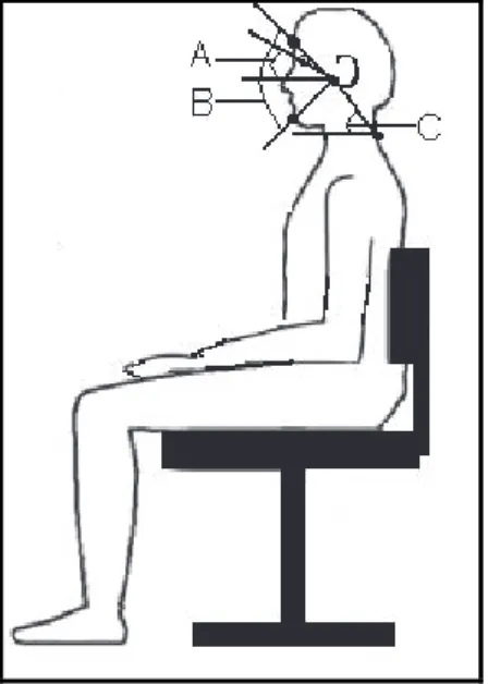
the onset of the chewing cycle, 2) Forward Head Posture at Maximum Mouth Opening (FHPMMO) expressed the angle value at the time of maximum mouth opening, 3) Forward Head Posture Amplitude (FHPA) calculated by the maximum extension of the head minus the minimum extention of head, 4) Forward Head Posture Mean (FHPM) calculated
by averaged to the angular values of the forward posture of the head.
Also was calculated the angle formed by the line connecting the tragus of the ear to the canthus of the eye and the horizontal, which gives the position of the upper cervical spine with increasing values
indicative of a more extended head13. From this
angle were analyzed variables: 1) Head Extension at Early Chewing (HEEC) expressed by the angle value at the onset of the chewing cycle, 2) Head Extension at Maximum Mouth Opening (HEMMO) expressed by the angle value at the time of maximum mouth opening, 3) Head Extension Amplitude (HEA) calculated by the maximum extension of the head minus the minimum extension of the head; 4) Head Extension Mean (HEM) averaged values of the angular extension of the head.
Also was calculated the opening of the mouth of a chewing cycle through the joint angle measured between the glabella, tragus and chin. The objective of this angle was to analyze the variables:
1) Mouth Opening Amplitude (MOA) calculated by the maximum opening of the mouth least the
minimum opening of the mouth (Figure 1). surface EMG of the AT and MA was performed
during chewing and during maximum voluntary
teeth clenching (MVC). The values of chewing were normalized by a MVC. All MVC were sustained for 5 s and repeated three times with an interval of 1 min between repetitions. A metronome with 60 beats per minute was used during the gathering of data, as
well as bars of parailm placed between the occlusal surface. The chewing task was repeated ive times,
with a duration of 10 s and 1 min intervals between each sampling.
Mandibular motion in 2-dimensional space was recorded in sagittal plane by the Canon Power Shot A710 IS® camera. This system tracks the
movements at a sample rate of 30 Hz. The subjects
were seated upright with camera on a tripod to 0.85 cm and a perpendicular distance of 1.2 m of volun-teers. The shooting occurred in the randomized side in DT group and the most affected side in CP group. For kinematic evaluation black spherical markers made on a white circular base were stuck with double-sided tape: glabella (midline of the face and 1 cm above the nose), canthus of the eye, tragus (cartilage above the ear), the tip of the chin, spinous process of C713. To calculate the real coordinates, a
system of two-dimensional calibration 1.0 x 1.0 was placed in the plane of the ilming.
Data Analysis
Of ive attempts to chewing performed for each subject, we analyzed only the irst three free of any
technical problem.
The kinematic analysis of head and jaw movements occurred in the medial chewing cycle cut from the electromyography signal through a
routine that runs through the already iltered EMG signal, using a ixed window size 200ms and deines the lowest RMS value of the signal. Having the
lowest RMS value and its standard deviation will
be deined the reference value to differentiate the
resting state and the activity muscular state14. The
reference value was used equal to 3σ (where σ is the standard deviation of the window of 200ms).
For scanning, which was performed visually, frame by frame, the system software used was the Ariel Performance Analysis System (APAS). The cycle synchronization between EMG and the
camera was made through a lash.
In kinematic analysis of chewing cycle was calcu-lated the angle formed by the line connecting C7 to the tragus of the ear and the horizontal, which gives the position of the head relative to the trunk. Its decreasing values are indicative of a more forward head posture13. From this angle were considered
other variables: 1) Forward Head Posture at Early Chewing (FHPEC) measured by the angle value at
RESULTS
The t test showed that there was no difference in the mean age, body mass and height between CP and TD groups (ρ> 0.05). In the group of children with TD, 50% (8/16) did not show any TMD signs or symptoms and 50% (8/16) are TMD, yet this same group 56.25% (9/16) have Angle class I, 37.50 % (6/16) Angle class II and 6.25% (1/ 16) Angle class III. In the group of children with CP, 56.25% (9/16) did not show any TMD signs or symptoms 43.75 % (7/16) are TMD; still in the CP group 43.75% (7/16) showed Angle class I, 37.50 % (6 /16) Angle class II and 18.75% (3/16) Angle class III. The Chi-square test showed that there is no association between TD and CP groups with TMD (p=0.72) and changes in occlusion (p=0.48). The cross-product ratio also showed that TD and CP and TMD/changes in occlusion are independent variables.
Table 1 shows the results of electromyography variables of the TD and CP groups. It was observed that the CP group has greater bilateral asymmetry between the right and left muscles MA, between the right and left AT muscles and a greater imbalance in
the activity of the four muscles analyzed. However, the only signiicant difference was considered on
ATS (p<0.05).
Table 2 shows the results of angular and
space-temporal kinematic variables to the extension of the head. CP children were higher extension of the head while chewing. However, the only signiicant difference was considered for the variable HEA (p<0.01) and the HEMMO (p<0.05).
Table 3 describes the results of angular and space-temporal kinematic variables of the forward head posture and the opening of the mouth. We observed greater forward head posture to the CP
group in all variables. For the angular FHPEC, FHPMMO and FHPM, the smaller the average
angular value the greater the forward head posture.
However, for space-temporal variable FHPA, the
higher the value of the displacement of the head the greater forward head posture, and it was the
only one that was statistically signiicant (p<0.05).
Also observed for the CP group, further opening the mouth during chewing cycle, however, this result
was not statistically signiicant.
Electromyography variables were analyzed:
Symmetry Index of AT and MA muscle (ATS and MAS) and the Antero– Posterior Coeficient
(APC)15 during a chewing cycle for each of the three
attempts. The symmetry index of muscle activity was calculated by temporal quantiication between
the normalized EMG waves of paired muscles and the common area between the two EMG waves
was identiied. The two EMG waves were super -imposed and the ratio between the super-imposed areas and the total area was calculated. The APC compare muscle activity between the MA and TA
muscles. In this index EMG waves were overlapped
and was calculated the ratio between the areas not overlapped and the overlapped MA and AT muscle areas of both sides. The activity of the muscles
analyzed is balanced, both the symmetry index as
the APC, when the value is 100 %.
The rectiication and iltering of the signals was also carried out with a cut-off frequency of 6HZ
to obtain the linear envelope, which was reduced to 100 points (root mean square– RMS EMG potentials). This processing was carried out using MATLAB software (Version 5.3 The MathWorks Inc.). For normalization, the EMG potentials were
expressed as a percentage of a maximum 1s RMS
value obtained across the three repetitions of the RVC for each muscle and subject.
Statistical Analysis
Participants were characterized using descriptive statistics (mean, standard deviation) and for each variable, the arithmetic mean of three attempts was considered. The difference between the mean age, body mass and height between the TD and CP groups was analyzed by using the Student’s T-test.
To verify the existence of the association and its
risk among TD and CP groups with TMD and the presence of changes in occlusion was applied, respectively, the chi-square test and odds ratio
(OR).
After checking the data normality by the Shapiro – Wilk test, we used the t test for independent data on parametric variables and the Mann-Whitney non– parametric variables. Analysis was performed using the program Statistical Package for Social Sciences (SPSS) version 17.0 for Windows, and for
all procedures was adopted signiicance level of 5%
Mean (SD) 95% CIM
P
TD CP TD CP
MAS a (%) 82,80 (7,73) 80,39 (9,37) 78,67- 86,92 75,40 - 85,39 0,49 ATS b (%) 85,10 (7,71) 81,44 (6,98) 80,99 - 89,21 77,72 - 85,16 0,04* CAP b (%) 85,82 (7,15) 84,16 (4,94) 82,01 - 89,64 81,53 - 86,80 0,62
Table 1 – Mean, standard deviation (SD) and conidence interval of the mean (95% CIM) of the
electromyographic variables of the groups with Typical Development (TD) (n = 16) and Cerebral Palsy (CP) (n = 16)
a T Test for independent data; b Mann-Whitney; Statistically signiicant difference: *p<0.05; MAS = Masseter Symmetry; TAS = Tempo-ral Symmetry; CAP = Coeficient antero-posterior.
Mean (SD) 95% CIM
P
TD CP TD CP
HEEC a (graus) 20,10 (6,59) 25,97 (14,09) 16,59 – 23,60 18,46 - 33,47 0,14
HEMMO a (graus) 21,18 (6,36) 29,93 (14,87) 17,79 - 24,57 22,00 – 37,85 0,04*
HEA b (graus) 4,21 (1,43) 8,54 (6,58) 3,45 – 4,97 5,04 – 12,05 0,00**
HEM a (graus) 20,65 (6,40) 27,24 (14,46) 17,24 – 24,06 19,54 – 34,95 0,11
Table 2 – Mean, standard deviation (SD) and conidence interval of the mean (95% CIM) of the
kinematic variables of angular head extension of the groups with Typical Development (TD) (n = 16) and Cerebral Palsy (CP) (n = 16)
a T Test for independent data; b Mann-Whitney; Statistically signiicant difference: * p<0.05, ** p<0.01; HEEC = Head Extension at Early Chewing; HEMMO = Head Extension at Maximum Mouth Opening; HEA = Head Extension Amplitude; HEM = Head Extension Mean.
Mean (SD) 95% CIM
P
TD CP TD CP
FHPEC (graus) 44,92 (7,55) 41,00 (9,26) 40,90 - 48,94 36,07 - 45,94 0,09 FHPMMO (graus) 45,14 (7,75) 41,41 (9,14) 41,01 - 49,27 36,54 - 46,28 0,05 FHPA (graus) 1,89 (0,82) 3,54 (2,35) 1,45 - 2,33 2,29 - 4,79 0,01* FHPM (graus) 44,99 (7,68) 41,78 (9,16) 40,90 - 49,09 36,90 - 46,66 0,09
MOA (graus) 9,73 (3,80) 12,80 (6,59) 7,71 - 11,76 9,28 - 16,31 0,11
Table 3 – Mean, standard deviation (SD) and conidence interval of the mean (95% CIM) kinematic
variables of angular head previous projection and mouth opening of the groups with Typical Development (TD) (n = 16) and Paralysis cerebral palsy (CP) (n = 16)
Test de Mann-Whitney; Difference statistically signiicant: * p<0,05; FHPEC = Forward Head Posture at Early Chewing; FHPMMO = Forward Head Posture at Maximum Mouth Opening; FHPA = Forward Head Posture Amplitude; FHPM = Forward Head Posture Mean; MOA = Mouth Opening Amplitude.
DISCUSSION
Among the changes commonly found on
neuro-logical examination of individuals with CP are
the asymmetries of posture, muscle tone and/or skills functional16. This asymmetry was found also
during chewing task. For Both muscles, AT and MA, the asymmetry of electrical activity during an
isotonic contraction was larger in the CP group.
However, the difference was signiicant only for the AT muscle. Corroborating these indings, Ries
and Bérzin (2009) found greater asymmetry in the activity of MA and AT muscles during mastication, both during isometric and isotonic contraction8.
Disorders of tone, posture and movement of the
CP child inluence also muscle activity involved in
lead to protrusion or retraction of the tongue and an inadequate jaw movement23. Likewise, the correct
head extension obtains biomechanical advantages
which promote coordination between the head and jaw and enhances force production during bite5.
Although it found greater forward posture and
extension of the head in CP children, the difference
in mouth opening amplitude between the groups
was not signiicant. Corroborating these indings,
Ries and Bérzin (2005)12 also found no signiicant
difference in maximal mouth opening in children
with CP and TD, and the same is presented in a similar manner and within the normal range for both groups.
These changes in head posture found in CP children could be etiologic factors of TMD, for
inluence in jaw rest position and cause dysfunction of the masticatory muscles. However, postural
changes of the head are not necessarily more frequent in subjects with TMD4. There are also
reports that the asymmetric electrical activity of masticatory muscles is higher in individuals with TMD24,25 and this abnormal activity can be inlu
-enced by changes oclusais15. It was considered
that CP children have a higher incidence of occlusal changes due to abnormalities of oromotor muscu-lature, with a higher proportion of Angle class II and minor of Angle class III10. Although in this study the
CP children had higher asymmetry of MA and AT muscles and major changes in head movement during jaw movement, these changes do not seem to have increased the risk of TMD and occlusal changes. The TMD and occlusal changes were not associated with the presence of CP.
The functional movements of the jaw are the result of the coordinated activation of the jaw and neck muscles, allowing simultaneous movements between the temporomandibular, atlanto-occipital and cervical spine joints5. The results of this
study show that motor and functional limitations of children with CP are associated with abnormalities in the control of jaw and head movements during the chewing task. It is important to evaluate cervical movements during the evaluation of orofacial myofunctional disorders in child with CP. The appro-priate development of masticatory function provides muscle balance preventing disturbances in
cranio-facial complex.
CONCLUSION
The greater asymmetry in muscle activity during the chewing cycle, with greater change in length
and forward head posture shows the greatest difi -culty in controlling the jaw and head movements of children with CP. These changes in jaw and In general, the jaw movement during mastication
may seem simple, but careful observation show
asymmetrical characteristic and exhibit large varia -tions from cycle to cycle17. Some authors admit the
usual an index of symmetry for healthy adults at
least 82 ± 1.34%18 and others determined the usual
parameters of MA symmetry of 87.11 ± 1.60% and AT 88,11 ± 1.45%19. Therefore, some degree of
asymmetrical activity should be considered in most individuals, since the skull is rarely symmetrical and muscular system to try to redress this skeletal imbalance generates also asymmetric forces 20.
If we consider as normal the parameters of symmetry Felicio, Sidequersky, Tartaglia, and Sforza (2009)19, the two groups could be within the
normality. Already parameters of Ferrario, Sforza, Miani Jr, D’ Addona, and Barbini (1993)18 show that
symmetry of both the AT muscle (81,44%) and MA muscle (80.39%) in the CP group, is outside the
normality. The characteristic exacerbate activity
asymmetry of these muscles in CP children can impair the performance of the chewing task.
The AT muscle has the function of elevation and retraction of the mandible during mastication and unlike MA muscle is more related to jaw movement than masticatory force21. Additionally, accounts for
balance and postural control of the jaw. Thus, the greatest imbalance in the activity of the AT muscle in CP children could be due to changes in muscle tone, posture and movements in this pathology, changing the balance and postural control of the jaw.
The limitation in muscular synergisms in CP children is considered one of the causes of disability and functional limitation of same1. However, this
limitation was not observed in balance of the EMG activity of four muscles analyzed. CP and TD groups showed similar values of the APC.
The abnormal tone found in children with CP can result in abnormal patterns of posture and movement such as, delay and decrease of the head control22.
The start of head extension generally precedes the
beginning of jaw opening, which indicates the antici-patory adjustment of the head position preparatory to jaw movement5. The muscles of the jaw and neck
have associated movements and changes in one of the structures can disrupt the other.
The HEMMO, the HEA and the FHPA in the CP group were signiicantly higher compared to the
TD. These changes in the forward head posture
and head extension could be explained by the
lower control of posture and movement of the head
and the presence of an extensor pattern present in children with spastic CP. Hyperextension of the
head in these children affect the elevation of the
occlusion, although this group exhibit greater motor
impairment and greater change in control of the head and jaw movements during chewing activity. cervical motor control can be causes of disorders
in oral motor function of CP children. The CP was not associated with TMD or the alteration of dental
RESUMO
Objetivo: analisar a atividade elétrica dos músculos Temporal e Masseter e o padrão de postura e movimento de cabeça e mandíbula de crianças com Paralisia Cerebral (PC). Métodos: a amostra deste estudo compreendeu 32 voluntários com PC espástica e com Desenvolvimento Típico, com a
faixa etária de 7 a 13 anos de idade, caracterizados com base na Classiicação de Angle e Critério
de Diagnóstico para Pesquisa das Disfunções Temporomandibulares (RDC/TMD). De forma simul-tânea, foram avaliadas a postura e movimentação da cabeça e mandíbula e a atividade elétrica dos
músculos Temporal e Masseter por meio da cinemática e eletromiograia. Resultados: a PC não foi associada a presença de DTM ou com a alteração da oclusão dentária. No grupo PC, foi observada
maior assimetria do músculo temporal (p<0.05), maior extensão da cabeça na máxima abertura da boca (p<0.05), maior amplitude de extensão da cabeça (p<0.01) e maior amplitude de projeção ante -rior da cabeça (p<0.05). Conclusão: a maior assimetria na atividade muscular, a maior extensão e projeção anterior da cabeça durante o ciclo mastigatório podem ser causas das desordens da função motora oral das crianças com PC.
DESCRITORES: Paralisia Cerebral; Músculos Mastigatórios; Eletromiograia
REFERENCES
1. Mayston MJ. People with cerebral palsy: effects of and perspectives for therapy. Neural Plast. 2001;8(1-2):51-69.
2. Cesa CC, Ecco CT, Bersch R, Chiappetta ALML. Functions of the stomatognathic system and
motor-speech relexes in children with chronic spastic
quadriparesis encephalopathy. Rev CEFAC. 2004;6(2):158-63.
3. Vivone GP, Tavares MMM, Bartolomeu RS, Nemr K, Chiappetta ALML. Analysis of alimentary consistency and deglutition time in children with spastic quadriplegic cerebral palsy. Rev CEFAC. 2007;9(4):504-11.
4. Visscher CM, De Boer W, Lobbezzo F, Habets
LL, Naeije M. Is there a relationship between head
posture in craniomandibular pain? J Oral Rehabil.
2002;29(11):1030-6.
5. Eriksson P-O, Häggman-Henrikson B, Nordh E, Zafar H. Co-ordinated Mandibular and Head-Neck
Movements during Rhythmic Jaw Activities in Man. J Dent Res. 2000;79(6):1378-84.
6. Igarashi N, Yamamura K, Yamada Y, Kohno
S. Head movements and neck muscle activities
associated with the jaw movement during mastication in the rabbit authors. Brain Res. 2000;871(1):151-5.
7. Val DC, Limongi SCO, Flabiano FC, Silva
KCL. Stomatognathic system and body posture
in children with sensoriomotor déicits. Pro Fono.
2005;17(3):345-54.
8. Ries LGK, Bérzin F. Asymmetric activation of temporalis and masseter muscles in children with cerebral palsy. Fisioter Mov. 2009;22(1):45-52. 9. Palisano R, Rosenbaum P, Walter S, Russell D, Wood E, Galuppi B. Development and reliability of a system to classify gross motor function in children with cerebral palsy. Dev Med Child Neurol. 1997;39:214-23.
10. Winter K, Baccaglini L, Tomar S. A review of malocclusion among individuals with mental and physical disabilities. Spec Care Dentist. 2008;28(1):19-26.
11. Dworkin SF, Leresche L. Diagnostic criteria for temporomandibular disorders: review, criteria,
examinations and speciications, critique. Cranio.
1992;6(4):301–55.
12. Ries LGK, Bérzin F. Signs and symptoms of temporomandibular disorders in children with cerebral palsy. Rev Bras Fisioter. 2005;9:341-6. 13. Silva AG, Punt TD, Sharples P, Vilas-Boas JP,
Johnson MI. Head posture and neck pain of chronic
20. Widmalm SE, Lee Y-S, Mckay D. Clinical use of qualitative electromyography in the evaluation of jaw muscle function: A practitioner’s guide. Cranio. 2007;25(1):63-73.
21. Gomes CF, Trezza EMC, Murade ECM, Padovani CR. Surface electromyography of facial
muscles during natural and artiicial feeding of
infants. J Pediatr. 2006;82(2):103-9.
22. Jones MW, Morgan E, Shelton JE, Thorogood C. Cerebral palsy: introduction and diagnosis (part
I). J Pediatr Health Care. 2007;21(3):146-52.
23. West JF, Redstone F. Alignment during feeding and swallowing does it matter? A review. Percept Mot Skills. 2004;98(1):349-58.
24. Ries LGK, Alves MC, Bérzin F. Asymmetric activation of temporalis, masseter, and sternocleidomastoid muscles in temporomandibular disorder patients. Crânio. 2008;26(1):59-64.
25. Tartaglia GM, Silva MAMR, Bottinia S, Sforza C, Ferrario VF. Mastigatory muscle activity during
maximum voluntary clench in different research
diagnostic criteria for temporomandibular disorders (RDC/TMD) groups. Man Ther. 2008;13(5):434-40.
14. Abbink JH, Van Der Bilt A, Van Der Glas HW.
Detection of onset and termination of muscle
activity in surface electromyograms. J Oral Rehabil.
1998;25(5):365-9.
15. Ferrario VF, Tartaglia GM, Galletta A, Grassi
GP, Sforza C. The inluence of occlusion on jaw and
neck muscle activity: a surface EMG study in healthy
young adults. J Oral Rehabil. 2006;33(5):341-8.
16. Aruin AS. The effect of asymmetry of posture on anticipatory postural adjustments. Neurosci Lett. 2006;401:150-3.
17. Green JR, Moore CA, Ruark JL, Rodda PR, Morvée WT, Vanwitzenburg MJ. Development of chewing in children from 12 to 48 months: longitudinal study of EMG patterns. J Neurophysiol. 1997;77(5):2704-16.
18. Ferrario VF, Sforza C, Miani Jr A, D’Addona A, Barbini E. Electromyographic activity of human masticatory in normal young people. Statistical evaluation of reference values for clinical
applications. J Oral Rehabil. 1993;20(3):271-80.
19. Felicio CM, Sidequersky FV, Tartaglia GM, Sforza C. Electromyographic standardized indices in healthy Brazilian young adults and data
reproducibility. J Oral Rehabil. 2009;36(8):577-83.
Received on: May 28, 2012 Accepted on: September 05, 2013
Mailing address: Lilian Gerdi Kittel Ries
Rua Pascoal Simone, 358 – Coqueiros Florianópolis – SC
CEP: 88080-350