2255-4823/$ – see front matter © 2013 Elsevier Editora Ltda. All rights reserved.
www.ramb.org.br ARTIGOS
ARTIGOS ORIGINAIS _____________Qualidade da informação da internet disponível para pacientes em páginas em português ___________________________________________________________645 Acesso a informações de saúde na internet: uma questão de saúde pública? ______650 Maus-tratos contra a criança e o adolescente no Estado de São Paulo, 2009_______659 Obesidade e hipertensão arterial em escolares de Santa Cruz do Sul – RS, Brasil ___666 Bone mineral density in postmenopausal women with and without breast cancer ___673 Prevalence and factors associated with thoracic alterations in infants born prematurely __________________________________________________679 Análise espacial de óbitos por acidentes de trânsito, antes e após a Lei Seca, nas microrregiões do estado de São Paulo ___________________________________685 Sobrevida e complicações em idosos com doenças neurológicas em nutrição enteral ______________________________________________________691 Infliximab reduces cardiac output in rheumatoid arthritis patients without heart failure ______________________________________________________698 Análise dos resultados maternos e fetais dos procedimentos invasivos genéticos fetais: um estudo exploratório em Hospital Universitário _______________703 Frequência dos tipos de cefaleia no centro de atendimento terciário do Hospital das Clínicas da Universidade Federal de Minas Gerais __________________709 ARTIGO DE REVISÃO ______________Influência das variáveis nutricionais e da obesidade sobre a saúde e o metabolismo __714
PONTO DE VISTAOs paradoxos da medicina contemporânea 634
IMAGEM EM MEDICINAObstrução duodenal maligna: tratamento endoscópico paliativo utilizando prótese metálica autoexpansível 636 Gossypiboma 638
tratamento cirúrgico 639
ACREDITAÇÃO
Atualização em perda auditiva: diagnóstico radiológico 644
www.ramb.org.br
Revista da
ASSOCIAÇÃO MÉDICA BRASILEIRA
Original article
High-frequency oscillatory ventilation in children with acute
respiratory distress syndrome: experience of a pediatric
intensive care unit
q
Anelise Dentzien Pinzon
a,b, Taís Sica da Rocha
a, Cláudia Ricachinevsky
a,
Jefferson Pedro Piva
c, Gilberto Friedman
b,c,*
aPediatric Intensive Care Unit, Hospital de Criança Santo Antonio, Complexo Hospitalar Santa Casa, Porto Alegre, RS, Brazil
bPostgraduate Program in Pneumology, Universidade Federal do Rio Grande do Sul (UFRGS), Porto Alegre, RS, Brazil
cMedical School, UFRGS, Porto Alegre, RS, Brazil
A RT I C L E I N F O
Article history:
Received 11 October 2012 Accepted 11 February 2013
Keywords:
Acute respiratory distress syndrome High frequency oscillatory
ventilation Respiratory failure Pediatrics
Protective mechanical ventilation
qStudy conducted at Universidade Federal do Rio Grande do Sul, Porto Alegre, RS, Brazil. * Corresponding author.
E-mail: [email protected] (G. Friedman).
A B S T R A C T
Objective: To describe the effects of high-frequency oscillatory ventilation (HFOV) as a rescue ventilatory support in pediatric patients with acute respiratory distress syndrome (ARDS).
Methods: Twenty-five children (1 month < age < 17 years) admitted to a university hospital pediatric intensive care unit (ICU) with ARDS and submitted to HFOV for a minimum of 48 hours after failure of conventional mechanical ventilation were assessed.
Results: 28 days after the onset of ARDS, the mortality rate was 52% (13/25). Over the course of 48 hours, the use of HFOV reduced the oxygenation index [38 (31-50) vs. 17 (10 - 27)] and increased the ratio of partial arterial pressure O2 and fraction of inspired O2 [65 [44-80) vs. 152 (106-213)]. Arterial CO2 partial pressure [54 (45-74) vs. 48 (39-58) mmHg] remained unchanged. The mean airway pressure ranged between 23 and 29 cmH2O. HFOV did not compromise hemodynamics, and a reduction in heart rate was observed (141 ± 32 vs. 119 ± 22 beats/min), whereas mean arterial pressure (66 ± 20 vs. 71 ± 17 mmHg) and inotropic score [44 (17-130) vs. 20 (16-75)] remained stable during this period. No survivors were dependent on oxygen.
Conclusion: HFOV improves oxygenation in pediatric patients with ARDS and severe hypoxemia refractory to conventional ventilatory support.
Ventilação oscilatória de alta frequência em crianças com síndrome da angústia respiratória aguda: experiência de um centro de tratamento intensivo pediátrico
R E S U M O
Objetivo: Descrever os efeitos da aplicação da ventilação de alta frequência oscilatória como suporte ventilatório de resgate em uma série de pacientes pediátricos com síndrome da angústia respiratória aguda (SARA).
Métodos: Participaram do estudo 25 crianças (> 1 mês e < 17 anos) internadas em uma UTI pediátrica universitária com SARA e submetidas à ventilação de alta frequência oscilatória (VAFO) por um mínimo de 48 horas, após falha da ventilação mecânica convencional.
Resultados: A taxa de mortalidade foi de 52% (13/25) 28 dias após o início da SARA. Ao longo de 48 horas, a aplicação da VAFO reduziu o índice de oxigenação [38 (31-50) vs. 17 (10-27)] e aumentou a relação pressão arterial parcial de O2/fração inspirada de O2 [65 (44-80) vs. 152 (106-213)]. A pressão arterial parcial de CO2 [54 (45-74) vs. 48 (39-58) mmHg] manteve-se inalterada. A pressão média de vias aéreas oscilou entre 23 e 29 cmH2O. A VAFO não comprometeu a hemodinâmica e observou-se uma redução da frequência cardíaca (141 ± 32 vs. 119 ± 22 bat/min), a pressão arterial média (66 ± 20 vs. 71 ± 17 mmHg) e o escore inotrópico [44 (17-130) vs. 20 (16-75)] mantiveram-se estáveis nesse período. Nenhum sobrevivente ficou dependente de oxigênio.
Conclusão: VAFO melhora a oxigenação de pacientes pediátricos com SARA grave e hipoxemia refratária ao suporte ventilatório convencional.
© 2012 Elsevier Editora Ltda. Todos os direitos reservados.
Palavras-chave:
Síndrome da angústia respiratória aguda
Ventilação de alta frequência oscilatória
Insuficiência respiratória Pediatria
Ventilação mecânica protetora
Introduction
The prevalence of acute respiratory distress syndrome (ARDS)
in pediatric intensive care units varies from 2% to 7.6%.1,2 In
pediatrics, ARDS is associated with high mortality rates, which vary according to the service, population studied, and risk factors. Clinical studies suggest that mechanical ventilation (MV) may modify inflammatory responses in patients with acute lung injury. In patients with prior pulmonary and
systemic inflammation, ventilation with tidal volumes (VT) of
10-15 mL/kg of ideal body weight (IBW) and moderate-to-low levels of positive end-expiratory pressure (PEEP) are associated with increased levels of intra-alveolar and systemic
inflammatory mediators.3 In contrast, mechanical ventilation
with moderate-to-high levels of PEEP and reduced VT of
approximately 6 mL/kg of IBW ensured proper gas exchange, reduced systemic and intra-alveolar inflammatory mediators,
and decreased mortality.3-6
The use of protective ventilatory strategies that prevent further lung injury associated with MV is a major concern in any patient, including those without acute
pathology.7 High-frequency oscillatory ventilation (HFOV)
is a protective ventilatory strategy, as it optimizes alveolar recruitment and lung volume, as well as improves oxygenation by applying high-flow rates and frequencies up to 900 cycles per minute with reduced tidal volumes (1-2 mL/kg) resulting from minor differences in inspiratory and expiratory pressures,
producing a high and persistent mean airway pressure.8
HFOV appears to represent an important therapeutic option in ventilatory support of children with respiratory failure.
Despite the increased use of HFOV in pediatric patients with acute respiratory failure, there have been few published studies, as well as few prospective studies and randomized clinical trials
involving children with ARDS.9-12 HFOV has been more often
used as a rescue therapy in children with severe respiratory failure after failure of conventional mechanical ventilation
(CMV) with lung protective strategies.12-15 However, to date
there is insufficient evidence to support its use.16,17 When HFOV
is shown to be effective as a rescue therapy, this ventilation
mode will become an extremely useful therapeutic option.18,19
The present study aimed to describe the effects of the use of HFOV as a rescue ventilatory support over oxygenation and ventilation in pediatric patients diagnosed with ARDS.
Methods
Study design
This was an observational and retrospective study performed through the analysis of medical records of children admitted
between 2005 and 2010 with ARDS,20 submitted to HFOV due
to treatment failure with conventional mechanical ventilation.
Patient selection
Committee of the Complexo Hospitalar Santa Casa de Porto Alegre (registration No.: 1935/08).
Patients were considered eligible for the study according to
the following criteria: a) 1 month < age < 17 years, b) used HFOV
for ARDS management (chest X-ray with bilateral infiltrates, ratio of partial pressure of arterial oxygen and inspired oxygen
fraction [PaO2/FiO2] ≤ 200, and no clinical evidence of left atrial
hypertension); c) failed protective conventional mechanical ventilation (CMV) (children: peak inspiratory pressure
[PIP] > 35 cmH2O, mean airway pressure [MAwP] > 15-18 cmH2O
and FiO2 ≥ 0.6; term infants: MAwP ≥ 10-12 cm H2O, FiO2 ≥ 0.6,
and failure to increase lung volume); and d) complete medical records. The decision to switch to HFOV considering the difficulty to maintain ventilatory parameters/oxygenation was made by the attending physician.
Patients were excluded from the study if HFOV was applied for less than 48 hours in the event of death or early weaning from HFOV in the same period.
Data were collected on diagnosis (primary and associated) and outcome variables (time in HFOV, time in CMV before and after HFOV, duration of ICU stay, in-hospital mortality, and mortality at day 28 after the ARDS diagnosis).
Ventilatory strategies
Conventional mechanical ventilation
Initially, all patients used pressure-controlled CMV (Servo 300, Siemens-Elema AB –Sweden; SERVOi, Maquet GmbH & Co, KG – Rastatt, Germany). The ventilation strategy
consisted of “protective ventilation” with FiO2 < 0.5, tolerating
a saturation of arterial hemoglobin oxygen (SaO2) > 85%,
permissive hypercapnia as long as pH > 7.2, and a tidal
volume < 7 mL/kg of ideal body weight. The ventilation mode
used was synchronized intermittent mandatory ventilation with controlled pressure and assisted pressure. The general support included sedation (continuous infusion of opioid and benzodiazepine), fluid maintenance, nutritional support, and antibiotics when indicated. Whenever necessary, a muscle relaxant (pancuronium) was used to facilitate mechanical ventilation. Hemodynamic support with vasopressors/inotropes and/or fluids was administered through a central venous catheter when necessary.
High-frequency oscillatory ventilation
All patients submitted to HFOV were ventilated with a High-Frequency Oscillator Sensor Medics 3100B (Sensor Medics – Yorba Linda, CA, USA). Until the year 2007, there was no protocol for the start of HFOV, and HFOV parameters were chosen at the discretion of the attending physician. From 2008 onward, the following protocol was adopted:
MAwP = 5 cmH2O above the MAwP in CMV, FiO2 = 1.0,
amplitude adjusted to achieve adequate power for chest wall vibration, and airflow maintained at 30 mL/min. The initial oscillatory frequency was adjusted between 10-15 Hz. To attain
HFOV weaning, FiO2 was kept between 0.4 and 0.6, followed
by a decrease of 1 to 2 cmH2O to decrease airway pressure.
Regarding ventilation, there were progressive reductions (3-5 cmH2O) in amplitude pressure. CMV would be resumed
when airway pressure was ≤ 20 cmH2O, FiO2 ≤ 0.4, and when
the patient tolerated the aspiration of the endotracheal tube
without oxygen saturation decrease.21,22
Monitoring
Arterial blood gases and ventilatory parameters were collected in CMV (peak inspiratory pressure [PIP], positive pressure at the end of exhalation, positive end-expiratory pressure
[PEEP], respiratory rate [RR], fraction of inspired oxygen [FiO2],
inspiratory time) at the beginning of HFOV use and after 6, 12, 24, and 48 hours (mean airway pressure [MAwP], amplitude
[AMP], RR, FIO2). The oxygenation index (OI = [MAwP X FIO2 X
100]/PaO2)23 and the PaO
2/FiO2 ratio were calculated in the
same time intervals. Hemodynamic parameters (heart rate [HR] and mean arterial pressure [MAP]) and inotropic score
(dopamine X 10 + adrenaline X 100) were obtained over
48 hours.24 Patient severity was evaluated through the Pediatric
Index of Mortality (PIM) score.25
Statistical analysis
Analysis of variance and Student’s t-test were used to analyze the data with normal distribution (Tukey’s test for comparisons). The nonparametric Mann-Whitney test and Friedman’s ANOVA (Dunn test for comparisons) were used for variables with non-normal distribution. Results were expressed
as mean ± standard deviation or median (25-75 percentile).
Results
Patient characteristics
Table 1 describes patient characteristics and mortality rates. There were 31 identified patients who were diagnosed with ARDS, submitted to HFOV during a five-year period. Six patients were excluded; five died within less than 24 hours, and one was weaned from HFOV before 48 hours, leaving 25 patients at the final analysis. The patients had high risk of death with high mortality rate and aggressive ventilatory support before the use of HFOV. The associated comorbidities were: postoperative of
congenital surgery (n = 6), Cushing’s syndrome (n = 1), anoxic
encephalopathy (n = 3), hematologic malignancies (n = 3), major
burn injury (n = 1), late complications of kidney transplantation
(n = 1), cytomegalovirus (n = 1), pulmonary lymphangioma (n = 1),
postoperative of late kidney transplantation (n = 1), prematurity
(n = 3), neonatal anoxia (n = 1), hyaline membrane disease (n = 1),
bronchopulmonary dysplasia (n = 1), postoperative of pulmonary
surgery (n = 2), and nonspecific immune deficiency (n = 1).
Ventilation and oxygenation parameters
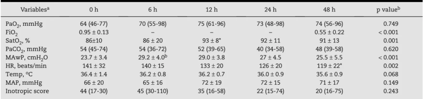
After 48 hours of HFOV, FiO2 decrease and a significant increase
the oxygenation index and increasing the PaO2/FiO2 ratio
(Fig. 1) over 48 hours. PaCO2 remained almost unchanged. The
MAwP necessary to maintain oxygenation with progressive
reduction of FiO2 during the 48 hours of HFOV ranged between
23 and 29 cmH2O.
Hemodynamic parameters
Before HFOV use, 20 patients were receiving one or a combination of vasoactive drugs; 24 hours after the start of HFOV, three other patients needed a drug or combination
of vasoactive drugs (dopamine, n = 22; noradrenaline, n = 6;
adrenaline, n = 10; milrinone, n = 3). The two main causes
of hemodynamic instability were septic shock (n = 17) and
postoperative of heart surgery. Only two patients did not need vasoactive drugs. Even with high mean airway pressures, hemodynamic performance was not impaired by HFOV; it was also observed that HR decreased significantly and MAP remained stable. Moreover, the inotropic score remained unchanged during the evaluation period.
In seven patients with bronchiolitis, the PaO2/FiO2 ratio
increased from 62 ± 25 to 193 ± 114 (p = 0.027), and OI decreased
from 48 ± 17 to 15 ± 7 (p = 0.001) over 48 hours. Furthermore,
the PaCO2 decreased (59 ± 17 vs. 42 ± 10 mmHg, p = NS) during
this same period.
Clinical outcomes
Table 1 shows the main clinical outcomes. The improvement of the parameters related to oxygenation was higher in survivors than in non-survivors (Table 2). No survivors were dependent on oxygen. Among the complications potentially related to ventilation and/or pulmonary disease, ten patients had nonhypertensive pneumothorax without additional hemodynamic involvement.
Comparison between the pre-protocol and post-protocol periods of high-frequency oscillatory ventilation implementation
Table 3 describes the comparison of the main physiological and clinical outcomes for the two periods. No significant differences were observed among the seven patients who were ventilated without an adjuvant protocol and the remaining 18 patients who were ventilated based on the established HFOV protocol since 2008.
Discussion
The present study, which involved a sample of patients with severe ARDS submitted to rescue HFOV, did not allow for the determination of the true effectiveness of this method. However, the results indicate that HFOV significantly improves gas exchange and allows reductions in oxygen supply. These findings are consistent with other studies that evaluated the use of HFOV in pediatric patients with ARDS, and suggest that the benefit would be greater with an earlier start of
Variables n = 25
Age (months) 9 (4-81)
Weight (kg) 7 (4-19)
Gender (M/F) 13/12
PIM 30 ± 24
Mortality rate 28 days after ARDS 52% (13/25)
Time in ICU (days) 19 (13-37)
Time of HFOV (h) 82 (72-144)
Time in ICU pre-death (days) 17 (12-37)
Time of CMV pre-HFOV (hours) 24 (19-144)
Time of CMV post-HFOV (hours) 72 (0-276)
PIP (mmHg) 37 ± 6
PEEP (cmH2O) 11 ± 4
RR (resp/min) 34 ± 9
FiO2 0.95 ± 0.13
Diagnosis
Pneumonia 9
Pneumonia (RSV+) 1
Bronchiolitis (RSV+) 5
Bronchiolitis 2
Extra-pulmonary ARDS 6
ARDS, acute respiratory distress syndrome; CMV, conventional
mechanical ventilation; F, female; FiO2, fraction of inspired
oxygen; ICU, intensive care unit; HFOV, high-frequency oscillatory ventilation; M, male; PEEP, positive end-expiratory pressure; PIM, pediatric index of mortality; PIP, peak inspiratory pressure; RR, respiratory rate; RSV, respiratory syncytial virus.
Data are shown as median (25-75 percentiles) or mean ± standard
deviation.
Each patient may have had more than one diagnosis.
Table 1 – Patient characteristics, respiratory failure severity, and clinical outcomes.
Time (hours)
0 6 12 24 48
Oxigenation index
0 20 40 60 80 100 120
Figure 1 – Changes in oxygenation index and arterial partial pressure of oxygen/fraction of inspired oxygen during the initial 48 hours of high-frequency oscillatory ventilation (HFOV). HFOV was established at time 0, which represents the values immediately before the HFOV. Values are expressed as median with 25-75% percentiles. ap < 0.001
(Friedman’s ANOVA); bp < 0.05, compared to the previous
HFOV, especially in the first 24 hours in cases associated with
refractory hypoxemia.10,16,26 Even though the median time of
CMV before HFOV was around 24 hours in this study, it must be concluded that the indication was delayed. It can be observed
that at the moment of transition, a mean FiO2 of 95% was used
in CMV, as well as a mean PIP of 37 mmHg, thus maintaining a high shunt fraction (refractory hypoxemia). Therefore, the use of HFOV should not be based on time of evolution, but on refractoriness to CMV.
The decision to indicate HFOV defined by a criterion of refractory response to CMV is reinforced by another observation from the present study. There were no differences between patients submitted to HFOV with no defined protocol (up to 2007) when compared with those in whom HFOV was used according to clear definitions of utilization. Patients did not differ in severity or ventilatory parameters at the beginning of HFOV implementation, and had the same clinical outcome. It can be speculated that the definition of decision criteria for changing the ventilatory method is more important than HFOV implementation using a strict protocol.
HFOV, even when started late, promoted significant
improvement in OI and PaO2/FiO2 ratio during the 48 hours.
Most studies have indicated HFOV as a rescue ventilatory
support for ARDS patients who had difficulties in CMV with
worsening of OI.10,12,17 A survey among 14 centers, which
included 232 pediatric patients, demonstrated a mean OI
of 27 before HFOV.12 In the present study, when HFOV was
indicated, the mean OI was almost 40, confirming that the decision to perform the transition was probably late for most cases. Several studies have focused on OI as a predictor of
mortality after the transition to HFOV.12,16 Sarnaik et al.
suggested that, in patients with initial OI < 20, the absence
of a decrease of at least 20% in OI within the first six hours of
HFOV may be considered a predictor of death.15
Classically, HFOV uses relatively high MAwP, allowing for a more effective maintenance of lung recruitment than
that promoted by the use of PEEP in CMV.22,27 In the present
study MAwP increased significantly after start of HFOV, with significant improvement in oxygenation indices, suggesting the opening of a major portion of alveolar units with improved
gas exchange (alveolar recruitment). The impact on PaCO2 was
not significant due to adjustments in the amplitude of the respirator, in order to prevent unnecessary and unwanted
alveolar hyperventilation.9,16,17,28
With the increase in MAwP during HFOV, hemodynamic impairment can occur, as pleural pressure elevation
Variablesa 0 h 6 h 12 h 24 h 48 h p valueb
PaO2, mmHg 64 (46-77) 70 (55-98) 75 (61-96) 73 (48-98) 74 (56-96) 0.749
FiO2 0.95 ± 0.13 – – – 0.55 ± 0.22 < 0.001
SatO2, % 86±10 86 ± 20 93 ± 8* 92 ± 11 91 ± 13 0.001
PaCO2, mmHg 54 (45-74) 54 (36-72) 52 (39-65) 40 (34-58) 48 (39-58) 0.620
MAwP, cmH2O 23.7 ± 3.4 29.2 ± 4.0b 29.0 ± 3.8 27 ± 4.5 25.5 ± 5.5 < 0.001
HR, beats/min 141 ± 32 140 ± 15 133 ± 20 126 ± 20 119 ± 22* 0.002
Temp, oC 36.4 ± 1.4 36.2 ± 0.8 36.2 ± 0.7 36.0 ± 0.9 35.6 ± 0.9 0.068
MAP, mmHg 66 ± 20 65 ± 16 72 ± 19 72 ± 15 71 ± 17 0.149
Inotropic score 44 (17-30) 45 (30-110) 35 (16-58) 22 (15-74) 20 (16-75) 0.243
FiO2, fraction of inspired oxygen; HR, heart rate; MAP, mean arterial pressure; MAwP, mean airway pressure; PaO2, arterial partial pressure of
oxygen; PaCO2, partial pressure of arterial carbon dioxide, SatO2, arterial oxygen saturation; Temp, temperature.
aDescribed by median (percentiles 25-75) or mean ± SD. Analysis of variance (ANOVA) or Friedman’s test.
bp < 0.05 comparatively to the previous level (Tukey’s or Dunn’s test).
Table 2 – Alterations in blood gas, oxygenation, and hemodynamic variables within the first 48 hours.
Variablesa 0 h 6 h 12 h 24 h 48 h
PaO2, mmHg NS 56 (46-73) 67 (42-86) 66 (48-79) 65 (43-90) 59 (52-76)
S 65 (47-77) 74 (61-115) 92 (77-109) 78 (62-108) 88 (71-118)
PaO2/FiO2 NS 56 (42-73) 69 (56-138) 109 (75-151)b 123 (75-145) 112 (82-139)
S 67 (47-88) 94 (73-171) 116 (92-175) 145 (123-271)b 197 (161-267)a
OI NS 41 (36-59) 39 (24-59) 27 (22-46) 22 (13-42) 17 (14-35)
S 35 (27-44) 34 (13-42) 28 (13-33) 19 (10-25) 13 (7-21)
PaCO2, mmHg NS 48 (43-63) 55 (38-67) 44 (36-66) 39 (34-51) 47 (44-52)
S 63 (48-76) 54 (31-76) 59 (42-69) 45 (34-68) 44 (37-52)
FiO2, fraction of inspired oxygen; OI, oxygenation index; PaO2, arterial partial pressure of oxygen; PaCO2, partial pressure of arterial carbon
dioxide.
ap < 0.005 comparatively to the previous level (Dunn’s test).
bp = 0.004 between groups.
causes a decrease in venous return and cardiac output. Most patients in the present study were already receiving inotropic-vasoactive drugs during CMV; the use of HFOV did not impair hemodynamic stability and there was a decrease in hemodynamic support throughout the 48 hours. A study by Mehta et al. in adult patients showed that HFOV can lead to increased filling pressures and significant decrease
in cardiac output.29 In contrast, Derdak et al. found no
significant differences in heart rate, mean arterial pressure, or cardiac output between adult patients undergoing HFOV versus those submitted to CMV within the first 72 hours of
treatment.30 Although cardiac output was not measured in
the present study, the observed hemodynamic performance suggests that there was no additional blood flow impairment in the present patients, as MAP remained stable and HR decreased.
The mortality from ARDS in children has been decreasing
to around 20%.31-33 Although some researchers have
estimated that it is higher,34 with explicit protocols in certain
populations of children with ARDS, mortality can be as low
as 8%.35 However, ARDS patients continue to be among those
at higher risk in pediatric ICUs, with prolonged mechanical ventilation time and increased risk for nosocomial infections, as well as increased risk for unknown respiratory morbidities and neurodevelopmental injuries. In the present study, a mortality rate of 52% after 28 days of ARDS diagnosis and treatment with HFOV was observed. When evaluating the high mortality rate from ARDS observed in this group, it should be noted that: a) this was a selected group of patients with
refractory hypoxemia in CMV (mean PIP of 30 and FIO2 = 95%),
b) a large number of patients presented septic shock and several co-morbidities, c) there was sample selection, which excluded from the study those patients in whom HFOV was used for less than 48 hours (milder cases), and d) there was a lack of an explicit protocol for conventional ventilatory support and transition to HFOV. It is known that the initial severity of the oxygenation defect, non-pulmonary organ failure, and the presence of neurological dysfunction are
independent predictors of mortality in children with ARDS.31 In
studies of populations with similar severity, severe sepsis and multiple-organ failure are common causes of death in patients
with ARDS, with a mortality rate that can reach 61%.36
The rate of pneumothorax after HFOV initiation was particularly high. However, no patient developed chronic lung disease, and no survivors remained more than 28 days on oxygen therapy. In the study by Arnold et al., the incidence of barotrauma was lower (25%), but the need for prolonged
supplemental oxygen was of 21%.10
One of the contraindications related to HFOV is in patients with increased airway resistance, such as asthma
and bronchiolitis.21 Seven patients with bronchiolitis were
ventilated through HFOV, of which three survived. Oxygenation improved significantly in these patients and there was a trend toward improved ventilation. The present results are similar to those obtained by Berner et al., who also demonstrated lower oxygen supplementation and improvement of other
gas exchange parameters.37 However, at the time of HFOV
implementation, as the patients met the criteria for ARDS, it is possible that the benefit may have been observed on
alveolar-interstitial alterations, characteristic of ARDS, and not on small airway obstruction, characteristic of bronchiolitis. This study has certain limitations related to its retrospective design, the data from medical records that were sometimes incomplete, and the size and heterogeneity of the studied population sample. In addition, the study was performed in a single center; these limitations, when considered together, make any extrapolation of results uncertain.
Conclusion
In patients with severe ARDS and severe hypoxemia refractory to conventional ventilatory support, HFOV promotes sustained improvement in oxygenation indices.
However, randomized controlled trials are still needed to identify whether HFOV can become an alternative ventilatory method to conventional ventilation modes, and to establish the optimal time for its use.
Conflicts of interest
The authors declare no conflicts of interest.
R E F E R E N C E S
1. Farias JA, Frutos F, Esteban A, Flores JC, Retta A, Baltodano A, et al. What is the daily practice of mechanical ventilation in pediatric intensive care units? A multicenter study. Intensive Care Med. 2004;30:918-25.
2. Silva DC, Shibata AR, Farias JA, Troster EJ. How is mechanical ventilation employed in a pediatric intensive care unit in Brazil? Clinics. (São Paulo). 2009;64:1161-6.
3. Ranieri VM, Suter PM, Tortorella C, De Tullio R, Dayer JM, Brienza A, et al. Effect of mechanical ventilation on inflammatory mediators in patients with acute respiratory distress syndrome: a randomized controlled trial. JAMA.
1999;282:54-61.
4. Parsons PE, Eisner MD, Thompson BT, Matthay MA, Ancukiewicz M, Bernard GR, et al. Lower tidal volume ventilation and plasma cytokine markers of inflammation in patients with acute lung injury. Crit Care Med. 2005;33:1-6.
5. Amato MB, Barbas CS, Medeiros DM, Magaldi RB, Schettino GP, Lorenzi-Filho G, et al. Effect of a protective-ventilation strategy on mortality in the acute respiratory distress syndrome. N Engl J Med. 1998;338:347-54.
6.Ventilation with lower tidal volumes as compared with traditional tidal volumes for acute lung injury and the acute respiratory distress syndrome. The Acute Respiratory Distress Syndrome Network. N Engl J Med. 2000;342:1301-8.
7. Pinheiro RO, Hetzel MP, Anjos SM, Dallegrave D, Friedman G. Mechanical ventilation with high tidal volume induces inflammation in patients without lung disease. Crit Care. 2010;14:R39.
8. Riphagen S, Bohn D. High frequency oscillatory ventilation. Intensive Care Med. 1999;25:1459-62.
patients with acute respiratory failure. Pediatr Crit Care Med. 2006;7:362-7.
10. Arnold JH, Hanson JH, Toro-Figuero LO, Gutierrez J, Berens RJ, Anglin DL. Prospective, randomized comparison of high-frequency oscillatory ventilation and conventional mechanical ventilation in pediatric respiratory failure. Crit Care Med. 1994;22:1530-9.
11. Clark RH, Gerstmann DR, Null DM Jr, Lemos RA. Prospective randomized comparison of high-frequency oscillatory and conventional ventilation in respiratory distress syndrome. Pediatrics. 1992;89:5-12.
12. Arnold JH, Anas NG, Luckett P, Cheifetz IM, Reyes G, Newth CJ, et al. High-frequency oscillatory ventilation in pediatric respiratory failure: a multicenter experience. Crit Care Med. 2000;28:3913-9.
13. Fessler HE, Brower RG. Protocols for lung protective ventilation. Crit Care Med. 2005;33:S223-S7.
14. Khemani RG, Newth CJ. The design of future pediatric mechanical ventilation trials for acute lung injury. Am J Respir Crit Care Med. 2010;182:1465-74.
15. Sarnaik AP, Meert KL, Pappas MD, Simpson PM, Lieh-Lai MW, Heidemann SM. Predicting outcome in children with severe acute respiratory failure treated with high-frequency ventilation. Crit Care Med. 1996;24:1396-402.
16. Slee-Wijffels FY, van der Vaart KR, Twisk JW, Markhorst DG, Plotz FB. High-frequency oscillatory ventilation in children: a single-center experience of 53 cases. Crit Care 2005;9:R274-R9. 17. Faqih NA, Qabba’h SH, Rihani RS, Ghonimat IM, Yamani YM,
Sultan IY. The use of high frequency oscillatory ventilation in a pediatric oncology intensive care unit. Pediatr Blood Cancer. 2012;58:384-9.
18. Randolph AG. Management of acute lung injury and acute respiratory distress syndrome in children. Crit Care Med. 2009;37: 2448-54.
19. Faria LS, Arneiro AH, Troster EJ. High-frequency ventilation in children and adolescents with acute respiratory distress syndrome (impact on the use of ECMO). Rev Assoc Med Bras. 2007;53:223-8.
20. International consensus conferences in intensive care medicine. Ventilator-associated lung injury in ARDS. American Thoracic Society, European Society of Intensive Care Medicine, Société de Réanimation de Langue Française. Intensive Care Med. 1999;25:1444-52.
21. Ventre KM, Arnold JH. High frequency oscillatory ventilation in acute respiratory failure. Paediatr Respir Rev. 2004;5:323-2. 22. Froese AB, Kinsella JP. High-frequency oscillatory ventilation:
lessons from the neonatal/pediatric experience. Crit Care Med. 2005;33:S115-S21.
23. Bayrakci B, Josephson C, Fackler J. Oxygenation index for extracorporeal membrane oxygenation: is there predictive significance? J Artif Organs. 2007;10:6-9.
24. Soares LC, Ribas D, Spring R, Silva JM, Miyague NI. Clinical profile of systemic inflammatory response after pediatric cardiac surgery with cardiopulmonary bypass. Arq BrasCardiol. 2010;94:127-33.
25. Shann F, Pearson G, Slater A, Wilkinson K. Paediatric index of mortality (PIM): a mortality prediction model for children in intensive care. Intensive Care Med. 1997;23:201-7.
26. Fedora M, Klimovic M, Seda M, Dominik P, Nekvasil R. Effect of early intervention of high-frequency oscillatory ventilation on the outcome in pediatric acute respiratory distress syndrome. Bratisl Lek Listy. 2000;101:8-13.
27. Bouchut JC, Godard J, Claris O. High-frequency oscillatory ventilation. Anesthesiology. 2004;100:1007-12.
28. Yildizdas D, Yapicioglu H, Bayram I, Yilmaz L, Sertdemir Y. High-frequency oscillatory ventilation for acute respiratory distress syndrome. Indian J Pediatr. 2009;76:921-7.
29. Mehta S, Granton J, MacDonald RJ, Bowman D, Matte-Martyn A, Bachman T, et al. High-frequency oscillatory ventilation in adults: the Toronto experience. Chest. 2004;126:518-27. 30. Derdak S, Mehta S, Stewart TE, Smith T, Rogers M, Buchman
TG, et al. High-frequency oscillatory ventilation for acute respiratory distress syndrome in adults: a randomized, controlled trial. Am J Respir Crit Care Med. 2002;166:801-8. 31. Flori HR, Glidden DV, Rutherford GW, Matthay MA. Pediatric
acute lung injury: prospective evaluation of risk factors associated with mortality. Am J Respir Crit Care Med.
2005;171:995-1001.
32. Willson DF, Thomas NJ, Markovitz BP, Bauman LA, DiCarlo JV, Pon S, et al. Effect of exogenous surfactant (calfactant) in pediatric acute lung injury: a randomized controlled trial. JAMA. 2005;293:470-6.
33. Prella M, Feihl F, Domenighetti G. Effects of short-term pressure-controlled ventilation on gas exchange, airway pressures, and gas distribution in patients with acute lung injury/ARDS: comparison with volume-controlled ventilation. Chest. 2002;122:1382-8.
34. Erickson S, Schibler A, Numa A, Nuthall G, Yung M, Pascoe E, et al. Acute lung injury in pediatric intensive care in Australia and New Zealand: a prospective, multicenter, observational study. Pediatr Crit Care Med. 2007;8:317-23.
35. Curley MA, Hibberd PL, Fineman LD, Wypij D, Shih MC, Thompson JE, et al. Effect of prone positioning on clinical outcomes in children with acute lung injury: a randomized controlled trial. JAMA. 2005;294:229-37.
36. Yu WL, Lu ZJ, Wang Y, Shi LP, Kuang FW, Qian SY, et al. The epidemiology of acute respiratory distress syndrome in pediatric intensive care units in China. Intensive Care Med. 2009;35:136-43.