The Diacylglycerol Kinase
a
/Atypical PKC/
b
1 Integrin
Pathway in SDF-1
a
Mammary Carcinoma Invasiveness
Elena Rainero1, Cristina Cianflone2, Paolo Ettore Porporato2¤b, Federica Chianale2¤a, Valeria Malacarne2, Valentina Bettio2, Elisa Ruffo2, Michele Ferrara2, Fabio Benecchia2, Daniela Capello2, Wolfgang Paster3, Irene Locatelli2¤c, Alessandra Bertoni2, Nicoletta Filigheddu2, Fabiola Sinigaglia2, Jim C. Norman1, Gianluca Baldanzi1*, Andrea Graziani1
1Integrin Biology Laboratory, Beatson Institute for Cancer Research, Glasgow, Scotland, United Kingdom, 2Department of Translational Medicine, Universita` del Piemonte Orientale, Novara, Italy,3Sir William Dunn School of Pathology, University of Oxford, Oxford, United Kingdom
Abstract
Diacylglycerol kinasea(DGKa), by phosphorylating diacylglycerol into phosphatidic acid, provides a key signal driving cell migration and matrix invasion. We previously demonstrated that in epithelial cells activation of DGKa activity promotes cytoskeletal remodeling and matrix invasion by recruiting atypical PKC at ruffling sites and by promoting RCP-mediated recycling ofa5b1 integrin to the tip of pseudopods. In here we investigate the signaling pathway by which DGKamediates SDF-1a-induced matrix invasion of MDA-MB-231 invasive breast carcinoma cells. Indeed we showed that, following SDF-1a
stimulation, DGKa is activated and localized at cell protrusion, thus promoting their elongation and mediating SDF-1a
induced MMP-9 metalloproteinase secretion and matrix invasion. Phosphatidic acid generated by DGKa promotes localization at cell protrusions of atypical PKCs which play an essential role downstream of DGKa by promoting Rac-mediated protrusion elongation and localized recruitment ofb1 integrin and MMP-9. We finally demonstrate that activation of DGKa, atypical PKCs signaling andb1 integrin are all essential for MDA-MB-231 invasiveness. These data indicates the existence of a SDF-1ainduced DGKa- atypical PKC -b1 integrin signaling pathway, which is essential for matrix invasion of carcinoma cells.
Citation:Rainero E, Cianflone C, Porporato PE, Chianale F, Malacarne V, et al. (2014) The Diacylglycerol Kinasea/Atypical PKC/b1 Integrin Pathway in SDF-1a Mammary Carcinoma Invasiveness. PLoS ONE 9(6): e97144. doi:10.1371/journal.pone.0097144
Editor:Donald Gullberg, University of Bergen, Norway
ReceivedNovember 27, 2013;AcceptedApril 15, 2014;PublishedJune 2, 2014
Copyright:ß2014 Rainero et al. This is an open-access article distributed under the terms of the Creative Commons Attribution License, which permits unrestricted use, distribution, and reproduction in any medium, provided the original author and source are credited.
Funding:This work was supported by: AIRC, Italian Association for Cancer Research, (IG 13524 and IG 5392 grants) www.airc.it, and CARIPLO Foundation (2010-0737 grant) www.fondazionecariplo.it. CC was supported by a mobility grant of CIB, Consorzio Interuniversitario Biotecnologie www.cibiotech.it. GB was supported by EMBO (short term fellowships) www.embo.org and University Piemonte Orientale (Young Investigators) www.unipmn.it. VM was supported by Compagnia di San Paolo www.compagnia.torino.it/. DC was supported by Fondo Di Solidarieta’ Edo Tempia Valenta Per Lotta Contro I Tumori www. fondoedotempia.it. The funders had no role in study design, data collection and analysis, decision to publish, or preparation of the manuscript.
Competing Interests:The authors have declared that no competing interests exist.
* E-mail: gianluca.baldanzi@med.unipmn.it
¤a Current address: Physical Biology of the Cancer Cell, IRCC Institute for Cancer Research and Treatment, Candiolo, Turin, Italy
¤b Current address: Unit of Pharmacology & Therapeutics, Angiogenesis and Cancer Research Group, University of Louvain Medical School, Brussels, Belgium ¤c Current address: Department of Health Sciences, Universita` del Piemonte Orientale, Novara, Italy
Introduction
Most cancer-associated mortality is caused by metastatic dissemination of primary tumors and the outgrowth of secondary tumors at distant sites. Among the microenvironment signals sustaining the invasive phenotype of cancer cells, stromal cell-derived factor-1a(SDF-1a, also named CXCL12), plays a major role in promoting cancer metastasis in several cancers, including breast cancer [1]. SDF-1a is a chemokine secreted by tumor-associated fibroblasts and bone marrow stromal cells, which through activation of its CXCR4 receptor, promotes migration and invasion of malignant cells and their homing to target organs [2,3]. Indeed CXCR4 is a poor prognosis predictor in several cancer types [4].
In breast cancer, the chemotactic and invasive activity of SDF-1a/CXCR4 is mediated by both Ga13-mediated activation of
RhoA and Gai-mediated activation of Rac1 via DOCK180/ ELMO, which regulate cytoskeletal remodeling [5,6]. In myeloid cells, Rac1 mediates SDF-1a-induced increase of integrin affinity,
while RhoA mediates formation of membrane protrusions and CXCR4 trafficking to the cell surface in Rab11+endosomes [7,8]. Moreover, in gastric cancer cells SDF-1ainvasive and proliferative activity is also stimulated by Gai- and PI3Kb-mediated activation of mTOR complex 1, which contributes to Rac1 activation as well [9]. Finally, atypical protein kinases C (PKCf and i, hereafter aPKCs), which do not bind diacylglycerol (DG), play a key role in mediating chemotaxis of bone marrow and muscle stem cells, and of lymphocytes [10,11]. However neither the mechanisms by which SDF-1astimulates aPKCs nor their role in SDF-1ainvasive signaling in breast cancer cells have been elucidated.
and as a putative novel therapeutic target in cancer: inhibition or silencing of DGKahas been shown to reduce tumor growth and mortality in glioblastoma and hepatic carcinoma xenograft models [13,14]. Moreover, we recently showed that DGKa activity sustains the pro-invasive activity of metastatic p53 mutations, by promoting the recycling of a5b1 integrin to the tip of invasive protrusions in tridimensional matrix [15]. DGKais activated and recruited to the membrane by growth factors, estrogen and tyrosine kinase oncogenes through Src-mediated phosphorylation. Upon growth factor stimulation, activation of DGKamediates cell migration, invasion and anchorage-independent growth [16–21]. Indeed, activation of DGKais a central element of a novel lipid signaling pathway involving PA-mediated recruitment at the plasma membrane and activation of aPKCs in a complex with RhoGDI and Rac1, thus providing a positional signal regulating Rac1 activation and association to the membrane [22,23].
Altogether these data suggest that DGKaand aPKCs may act as signaling nodes in the molecular crosstalk between soluble chemotactic factors and the extracellular matrix, thus prompting us to investigate the involvement of DGKain cell migration and invasion induced by SDF-1a in breast cancer cells. In here we show that upon SDF-1astimulation of breast cancer cells, DGKa activity mediates aPKCs localization at protrusion sites and the subsequent recruitment of b1 integrin and MMP-9 secretion. Conversely over-expression of DGKa is sufficient to induce aPKCs-dependent cell elongation. Finally, we observed that the DGKa– aPKCs –b1 integrin pathway is an essential mediator of chemokine-promoted cell migration and matrix invasion.
Materials and Methods
Cells Culture and Reagents
MDA-MB-231 cells were from ATCC, 293FT were from Life Technologies. Cells were cultured in DMEM (Life Technologies) with 10% FCS (LONZA) and antibiotics/antimycotics (Sigma-Aldrich) in humidified atmosphere 5% CO2at 37uC.
R59949 (Sigma-Aldrich) was dissolved in DMSO; equal amounts of DMSO were used in the control samples. All reagents are from Sigma-Aldrich apart: matrigel growth factor reduced (BD Bioscences), human recombinant SDF-1aand HGF (Peprotech), Myr-PKCf/i peptide inhibitor (BIOMOL) and NSC23766 (Tokris bioscience).
Antibodies: myc (clone 9E10 Santa Cruz), MMP-9 (2C3 Santa Cruz for western blotting and immunofluorescence or IC9111F RDsystems); PKCf/i(P0713 Sigma);b1 integrin (cat. 610467 BD Transduction Laboratories for western blotting and immunoflu-orescence or BV7 Abcam for cytofluorimetry); StrepMab-tag II (2-1507-001 IBA); actin (C-2 Santa Cruz); tubulin (DM1A Sigma-Aldrich); DGKa(Shaap et al., 1993), human RCP (rabbit in-house Ab raised against RCP residues 379–649); Cdc42 (2462 Cell signaling). Secondary antibodies HRP-mouse and HRP-rabbit were from Perkin Elmer. Secondary antibodies anti-rabbit Ig Alexa Flour-488 and anti-mouse Ig Alexa Flour-488 were from Life Technologies as well as Alexa Flour 546-phalloidin, TO-PRO-3 is from Life Technologies.
Invasion Assay
Invasion assay were performed in BD BioCoat Matrigel Chambers. 50,000 cells/well were plated in the upper chamber whereas SDF-1a (100 ng/ml) or 10% FCS were added to the lower chamber in serum free medium. After 22 hours of incubation in a humidified atmosphere 5% CO2 at 37uC, non
invading cells were removed from the upper surface of the
membrane and invading cells were fixed and stained with Diff-Quik (Medion Diagnostic) before counting.
Wound Healing Assay
Cells were grown to confluence in 12 wells plates and the monolayer wounded with a pipet tip. Cell debris were removed and monolayer maintained in serum free medium for 24 hours with or without HGF (50 ng/ml). The cells were stained with Diff-Quik (Medion Diagnostic) and for each experimental point 8 fields photographed (Axiovert inverted microscope with a 4x objective and a digital camera). Cells migrating inside 2.3 mm of wound were counted.
DGKa Activation Assay
Cells homogenates were prepared by collecting the cells with a rubber scraper in buffer B (25 mM Hepes (pH 8), 10% glycerol, 150 mM NaCl, 5 mM EDTA, 2 mM EGTA, 1 mM ZnCl2, 50 mM ammonium molibdate, 10 mM NaF, 1 mM sodium orthovanadate and Protease Inhibitor Cocktail), homogenizing them with a 23 G syringe and by spinning at 500 g for 15 min. Protein concentration was determined by the bicinchoninic acid method (Pierce) and equalized for each point with buffer.
DGKa activity in cell homogenates (25ml) was assayed by measuring initial velocities (5 min at 30uC) in presence of saturating substrates concentration (1 mg/ml diolein, 5 mM ATP, 3mCi/ml c32
P-ATP (Perkin Elmer), 10 mM MgCl2, 1 mM ZnCl2, 1 mM EGTA in 25 mM Hepes pH 8, final reaction volume 50 ml). Reaction was terminated with 0.1 M HCl and lipids were extracted with cloroform methanol (1:1). PA was separated by TLC in chloroform:methanol:water:25% ammonium hydroxide (60:47:11:4). 32P-PA was identified by co-migration with PA standards stained by incubation in iodine chamber. Radioactive signals were detected and quantified by Molecular Imager (Bio-Rad).
Immunofluorescence
Cells (30,000/well) were plated on matrigel coated coverlips in 24 wells cell culture plate and serum deprived for 16–24 hours before stimulation. After stimulation cells were washed with PBS, fixed in PBS containing 3% paraformaldehyde and 4% sucrose and permeabilized in cold Hepes-Triton buffer (20 mM Hepes, 300 mM sucrose, 50 mM NaCl, 3 mM MgCl2, 0.5% Triton X-100, pH 7.4). PBS containing 2% BSA was used as blocking reagent for 15 minutes and as diluting agent for primary and secondary antibodies (incubated for at least 1 hour). Intermediate washing was performed with PBS containing 0.2% BSA.
Antibodies were added directly onto each glass coverslip in a humidified chamber. Finally, each glass coverslip was washed briefly in water and mounted onto a glass microscope slide using Mowiol (20% Mowiol 4–88, 2.5% 1, 4-diazabicyclo [2.2.2] octane in PBS, pH 7.4).
Confocal images were acquired with Leica confocal microscope TCS SP2 using a 63x objective, NA = 1.32, equipped with LCS Leica confocal software. Basal planes are shown. Each experi-mental point was performed in duplicate. Depending on preparation quality in each replicate roughly 30 images were taken, containing between 70 and 100 cells.
Morphometry
Image-Pro Plus software (MediaCybernetics). Alternatively in Fig. 6D and Fig. S5B we used a 10x Plan Fluor objective, NA 0.3, and an inverted microscope (TE200; Nikon) with a digital camera (CoolSNAP HQ; Photometrics) and Metamorph software (Molecular Devices). For each experimental condition 5 random fields were photographed containing more than 100 cells.
Cytofluorimetry
Cells were detached with ice could PBS 4 mM EDTA, fixed with PBS containing 3% paraformaldehyde and stained as indicated for 30 min. After washing with PBS containing 0.2%
BSA cells were analyzed with a FACScalibur instrument an CellQuest software (BD) or Flowing software (Turku Bioimaging).
siRNA for Transient Silencing
Transient silencing was obtained by transfection of siRNA (Sigma Genosys or Life Technologies). Briefly were plated on matrigel coated coverlips to 30–50% confluence the day before transfection and transfected using lipofectamine 2000 (Life Technologies) according to manufacturer’s instructions. The day after transfection cells were serum deprived for further 18 hours before immunofluorescences or western blotting.
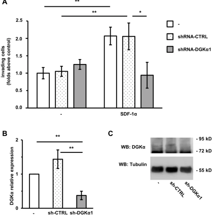
Figure 1. DGKais necessary for SDF-1a-induced cell invasion.MDA-MB-231 cells were infected with lentiviral vectors expressing an inducible shRNA against DGKa(shRNA-DGKa1) or an inducible control shRNA (shRNA-CTRL). Parental and infected cells were treated with 1mg/ml doxycycline for 72 hours to promote shRNA transcription. A) 50,000 cells were plated on matrigel invasion chamber and incubated for 24 hours in presence or in absence of SDF-1a(100 ng/ml). Histogram reports mean6SE of fold over control values from 3 independent experiments with *t-test p,0.05, **t-test p,0.01. B) The efficiency of DGKadown–regulation by shRNA was verified by quantitative RT-PCR. **t-test p,0.01. A) Cells were lysed and the efficiency of DGKadown–regulation by shRNA was verified by western blot, tubulin was used as a loading control.
doi:10.1371/journal.pone.0097144.g001
Validated siRNA DGKa [17] sense 59 GGAUGGCGA-GAUGGCUAAAtt 39 antisense 59 UUUAGCCAUCUCGC-CAUCCgg 39.
siRNA PKCfsense 59CGUUCGACAUCAUCACCGAtt39 an-tisense 59UCGGUGAUGAUGUCGAACGgg39.
siRNA PKCi sense 59CGUUCGACAUCAUCACCGAtt39 antisense 59UCGGUGAUGAUGUCGAACGgg39.
siRNAb1 integrin sense 59GGAGGAAUGUUACACGGCU39 antisense 59AGCCGUGUAACAUUCCUCCag 39.
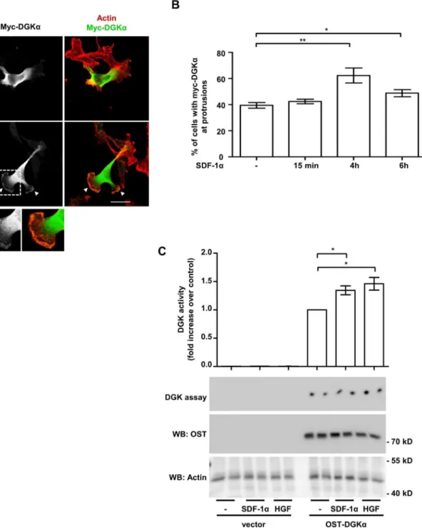
Figure 2. SDF-1astimulates DGKaactivity and localization at protrusions site.A) MDA-MB-231 cells, stably expressing myc-DGKa, were plated on matrigel-coated coverslips for 20 hours in FCS containing medium and cultured for further 20 hours in serum free medium. Cells were then stimulated with 50 ng/ml of SDF-1afor the indicated times, fixed and stained for actin (red) and myc-DGKa(green). Representative images at 4 hours after stimulation. Arrowheads indicate DGKaat protrusions. Histogram (B) reports the percentage of cells displaying myc-DGKaat protrusion as mean 6SE of 5 independent experiments, *t-test p,0.05, **t-test p,0.005. Scale bar 24mm. C) MDA-MB-231 cells were infected with a lentiviral vector expressing inducible OST-tagged DGKaor an empty vector. To induce DGKaexpression, cells were treated overnight with doxycycline (1mg/ml) in serum free medium. Cell were homogenized with buffer B in absence of detergent and analysed for DGK activity (upper panel). Values are mean6SE of 4 independent experiments with *t-test p,0.05. OST-DGKaand actin protein expression was verified by anti-OST and anti-actin western blot (lower panel).
siRNA RCP: ON-TARGETplus RAB11FIP1 siRNA L-015968-00-0005 (Dharmacon). Silencer negative control siRNA AM4611 (Life Technologies) was used as negative control.
Generation of Tet-inducible Strep-tagged DGKa Construct and Cell Infection
Human DGKa was amplified from pMT2- DGKa [24] by PCR using the primers DGKa_ScII_fw (59 -CCGCGGGCAG-CATGGCCAAGGAGAGGGGC-39) and DGKa_H3_rv (59 -AAGCTTTTAGCTCAAGAAGCCAAA-39) and cloned into pEXPR-IBA-105 (IBA GmbH) via SacII and HindIII to generate pEXPR-Strep-DGKa. In a further step Strep-DGKa was amplified by PCR using primers IBA_fw_N1 (59 -GCGGCCGCA-GACCCACCATGGCTAGC-39) and 105DGKa_MluI_rv (59 -ACGCGTTTAGCTCAAGAAGCCAAA-39) and cloned via NotI and MluI to pLVX-Tight-Puro (Clontech). All constructs were verified by DNA sequencing.
The resulting pLVX-Tight-PURO-OST-DGKapresents OST-DGKaafter a tetracycline controlled promoter and was used with the Lenti-X Tet-On Advanced Inducible Expression System (Clontec) according to manufacturer’s instruction. Lentiviral particles were obtained in 293FT packaging cells co-transfected with helper vectors. After double infection and selection we obtained a polyclonal population of MDA-MB-231 cells express-ing OST-DGKain a tetracycline inducible manner. A control cell line was also generated with an empty vector.
Generation of MDA-MB-231 Stably Expressing Myc-DGKa Myc-DGKa was amplified from PMT2-myc-DGKa [16] by PCR using the primers sense.
59 CTCGAGACCAATGGAACAAAAGTTGATTTCAGAA-GAAGATTTATTAATGGCCAAGGAGG39, antisense 59 GCCCCTCTCCTTGGCCATTAATAAATCTTCTTCT-GAAACAACTTTTGTTCCATGGCTCGAGTGCA39 and cloned in the pDONOR211 vector using the Gateway system (Life Technologies) according to manufacturer’s instructions. The Gateway Technology (Life Technologies) was also used to subclone myc-DGKa into pLenti4/V5-DEST lentiviral vector. Lentiviral particles were obtained in 293FT packaging cells co-transfected with helper vectors. After infection and selection we obtained a polyclonal population of MDA-MB-231 cells constitu-tively expressing myc-DGKa.
Inducible Silencing of DGKa in MDA-MB-231
We used the commercial pTRIPZ Inducible Lentiviral Human DGKA shRNA Clone ID: V3THS_340705 (shRNA-DGKa1) or pTRIPZ Inducible Lentiviral Non-silencing shRNA Control RHS4743 (shRNA-CTRL). Those vectors express shRNA and turboRFP under a doxycycline regulated promoter (Thermo Scientific Open Biosystems). Lentiviral particles were obtained in 293FT packaging cells co-transfected with helper vectors. After infection and selection we obtained a polyclonal population of MDA-MB-231 cells which upon induction with doxycycline (1mg/ml, 72 hours) are 100% RFP positive.
Stable Silencing DGKain MDA-MB-231
The shRNA for DGKa(forward: 59 GATCCCCGGTCAGT- GATGTCCTAAAGTTCAAGAGACTTTAGGACATCACT-GACCTTTTTGGAAA reverse: 59 AGCTTTTC- CAAAAAGGTCAGTGATGTCCTAAAGTCTCTTGAACTT-TAGGACATCACTGACCGGG) was cloned with H1-Promoter within the lentiviral vector pCCL.sin.PPT.hPGK.GFPWpre [25]. The resulting vector co-express shRNA-DGKaand GFP (shRNA-DGKa2). Empty vector was used as a control. Lentiviral particles were obtained in 293FT packaging cells co-transfected with helper vectors (Life Technologies). At 1 week after infection nearly 100% of cells were GFP+
.
Generation of ShRNA-b1 Integrin MDA-MB-231
ShRNA-b1integrin in pLKO were a kind gift of P. Defilippi [26]. Lentiviral particles were generated with Sigma Mission Lentivaral packaging mix according to manufacturer’s instruction in 293FT cells and selected with puromycin. Empty pLKO was used as a control.
Western Blotting
To verified protein down-regulation cells were lysed 48 hours after transfection. Cell were washed with ice cold PBS, scraped on ice in lysis buffer (25 mM Hepes, pH 8, 150 mMNaCl, 0.5/1% Nonidet P-40, 5 mM EDTA, 2 mM EGTA, 1 mM ZnCl2, 50 mM NaF, 10% glycerol supplemented with fresh 1 mM Na3VO4, and protease inhibitors) and clarified after centrifugation
of 15 minutes at 12000 rpm at 4uC. Samples were then resuspended in Laemmli buffer, heat denatured, and separated by SDS/PAGE. Proteins were then transferred on PVDF membrane by using semi-dry system. Membrane was then blocked with 5% BSA in PBS and incubated at 4uC overnight with primary antibodies diluted in TBS tween 0.1%, BSA 2%, 0.01% azide. After 4 washes with TBS-Tween 0.1%, membranes were incubated with secondary antibodies and washed again. Western blot were visualized using Western Lightning Chemiluminescence Reagent Plus (Perkin Elmer).
Quantitative RT-PCR
RNA was extracted by TRI-Reagent Solution (Life Technol-ogies) retrotrascribed with High-Capacity cDNA Reverse Tran-scription Kits (Life Technologies) and cDNA quantified by real time PCR using GUSB as normalizer. TaqMan gene expression assays we from Life Technologies: b1 integrin (Hs 00559595), GUSB (Hs 00939627), DGKa (Hs 00176278) and MMP-9 (Hs 00234579).
MMP-9 Secretion
MDA-MB-231 cells (250,000 cells/well) were plated in 6-well cell culture plate and transfected with the indicated siRNA. After 24 hours in serum free media cells were treated with SDF-1a (100 ng/ml in 500ml serum-free medium). After 24 hours the MMP-9 concentration in the supernatants was determined by ELISA assay (Life Technologies).
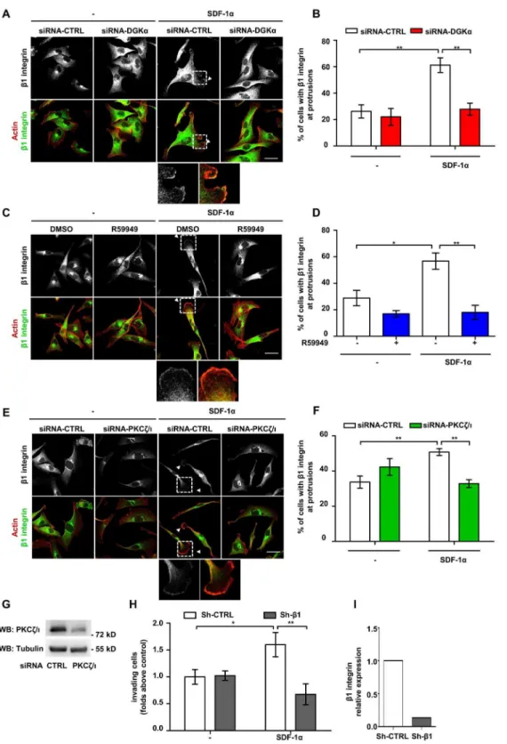
specific siRNA and lysed. The efficiency of DGKadown–regulation by siRNA was verified at 48 hours after transfection by western blot, tubulin was used as loading control. D) MDA-MB-231 cells were plated on matrigel-coated coverslips for 20 hours in FCS containing medium and cultured for further 20 hours in serum free medium. Cells were then stimulated for 6 hours with 50 ng/ml SDF-1a, in presence or in absence of 1mM R59949, fixed and stained for actin (red) and aPKCs (green). Arrowheads indicate aPKCs at protrusions. Scale bar 24mm. E) Histogram reports the percentage of cells displaying aPKCs at protrusions as mean6SE of 3 independent experiments with ***t-test p,0.0005. F) MDA-MB-231 cells (106/well) were plated on matrigel invasion chamber and stimulates for 24 hours with SDF-1a(50 ng/ml) in presence or absence of PKCfpseudosubstrate (PS-PKCf, 10mM). Histogram reports mean6SE of folds over control values from 3 independent experiments with *t-test p,0.05.
Statistical Analysis
Data are shown as the mean6SEM. For statistical analysis, Student’s t-test or ANOVA were used. Experiments shown are representative at least 3 independent experiments.
Results
DGKaIs Necessary for SDF-1a-induced Cell Invasion We previously showed that DGKa is necessary for matrix invasion promoted by Epidermal Growth Factor (EGF) [15] or Hepatocyte Growth Factor (HGF) in MDA-MB-231 breast carcinoma cells [27]. In order to investigate the role of DGKa in chemokine invasive signaling in breast cancer, we knocked down DGKa in MDA-MB-231 using a lentiviral construct expressing a DGKa-specific shRNA under an inducible promoter (shRNA-DGKa1). This construct strongly downregulated DGKa expression when compared with parental cells or a non-targeting control sequence (shRNA-CTRL, Fig. 1 B and C). The invasive ability of parental, DGKa-knocked down and control cells were evaluated in a Matrigel invasion assay. SDF-1a (100 ng/ml) doubles the number of parental as well as shRNA-CTRL MDA-MB-231 invading across the matrigel insert (Fig. 1 A). Conversely, shRNA-DGKa1 cells were unresponsive to SDF-1a stimulation. We confirmed this finding with an independent shRNA (shRNA-DGKa2) giving a comparable inhibition of SDF-1a stimulated matrix invasion (Fig. S1), making off-target effects unlikely.
Those findings indicates that DGKamediates the pro-invasive signaling promoted not only by tyrosine kinase receptors [22] but also by chemokine receptors involved in tumor cells metastatiza-tion, such as those of SDF-1a.
SDF-1aStimulates DGKa Activity and Localization at Protrusions Sites
The previous findings that HGF, EGF and VEGF activate DGKaand promote its recruitment to the plasma membrane in epithelial and endothelial cells [15,17,22] suggest that SDF-1a may promote localized DGKaactivation at ruffling sites. Despite its biological significance, the low level of DGKa expression in MDA-MB-231 cells hampers activation and localization studies of the endogenous protein with currently available antibodies.
Thus, for localization studies, MDA-MB-231 cells were stably infected with a lentiviral vector expressing myc-DGKaand plated on matrigel-coated coverslip to mimic the epithelial microenvi-ronment. In unstimulated serum-deprived cells, myc–DGKawas mainly cytoplasmic, with some cells displaying very little accumu-lation at cell protrusions (Fig. 2A). Prolonged SDF-1astimulation (50 ng/ml; 4 to 6 hours) resulted in the localization of DGKa at the tip of large protrusions (Fig. 2A and B). No detectable changes were observed at earlier time points (15 minutes, Fig. 2B).
For enzymatic activation assays, we infected MDA-MB-231 with a lentiviral vector expressing OneStrep-Tagged DGKa (OST-DGKa) under the control of a doxycycline-inducible promoter. Upon 48 hours doxycycline treatment (1mg/ml), OST-DGKa was strongly overexpressed as compared to endog-enous protein (Fig. S2A). Under these conditions the enzymatic activity of OST-DGKawas responsible for almost the entire DGK activity measured in cell homogenates. Both SDF-1aand HGF (a
well known DGKaactivator) induced a further moderate increase of OST-DGKaactivity within 15 minutes of stimulation (Fig. 2C). Altogether these data indicate that SDF-1a regulates DGKa activity and localization and suggest that DGKaplays a role in the formation and/or extension of cell protrusions induced by SDF-1a.
DGKa Mediates SDF-1a-induced Cell Invasion by Regulating aPKCs Recruitment to Cell Protrusions
DGKa, by producing PA, mediates aPKCs activation and recruitment to the cell surface induced by growth factors [23,28]. Thus, we set to investigate whether DGKa mediates SDF-1a-induced cell invasion by regulating aPKCs. To investigate the role of DGKain regulating aPKCs localization, MDA-MB-231 cells were transiently transfected with control (siRNA-CTRL) or DGKa-specific siRNA (siRNA-DGKa). Upon 48 hours from transfection with siRNA-DGKa, the expression of DGKa was nearly undetectable as compared to its expression in cells transfected with control siRNA (Fig. 3C). Then, MDA-MB-231 cells were plated on matrigel-coated coverslips, serum starved and stimulated with 50 ng/ml SDF-1afor 6 hours. In control siRNA transfected cells, SDF-1a treatment significantly increased the percentage of cells displaying aPKCs at protrusions, while DGKa silencing strongly impaired aPKCs recruitment to the membrane (Fig. 3A and B). In order to verify the requirement for DGKa enzymatic activity, we carried out aPKCs localization assays in presence or in absence of 1mM R59949, a rather specific DGKa inhibitor [16,29]. R59949 treatment completely abrogated aPKCs localization at protrusions induced by SDF-1a, while it did not affect aPKCs localization in unstimulated cells (Fig. 3D and E).
In order to investigate the role of aPKCs in SDF-1a-induced invasion through extracellular matrix, MDA-MB-231 cells were treated with 10mM cell permeable PKCf pseudosubstrate (PS-PKCf). In a matrigel invasion assay aPKCs inhibition significantly reduced SDF-1a-induced invasion, while basal invasion was unaffected in unstimulated cells (Fig. 3F).
Altogether, these data demonstrate that in SDF-1a-stimulated breast carcinoma cells, localized activity of DGKaat pseudopodial tips provides a crucial localization lipid signal for aPKCs recruitment, thus mediating SDF-1a-induced invasive signaling.
DGKa and aPKCs Mediate SDF-1a-induced Recruitment ofb1 Integrin to Protrusions Sites
Recycling and clustering of b1 integrin at the tip of invasive pseudopods is a key event sustaining the invasive properties of malignant cells [30]. Conversely, growth factors stimulate invasion both by inducing integrin clustering at actin-rich adhesive sites and lamellipodia and by stimulating integrin recycling [26,31]. Thus, we set to investigate whether the DGKaand aPKCs at protrusions promote local accumulation of b1 integrin. In serum starved MDA-MB-231 cells plated on matrigel-coated coverslips b1 integrin is mostly localized in intracellular vesicles in the perinuclear/Golgi area. Upon SDF-1a stimulation, b1 integrin also localized in clusters at the tip of cell protrusions (Fig. 4A, C and E). However, either siRNA-mediated silencing of DGKa or R59949-mediated inhibition of its enzymatic activity impaired SDF-1a-induced localization of b1 integrin at cell extensions were transfected with CTRL and PKCf/i–specific siRNA and lysed. The efficiency of PKCf/idown–regulation by siRNA was verified by western bloting, tubulin was used as a loading control. H) MDA-MB-231 cells were infected with lentiviral vectors expressing a shRNA againstb1-integrin (shRNA-b1) or a control sequence (shRNA-CTRL). 50,000 cells were plated on matrigel invasion chamber and incubated for 24 hours in presence or in absence of SDF-1a(100 ng/ml). Histogram reports mean6SE of fold over control values from 3 independent experiments with *t-test p,0.05, **t-test p,0.01. I) The efficiency ofb1-integrin down–regulation by shRNA was verified by quantitative RT-PCR.
(Fig. 4A, B, C and D). Interestingly SDF-1a stimulation and DGKainhibition did not affect the expression ofb1 integrin at the cell surface, as measured by FACS analysis (Fig. S4A). Since DGKa promotes Rac1 activation and membrane ruffles by regulating aPKCs [15] and as DGKamediates SDF-1a-induced aPKCs recruitment to the membrane protrusions, we assessed whether aPKCs controlsb1 integrin localization. Indeed, siRNA-mediated silencing of aPKCs (Fig. 4G) impaired SDF1-a-induced localization ofb1 integrin at cell protrusions (Fig. 4E and F).
Altogether these data suggest that SDF-1a, by activating the DGKa/aPKCs pathway, stimulates the clustering ofb1 integrin at cell protrusions, rather than stimulating its bulk translocation at the plasma membrane.
Since the expression of constitutively-membrane bound myr-DGKa stimulates cell invasion by triggering RCP-mediated recycling of integrina5b1 [15], we set to investigate the role of b1 integrin in SDF-1a-promoted cell invasion. To this purpose we used shRNA mediated knockdown ofb1 integrin which resulted in an 80% reduction of its expression in MDA-MB-231 cells (Fig. 4I). We found that, b1 integrin knock down severely impaired the ability of MDA-MB-231 cells to invade through matrigel in response to SDF-1astimulation (Fig. 4H).
Altogether these data indicate that DGKa, by regulating aPKCs, controls chemokine-induced b1 integrin localization at protrusion sites in breast carcinoma cells, thus confirming the pivotal role ofb1 integrin in SDF-1a-promoted matrix invasion.
DGKaand aPKCs Mediate SDF-1a-induced MMP-9 Secretion and Localization at Protrusions
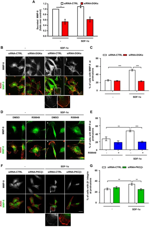
Secretion of matrix metalloproteinases (MMPs) is involved in the extracellular matrix degradation required for invasion of cancer cells [32,33]. SDF-1astimulates the secretion of MMP-9 in several cancer cells, including MDA-MB-231 cells [34,35]. In migrating cells, MMP-9 is addressed to the cellular extensions involved in cell migration and accumulates at their tips [36]. Thus, we investigated whether SDF1-a regulates intracellular localiza-tion and secrelocaliza-tion of MMP-9 through the DGKa/aPKCs axis.
MDA-MB-231 cells presented a low, constitutive secretion of MMP-9 (40–80 pg/ml in the supernatant), which was not affected by SDF-1abut was severely reduced by siRNA-mediated silencing of DGKa(Fig. 5A). However, the mRNA levels of MMP-9 were not affected by either SDF-1a stimulation or DGKa inhibition, suggesting that this pathway does not regulate MMP-9 at the transcriptional level in these cells (Fig. S4C). Conversely, SDF-1a stimulated MMP-9 accumulation at protrusions of serum-starved MDA-MB-231 plated on matrigel-coated coverslips (Fig. 5B to E). We cannot rule out that MMP-9 staining may be associated to the plasma membrane, indeed FACS analysis of these cells detected low amounts of membrane-bound MMP-9 with a small increase in MMP-9 surface positive cells following SDF-1a stimulation (Fig. S4B). Silencing of DGKaimpaired MMP-9 translocation induced
by SDF-1a, while it did not affect its localization in unstimulated cells (Fig. 5B and C). Similarly, DGKapharmacological inhibition with R59949, completely impaired MMP-9 recruitment induced by SDF-1a(Fig. 5D and E).
Altogether these data suggest that DGKais essential for MMP-9 accumulation at protrusions and subsequent release in the extracellular space. Given the role of DGKain regulating aPKCs, we investigated whether aPKCs mediates SDF-1a-induced regu-lation of MMP-9 localization. Indeed, siRNA-mediated silencing of aPKCs blunted SDF-1a induced MMP-9 localization at pseudopodial tips (Fig. 5F and G).
Altogether these data demonstrate that activation of the DGKa/aPKCs pathway drives both MMP-9 and b1 integrin localization at the pseudopodial tips, thus regulating the extension of invasive protrusions and sustaining the invasive behavior of MDA-MB-231 cells.
DGKa Overexpression Promotes aPKC/Rac Dependent Cell Elongation
We observed that prolonged SDF1atreatment (6 hours, 50 ng/ ml) of matrigel plated MDA-MB-231 promotes the transition to an elongated shape with the extension of long protrusions. Interest-ingly both siRNA downregulation of DGKa and R59949-mediated inhibition impairs this change in shape (Fig. S3A to C) indicating the crucial requirement of DGKaactivity.
Since the over-expression of membrane-bound myr-DGKa stimulates cell migration in untransformed cells [18] and pseudopod extension and invasion in A2780 ovarian cancer cells [15], we investigated whether wild type DGKa over-expression was sufficient to further stimulate invasion in MDA-MB-231 cells. The previously described inducible OST-DGKa construct in MDA-MB-231 cells allowed us to verify this issue as doxycycline treatment induced a 30-fold increase in DGKaexpression (Fig. 6A and Fig. S2A), with an increase of about 300-fold of the enzymatic activity (Fig. 2C). However, over-expression of OST-DGKawas not sufficient to enhance migration of MDA-MB-231 in wound-healing assay or to increase invasion through matrigel (Fig. S2B and C). Nevertheless, over-expression of OST-DGKa led to elongation of serum-starved MDA-MB-231 cells, while doxycy-cline did not affect the cell length of empty vector-infected MDA-MB-231 cells (Fig. 6B and D). Both in elongated and in shorter cells, OST-DGKais localized at the tip of cell protrusions (Fig. 6C) suggesting that despite the absence of cytokines and growth factors the strong up-regulation of DGKaactivity is sufficient to recruit the signaling machinery for membrane extension and to establish a feed forward loop recruiting further DGKa.
Consistently, with the reported role of the aPKCs in mediating DGKa-dependent Rac activation and membrane protrusions [23], we observed that siRNA-mediated silencing of aPKCs (Fig. 6G) blunted cell elongation induced by OST-DGKa over-expression (Fig. 6E). Also the Rac inhibitor NSC23766 completely experiments normalized for control, with *t-test p,0.05. B) MDA-MB-231 cells were plated on matrigel-coated coverslips for 20 hours in FCS containing medium, transfected with CTRL or DGKa–specific siRNA and cultured for further 20 hours in serum free medium. Cells were stimulated for 6 hours with 50 ng/ml SDF-1a, fixed and stained for actin (red) and MMP-9 (green). Arrowhead indicates MMP-9 at protrusions. Scale bar 24mm. C) Histogram reports the percentage of cells displaying MMP-9 at protrusions as mean6SE of 3 independent experiments with ***t-test p,0.0005. D) MDA-MB-231 cells were plated on matrigel-coated coverslips for 20 hours in FCS containing medium and cultured for further 20 hours serum free medium. Cells were stimulated for 6 hours with 50 ng/ml SDF-1a, in presence or in absence of 1mM R59949, fixed and stained for actin (red) and MMP-9 (green). Arrowhead indicates MMP-9 at protrusions. Scale bar 24mm. E) Histogram reports the percentage of cells displaying MMP-9 at protrusions as mean6SE of 3 independent experiments with **t-test p,0.005, ***t-test p,0.01. F) MDA-MB-231 cells were plated on matrigel-coated coverslips for 20 hours in FCS containing medium, transfected with CTRL or PKCf/i–specific siRNA and cultured for further 20 hours in serum free medium. Cells were then stimulated for 6 hours with 50 ng/ml SDF-1a, fixed and stained for actin (red) and MMP-9 (green). Arrowhead indicates MMP-9 at protrusions. Scale bar 24mm. G) Histogram reports the percentage of cells displaying MMP-9 at protrusions as mean6SE of 3 independent experiments with *t-test p,0.05, **t-test p,0.005.
blunted OST-DGKa induced elongation indicating the involve-ment of Rac family GTPases (Fig. 6F). Those findings confirm the relevance of aPKCs and Rac as DGKa downstream effectors promoting cytoskeletal remodeling and extension of membrane protrusions.
The expression of myr-DGKadrives pseudopodial extension by stimulating RCP-mediated recycling of b1 integrin in A2780 carcinoma cells [15]. However, siRNA-mediated silencing of either b1 integrin or RCP (Fig. S5C and D) did not affect protrusion elongation induced by wild type DGKa in serum starved MDA-MB-231 cells (Fig. S5A and B), suggesting that in this experimental model b1 integrin and its RCP-mediated recycling are not required for protrusion elongation.
These data indicate that up-regulation of DGKa activity by SDF-1a is sufficient to promote the extension of membrane protrusions through the aPKCs – RhoGDI – Rac pathway [22,23], but that additional signaling pathways and/or its localization at specific myrstyoilation-directed membrane com-partment are required to trigger cells invasion.
Discussion
We and others established the relevance of DGKa activation and membrane recruitment in growth factors signaling [37]. In normal epithelia, endothelia and lymphocytes DGKa activity is required to convey proliferative [17,38,39] and migratory [16– 18,22,23] signaling. Several studies pointed out DGKa involve-ment in cancer showing that its activity is necessary in vivo for glioblastoma and hepatocellular carcinoma progression [13], and in vitro for proliferation and survival of endometrial carcinoma [21], anaplastic large cell lymphoma [19], and melanoma [40]. Moreover, DGKaactivity mediates matrix invasion sustained by p53 pro-metastatic mutations in cancer cells [15]. However, the molecular pathways by which DGKa controls carcinoma forma-tion and metastatizaforma-tion are poorly known.
Inhere we investigated the role of DGKain invasive signaling of SDF-1a, one of the key signals driving metastasis [41], whose receptor, CXCR4, is strongly associated to tumor growth and spontaneous metastasis formation [1]. We used MDA-MB-231 cells, a highly invasive human breast cancer cell line, whose invasiveness and tumorigenicity are dependent on the expression of SDF-1a receptor, CXCR4 [42–44]. In these cells we had previously shown that DGKais required for EGF- [15] and HGF-induced [27] migration in a tridimensional environment.
Interestingly, we show here that DGKa is also regulated by SDF-1a, which stimulates its enzymatic activity and promotes its recruitment at ruffling sites (Fig. 2). Moreover, we show that activation of DGKaprovides a key lipid signal required for SDF-1apro-invasive activity in MDA-MB-231 cells (Fig. 1).
We previously showed that the PA generated by HGF-induced activation of DGKa recruits to the plasma membrane and activates aPKCs in a complex with RhoGDI and Rac1, thus
mediating the release of Rac1 from RhoGDI, and its localization and activation at ruffle sites [23]. The aPKCs subfamily comprises thefandiisoforms, which are activated by PA [28] but insensitive to DG.
Several pieces of evidence show that aPKCs and in particular PKCi, play a key role in cancer cell invasion and tumor progression [45]. Interestingly, PKCiis essential for K-Ras-driven invasion in colon cancer by regulating Rac1 [46], while aPKCs mediates EGF-induced cell migration of MDA-MB-231 breast cancer cells [47]. Altogether these data further suggest that the DGKa/aPKCs signaling axis contributes to pro-invasive signaling. Accordingly, the finding that SDF-1ainduces aPKCs localiza-tion at protrusion sites through activalocaliza-tion of DGKa, indicates that the DGKa/aPKCs signaling axis mediates chemokine-driven mammary carcinoma invasiveness (Fig. 3). DGKa-dependent recruitment of aPKCs at protrusion is an essential signaling event, since the silencing of either DGKaor aPKCs impairs downstream events such as accumulation of b1 integrin and MMP-9 at the plasma membrane (Fig. 4 and 5). The functional relevance of aPKCs as a DGKaeffector is further proved by the observation that its silencing impairs DGKa-induced cell elongation (Fig. 6E) and that its inhibition blocks SDF-1a-induced matrix invasion (Fig. 3F).
The findings that aPKCs, RCP andb1 integrin are all required for the invasiveness of MDA-MB-231 (Fig. 3F, 4H and ref. [15]), and that upon SDF-1astimulationb1 integrin is concentrated at protrusion tips in a DGKa and aPKCs-dependent manner, are consistent with our previous data showing that DGKa-generated PA, through binding to RCP, docksa5b1 recycling vesicles to the tips of invasive pseudopods. Altogether these findings suggest that activation of aPKCs may also contribute to integrin recycling induced by chemokines and growth factors, although there is no experimental evidence for it.
Several pieces of evidence in different cell types indicate that activation of aPKCs regulates MMPs production and secretion [48]. For instance, PKCf activation mediates MMP-9 secretion induced by SDF-1ain hematopoietic progenitors [11]. MMPs are key players in the tumor microenvironment and play a major role in invasion of extracellular matrix [49]. While some MMPs are transmembrane proteins, most of them are soluble and bind to the extracellular cell surface by interaction with several membrane proteins, includingb1 integrin and CD44v [50–54].
Our finding that both DGKaand aPKCs are required for SDF-1a-induced release of MMP9 in the cell medium and for its accumulation at protrusions, provides further strength to our thesis that DGKa/aPKCs axis is a major component of chemokine pro-invasive signaling. Interestingly, in SDF-1a-stimulated cells, MMP-9 localization at cell surface superimposes with that ofb1 integrin, suggesting that their function at protrusion tips is coordinately regulated by activation of DGKa/aPKCs signaling. extent of overexpression was verified with anti DGKaantibodies. Tubulin was used as loading control. B) Phase contrast images of control and OST-DGKacells cultured in presence or absence of doxycycline. Arrows indicate cells with long protrusions. Scale bar 50mm. C) Confocal images of doxycycline induced cells showing OST-DGKalocalization, cells were stained for actin (red) and OST (green). Scale bar 24mm. D) Time course of cell elongation at 2, 10 and 18 hours with or without doxycycline treatment. Time lapse videos were recorded and total cell length measured. Box and whiskers plots (black lines show median, whiskers: 5–95 percentile) of data from 3 independent experiments are shown, ***p,0.0001, 1 way ANOVA. E) MDA-MB-231 cells expressing OST-DGKawere transiently transfected with control or PKCf/i-specific siRNA. After 48 hours DGKaexpression was induced by overnight treatment with doxycycline (1mg/ml) in serum free medium. Images were acquired with a phase contrast microscope, representative images are shown. Scale bar 50mm. Total cell length was measured for at least 100 cells and reported as box and whiskers plot. F) MDA-MB-231 cells expressing OST-DGKawere induced by overnight treatment with doxycycline (1mg/ml) in serum free medium with or without NSC23766 (100mM). Images were acquired with a phase contrast microscope, representative images are shown. Scale bar 50mm. Total cell length was measured for at least 100 cells and reported as box and whiskers plot. MDA-MB-231 cells were transfected with CTRL and PKCf/i–specific siRNA and lysed. The efficiency of PKCf/idown–regulation by siRNA was verified by western blotting, tubulin was used as a loading control.
Finally, the observation that DGKa over expression drives by itself elongation of cell protrusions by regulating aPKCs is consistent with active PKCfpromoting wide cytoskeletal remod-eling and protrusions in untrasformed cells [23]. The molecular mechanisms by which aPKCs induces cell elongation downstream to DGKa is still partially known. In line with our previous demonstration that activation of the DGKa/aPKCs signaling module stimulates the RhoGDI driven localization of both Rac1 and Cdc42 at membrane ruffles, we observed that the Rac inhibitor NSC23766 blunts DGKa induced cell elongation (Fig. 6G) and that SDF-1a-induced localization of Cdc42 at protrusions of MDA-MB-231 cells is significantly reduced by DGKa inhibition (Fig. S3D and E). Conversely, protrusion extension occurs even in the absence of b1 integrin and RCP, suggesting that DGKa-dependent activation of aPKCs regulates cytoskeletal remodeling independently fromb1 integrin recycling and function, which are required, however, to enable cell migration through a 3D matrix (Fig. 4H). While it is clear that DGKa/aPKCs activity on cell elongation is independent onb1 integrin recycling, these data cannot rule out that accumulation of b1 integrin and MMP-9 at protrusion tips depends on DGKa/ aPKCs-induced regulation of Rac1 or Cdc42 and cytoskeletal contractility [31].
Altogether we showed that activation of the DGKa/aPKCs/b1 integrin pathway plays a key role in chemokine-driven matrix invasion in breast cancer cells. Those observations suggest that DGKa inhibition or silencing could be effective not only in reducing primary tumor growth in vivo [13,14] but could potentially also reduce the metastatic potential of carcinoma cells.
Supporting Information
Figure S1 DGKa is necessary for SDF-1a-induced cell invasion. MDA-MB-231 cells were infected with lentiviral vectors expressing a shRNA against DGKa(shRNA-DGKa2) or an empty vector. A) Cells were lysed and the efficiency of DGKa down–regulation by shRNA was verified by western blot, tubulin was used as a loading control. B) 50,000 cells were plated on matrigel invasion chamber and incubated for 24 hours in presence or in absence of SDF-1a(100 ng/ml). Histogram reports mean6 SE of fold over control values from 3 independent experiments with *t-test p,0.05, ***t-test p,0.0005.
(TIF)
Figure S2 DGKaoverexpression does not affect migra-tion and invasion of MDA-MB-231 cells. MDA-MB-231 cells were infected with lentiviral vector expressing inducible OST-tagged DGKaor an empty vector. To induce DGKaexpression, cells were treated overnight with doxycycline (1mg/ml) in serum free medium. A) After cell lysis, the extent of DGKa overexpres-sion was verified with anti DGKa antibodies, long and short exposures are shown. Actin was used as loading control. B) Cells were grown to confluence in 12 well plates and subjected to a wound healing assay for 24 hours in serum free medium. HGF (50 ng/ml) was used as a positive control. The cells were stained and those migrating inside 2.3 mm of wound counted. Histogram reports mean6SE of fold over control values from 3 independent experiments with *t-test p,0.05. C) 50,000 cells were plated on matrigel invasion chamber and incubated for 24 hours in serum free medium. Medium with 10% FCS was used as positive control. Histogram reports mean6SE of fold over control values from 3 independent experiments with *t-test p,0.05.
(TIF)
Figure S3 DGKa is required for SDF-1a-induced pseu-dopod elongation. A) MDA-MB-231 cells were plated on matrigel-coated coverslips for 20 hours in FCS containing medium, transfected with CTRL or DGKa -specific siRNA and cultured for further 20 hours in serum free medium. Cells were then stimulated for 6 hours with 50 ng/ml SDF-1a, fixed and photographed at phase contrast. B) Histogram reports protrusions length inmm as mean6SE values of 4 independent experiments with *t-test p,0.005. C) MDA-MB-231 cells were plated on matrigel-coated coverslips for 20 hours in FCS containing medium and cultured for further 20 hours in serum free medium. Cells were then stimulated for 6 hours with 50 ng/ml SDF-1a, in presence or in absence of 1mM R59949, fixed and photographed at phase contrast. Histogram reports protrusions length inmm as mean6SE of 3 independent experiments with *t-test p,0.005. D) MDA-MB-231 cells were plated on matrigel-coated coverslips for 20 hours in FCS containing medium and cultured for further 20 hours serum free medium. Cells were stimulated for 6 hours with 50 ng/ml SDF-1a, in presence or in absence of 1mM R59949, fixed and stained for actin (red) and Cdc42 (green). Arrowhead indicates Cdc42 at protrusions. Scale bar 24mm. E) Histogram reports the percentage of cells displaying Cdc42 at protrusions as mean6SE of 3 independent experiments with *t-test p,0.05.
(TIF)
Figure S4 SDF-1a is not affecting surface exposition of b1-integrin and MMP-9.A) Surface expression ofb1 integrin was analyzed before (turquoise) and after (red) SDF-1a stimula-tion. Flow cytometry histogram overlay comparing the level ofb1 integrin expression before and after SDF-1a expression. Isotype-matched controls mAb staining are given as dashed lines. MFI, median fluorescence intensity. B) Surface expression of MMP-9 was analyzed before (turquoise) and after (red) SDF-1a stimula-tion. Flow cytometry histogram overlay comparing the level of MMP-9 expression before and after SDF-1a expression. Isotype-matched controls mAb staining are given as dashed lines. MFI, median fluorescence intensity. C) MDA-MB-231 cells were plated on 6 wells dish for 20 hours in FCS containing medium and cultured for further 20 hours serum free medium. Cells were stimulated for 24 hours with 100 ng/ml SDF-1a, in presence or in absence of 1mM R59949. MMP-9 mRNA was quantified by quantitative RT-PCR. Histogram reports the mean6 SE of 3 independent experiments.
(TIF)
loading control. D) MDA-MB-231 cells were transfected with CTRL and RCP-specific siRNA and lysed. The efficiency of RCP down–regulation by siRNA and of OST-DGKa induction was verified by western blotting, actin was used as a loading control. (TIF)
Acknowledgments
ShRNA-b1 integrin in pLKO were a kind gift of P. Defilippi [26]. We thank O. Acuto (Oxford, UK) for helpful discussions.
Author Contributions
Conceived and designed the experiments: E. Rainero GB AG JCN. Performed the experiments: E. Rainero CC PEP FC VM VB E. Ruffo MF FB DC WP IL. Analyzed the data: E. Rainero CC FC PEP VM VB E. Ruffo MF DC IL AB NF FS GB AG. Contributed reagents/materials/ analysis tools: E. Rainero WP GB AG. Wrote the paper: E. Rainero GB AG.
References
1. Mu¨ller A, Homey B, Soto H, Ge N, Catron D, et al. (2001) Involvement of chemokine receptors in breast cancer metastasis. Nature 410: 50–56. 2. Korkaya H, Liu S, Wicha MS (2011) Breast cancer stem cells, cytokine networks,
and the tumor microenvironment. J Clin Invest 121: 3804–3809.
3. Teicher BA, Fricker SP (2010) CXCL12 (SDF-1)/CXCR4 pathway in cancer. Clin Cancer Res 16: 2927–2931.
4. Burger JA, Kipps TJ (2006) CXCR4: a key receptor in the crosstalk between tumor cells and their microenvironment. Blood 107: 1761–1767.
5. Li H, Yang L, Fu H, Yan J, Wang Y, et al. (2013) Association between Gai2 and ELMO1/Dock180 connects chemokine signalling with Rac activation and metastasis. Nat Commun 4: 1706.
6. Yagi H, Tan W, Dillenburg-Pilla P, Armando S, Amornphimoltham P, et al. (2011) A synthetic biology approach reveals a CXCR4-G13-Rho signaling axis driving transendothelial migration of metastatic breast cancer cells. Sci Signal 4: ra60.
7. Azab AK, Azab F, Blotta S, Pitsillides CM, Thompson B, et al. (2009) RhoA and Rac1 GTPases play major and differential roles in stromal cell-derived factor-1-induced cell adhesion and chemotaxis in multiple myeloma. Blood 114: 619– 629.
8. Kumar A, Kremer KN, Dominguez D, Tadi M, Hedin KE (2011) Ga13 and Rho mediate endosomal trafficking of CXCR4 into Rab11+ vesicles upon stromal cell-derived factor-1 stimulation. J Immunol 186: 951–958.
9. Chen G, Chen SM, Wang X, Ding XF, Ding J, et al. (2012) Inhibition of chemokine (CXC motif) ligand 12/chemokine (CXC motif) receptor 4 axis (CXCL12/CXCR4)-mediated cell migration by targeting mammalian target of rapamycin (mTOR) pathway in human gastric carcinoma cells. J Biol Chem 287: 12132–12141.
10. Odemis V, Boosmann K, Dieterlen MT, Engele J (2007) The chemokine SDF1 controls multiple steps of myogenesis through atypical PKCzeta. J Cell Sci 120: 4050–4059.
11. Petit I, Goichberg P, Spiegel A, Peled A, Brodie C, et al. (2005) Atypical PKC-zeta regulates SDF-1-mediated migration and development of human CD34n+
progenitor cells. J Clin Invest 115: 168–176.
12. Me´rida I, Avila-Flores A, Merino E (2008) Diacylglycerol kinases: at the hub of cell signalling. Biochem J 409: 1–18.
13. Takeishi K, Taketomi A, Shirabe K, Toshima T, Motomura T, et al. (2012) Diacylglycerol kinase alpha enhances hepatocellular carcinoma progression by activation of Ras-Raf-MEK-ERK pathway. J Hepatol 57: 77–83.
14. Dominguez CL, Floyd DH, Xiao A, Mullins GR, Kefas BA, et al. (2013) Diacylglycerol kinaseais a critical signaling node and novel therapeutic target in glioblastoma and other cancers. Cancer Discov 3: 782–797.
15. Rainero E, Caswell PT, Muller PA, Grindlay J, McCaffrey MW, et al. (2012) Diacylglycerol kinaseacontrols RCP-dependent integrin trafficking to promote invasive migration. J Cell Biol 196: 277–295.
16. Cutrupi S, Baldanzi G, Gramaglia D, Maffe` A, Schaap D, et al. (2000) Src-mediated activation of alpha-diacylglycerol kinase is required for hepatocyte growth factor-induced cell motility. EMBO J 19: 4614–4622.
17. Baldanzi G, Mitola S, Cutrupi S, Filigheddu N, van Blitterswijk WJ, et al. (2004) Activation of diacylglycerol kinase alpha is required for VEGF-induced angiogenic signaling in vitro. Oncogene 23: 4828–4838.
18. Baldanzi G, Cutrupi S, Chianale F, Gnocchi V, Rainero E, et al. (2008) Diacylglycerol kinase-alpha phosphorylation by Src on Y335 is required for activation, membrane recruitment and Hgf-induced cell motility. Oncogene 27: 942–956.
19. Bacchiocchi R, Baldanzi G, Carbonari D, Capomagi C, Colombo E, et al. (2005) Activation of alpha-diacylglycerol kinase is critical for the mitogenic properties of anaplastic lymphoma kinase. Blood 106: 2175–2182.
20. Baldanzi G, Pietronave S, Locarno D, Merlin S, Porporato P, et al. (2011) Diacylglycerol kinases are essential for HGF-dependent proliferation and motility of Kaposi’s Sarcoma cells. Cancer Sci.
21. Filigheddu N, Sampietro S, Chianale F, Porporato PE, Gaggianesi M, et al. (2011) Diacylglycerol kinaseamediates 17-b-estradiol-induced proliferation, motility, and anchorage-independent growth of Hec-1A endometrial cancer cell line through the G protein-coupled estrogen receptor GPR30. Cell Signal 23: 1988–1996.
22. Chianale F, Cutrupi S, Rainero E, Baldanzi G, Porporato PE, et al. (2007) Diacylglycerol kinase-alpha mediates hepatocyte growth factor-induced
epithe-lial cell scatter by regulating Rac activation and membrane ruffling. Mol Biol Cell 18: 4859–4871.
23. Chianale F, Rainero E, Cianflone C, Bettio V, Pighini A, et al. (2010) Diacylglycerol kinase alpha mediates HGF-induced Rac activation and membrane ruffling by regulating atypical PKC and RhoGDI. Proc Natl Acad Sci U S A 107: 4182–4187.
24. Schaap D, de Widt J, van der Wal J, Vandekerckhove J, van Damme J, et al. (1990) Purification, cDNA-cloning and expression of human diacylglycerol kinase. FEBS Lett 275: 151–158.
25. Taulli R, Accornero P, Follenzi A, Mangano T, Morotti A, et al. (2005) RNAi technology and lentiviral delivery as a powerful tool to suppress Tpr-Met-mediated tumorigenesis. Cancer Gene Ther 12: 456–463.
26. Morello V, Cabodi S, Sigismund S, Camacho-Leal MP, Repetto D, et al. (2011) b1 integrin controls EGFR signaling and tumorigenic properties of lung cancer cells. Oncogene 30: 4087–4096.
27. Filigheddu N, Cutrupi S, Porporato PE, Riboni F, Baldanzi G, et al. (2007) Diacylglycerol kinase is required for HGF-induced invasiveness and anchorage-independent growth of MDA-MB-231 breast cancer cells. Anticancer Res 27: 1489–1492.
28. Limatola C, Schaap D, Moolenaar WH, van Blitterswijk WJ (1994) Phosphatidic acid activation of protein kinase C-zeta overexpressed in COS cells: comparison with other protein kinase C isotypes and other acidic lipids. Biochem J 304 (Pt 3): 1001–1008.
29. Sato M, Liu K, Sasaki S, Kunii N, Sakai H, et al. (2013) Evaluations of the selectivities of the diacylglycerol kinase inhibitors r59022 and r59949 among diacylglycerol kinase isozymes using a new non-radioactive assay method. Pharmacology 92: 99–107.
30. Desgrosellier JS, Cheresh DA (2010) Integrins in cancer: biological implications and therapeutic opportunities. Nat Rev Cancer 10: 9–22.
31. Trusolino L, Cavassa S, Angelini P, Ando´ M, Bertotti A, et al. (2000) HGF/ scatter factor selectively promotes cell invasion by increasing integrin avidity. FASEB J 14: 1629–1640.
32. Nabeshima K, Inoue T, Shimao Y, Sameshima T (2002) Matrix metallopro-teinases in tumor invasion: role for cell migration. Pathol Int 52: 255–264. 33. Itoh Y, Nagase H (2002) Matrix metalloproteinases in cancer. Essays Biochem
38: 21–36.
34. Yuecheng Y, Xiaoyan X (2007) Stromal-cell derived factor-1 regulates epithelial ovarian cancer cell invasion by activating matrix metalloproteinase-9 and matrix metalloproteinase-2. Eur J Cancer Prev 16: 430–435.
35. Fernandis AZ, Prasad A, Band H, Klo¨sel R, Ganju RK (2004) Regulation of CXCR4-mediated chemotaxis and chemoinvasion of breast cancer cells. Oncogene 23: 157–167.
36. Legrand C, Gilles C, Zahm JM, Polette M, Buisson AC, et al. (1999) Airway epithelial cell migration dynamics. MMP-9 role in cell-extracellular matrix remodeling. J Cell Biol 146: 517–529.
37. Me´rida I, Avila-Flores A, Garcı´a J, Merino E, Almena M, et al. (2009) Diacylglycerol kinase alpha, from negative modulation of T cell activation to control of cancer progression. Adv Enzyme Regul.
38. Flores I, Casaseca T, Martinez-A C, Kanoh H, Merida I (1996) Phosphatidic acid generation through interleukin 2 (IL-2)-induced alpha-diacylglycerol kinase activation is an essential step in IL-2-mediated lymphocyte proliferation. J Biol Chem 271: 10334–10340.
39. Flores I, Jones DR, Cipre´s A, Dı´az-Flores E, Sanjuan MA, et al. (1999) Diacylglycerol kinase inhibition prevents IL-2-induced G1 to S transition through a phosphatidylinositol-3 kinase-independent mechanism. J Immunol 163: 708–714.
40. Yanagisawa K, Yasuda S, Kai M, Imai S, Yamada K, et al. (2007) Diacylglycerol kinase alpha suppresses tumor necrosis factor-alpha-induced apoptosis of human melanoma cells through NF-kappaB activation. Biochim Biophys Acta 1771: 462–474.
41. Luker KE, Luker GD (2006) Functions of CXCL12 and CXCR4 in breast cancer. Cancer Lett 238: 30–41.
42. Kang H, Mansel RE, Jiang WG (2005) Genetic manipulation of stromal cell-derived factor-1 attests the pivotal role of the autocrine SDF-1-CXCR4 pathway in the aggressiveness of breast cancer cells. Int J Oncol 26: 1429–1434. 43. Kang H, Watkins G, Parr C, Douglas-Jones A, Mansel RE, et al. (2005) Stromal
cells in vitro, and its association with prognosis and survival in human breast cancer. Breast Cancer Res 7: R402–410.
44. Lapteva N, Yang AG, Sanders DE, Strube RW, Chen SY (2005) CXCR4 knockdown by small interfering RNA abrogates breast tumor growth in vivo. Cancer Gene Ther 12: 84–89.
45. Murray NR, Kalari KR, Fields AP (2011) Protein kinase Ciexpression and oncogenic signaling mechanisms in cancer. J Cell Physiol 226: 879–887. 46. Murray NR, Jamieson L, Yu W, Zhang J, Go¨kmen-Polar Y, et al. (2004) Protein
kinase Ciota is required for Ras transformation and colon carcinogenesis in vivo. J Cell Biol 164: 797–802.
47. Sun R, Gao P, Chen L, Ma D, Wang J, et al. (2005) Protein kinase C zeta is required for epidermal growth factor-induced chemotaxis of human breast cancer cells. Cancer Res 65: 1433–1441.
48. Frederick LA, Matthews JA, Jamieson L, Justilien V, Thompson EA, et al. (2008) Matrix metalloproteinase-10 is a critical effector of protein kinase Ciota-Par6alpha-mediated lung cancer. Oncogene 27: 4841–4853.
49. Kessenbrock K, Plaks V, Werb Z (2010) Matrix metalloproteinases: regulators of the tumor microenvironment. Cell 141: 52–67.
50. Brooks PC, Stro¨mblad S, Sanders LC, von Schalscha TL, Aimes RT, et al. (1996) Localization of matrix metalloproteinase MMP-2 to the surface of invasive cells by interaction with integrin alpha v beta 3. Cell 85: 683–693. 51. Yu WH, Woessner JF, McNeish JD, Stamenkovic I (2002) CD44 anchors the
assembly of matrilysin/MMP-7 with heparin-binding epidermal growth factor precursor and ErbB4 and regulates female reproductive organ remodeling. Genes Dev 16: 307–323.
52. Redondo-Mun˜oz J, Escobar-Dı´az E, Samaniego R, Terol MJ, Garcı´a-Marco JA, et al. (2006) MMP-9 in B-cell chronic lymphocytic leukemia is up-regulated by alpha4beta1 integrin or CXCR4 engagement via distinct signaling pathways, localizes to podosomes, and is involved in cell invasion and migration. Blood 108: 3143–3151.
53. Redondo-Mun˜oz J, Ugarte-Berzal E, Garcı´a-Marco JA, del Cerro MH, Van den Steen PE, et al. (2008) Alpha4beta1 integrin and 190-kDa CD44v constitute a cell surface docking complex for gelatinase B/MMP-9 in chronic leukemic but not in normal B cells. Blood 112: 169–178.
54. Redondo-Mun˜oz J, Ugarte-Berzal E, Terol MJ, Van den Steen PE, Herna´ndez del Cerro M, et al. (2010) Matrix metalloproteinase-9 promotes chronic lymphocytic leukemia b cell survival through its hemopexin domain. Cancer Cell 17: 160–172.