RESEARCH ARTICLE
Effect of Vitamin D on Peripheral Blood
Mononuclear Cells from Patients with
Psoriasis Vulgaris and Psoriatic Arthritis
Susana Cubillos*, Nadine Krieg, Johannes Norgauer
Department of Dermatology, Jena University Hospital, Jena, Thüringen, Germany
Abstract
Background
Psoriasis, a chronic skin disease with or without joint inflammation, has increased circulat-ing proinflammatory cytokine levels. Vitamin D is involved in calcium homeostasis, bone for-mation, osteoclastogenesis and osteoclast activity, as well as regulation of immune
response. We aimed to study osteoclast differentiation and cytokine secretion of peripheral blood mononuclear cells (PBMCs) from patients with psoriasis vulgaris and psoriatic arthri-tis, in response to 1,25(OH)2D3.
Methods
Serum levels of bone turnover markers were measured by ELISA in patients with psoriasis vulgaris and psoriatic arthritis, and healthy controls. PBMCs were isolated and cultured with or without RANKL/M-CSF and 1,25(OH)2D3. Osteoclast differentiation and cytokine
secre-tion were assessed.
Results
Psoriatic arthritis patients had lower osteocalcin, as well as higher C-telopeptide of type I collagen and cathepsin K serum levels compared with psoriasis vulgaris patients and con-trols. RANKL/M-CSF-stimulated PBMCs from psoriatic arthritis patients produced higher proinflammatory cytokine levels and had a differential secretion profile in response to 1,25 (OH)2D3, compared with psoriasis vulgaris and control PBMCs.
Conclusions
Our data confirmed altered bone turnover in psoriatic arthritis patients, and demonstrated increased osteoclastogenic potential and proinflammatory cytokine secretion capacity of these PBMCs compared with psoriasis vulgaris and controls. 1,25(OH)2D3abrogated these
effects. OPEN ACCESS
Citation:Cubillos S, Krieg N, Norgauer J (2016) Effect of Vitamin D on Peripheral Blood Mononuclear Cells from Patients with Psoriasis Vulgaris and Psoriatic Arthritis. PLoS ONE 11(4): e0153094. doi:10.1371/journal.pone.0153094
Editor:Dominique Heymann, Faculté de médecine de Nantes, FRANCE
Received:January 19, 2016
Accepted:March 23, 2016
Published:April 6, 2016
Copyright:© 2016 Cubillos et al. This is an open access article distributed under the terms of the
Creative Commons Attribution License, which permits unrestricted use, distribution, and reproduction in any medium, provided the original author and source are credited.
Data Availability Statement:All relevant data are within the paper and its Supporting Information files.
Funding:The authors have no support or funding to report.
Introduction
Psoriasis is a chronic inflammatory skin disease with or without joint inflammation. Photo-therapy and topical application of vitamin D analogs are widely used in the treatment of psoria-sis vulgaris. Topical vitamin D regulates serum calcium levels and phototherapy alters systemic levels of vitamin D, a crucial factor in the regulation of extracellular calcium homeostasis and bone metabolism [1,2]. Osteoclasts resorb mineralized bone and osteoblasts are responsible for new bone formation. Osteoclasts are multinucleated cells derived from the monocyte/macro-phage lineage [3]. Osteoclast differentiation is supported by osteoblasts through cell-to-cell interactions and two major cytokines—receptor activator of NF-ϰB ligand (RANKL) and
mac-rophage-colony stimulating factor (M-CSF) [4–6]. Activation of the receptors RANK and c-Fms on osteoclast precursors by these ligands induces calcium signalling pathways linked to activation of the nuclear factor of activated T cells cytoplasmic 1 (NFATc1) [7,8], which regu-lates the expression of osteoclast-specific markers such as the type I collagen degrading cathep-sin K (CTSK), the osteopontin dephosphorylating tartrate-resistant acid phosphatase (TRAP) and calcitonin receptor [9–11]. This receptor binds to calcitonin (CT), a hormone produced primarily by thyroid C-cells in response to elevated serum calcium levels [10]. CT reduces blood calcium, through inhibition of bone resorption [12] and regulation of 1,25(OH)2D3 pro-duction in the kidney [13]. In addition to the hormonal control of calcium homeostasis, the vitamin D active form 1,25(OH)2D3functions on cellular growth, proliferation and differentia-tion. Locally produced 1,25(OH)2D3by osteoblasts is involved in the regulation of osteoclasto-genesis and osteoclast activity [14] increasing the expression of RANKL as well as decreasing the expression of its antagonist osteoprotegerin in osteoblasts [15]. Cells of the monocyte/mac-rophage lineage hydroxylate 25(OH)D3into 1,25(OH)2D3[16,17]. In particular, PBMCs-derived osteoclasts also respond to vitamin D through vitamin D receptor with increased NFATc1 expression [18].
Bone and immune cells share bone marrow progenitors and are affected by the same cyto-kines and metabolites such as 1,25(OH)2D3.In fact, 1,25(OH)2D3inhibits the expression of cytokines such as IL-1, IL-2, IL-6, IL-12, IL-23, interferonγ(IFN-γ), tumour necrosis factorα
(TNF-α) and chemokines such as IL-8 and chemokine (C-C motif) ligand 5 (CCL5 or
RANTES) by monocytes, T and B cells [19–22]. Conversely, 1,25(OH)2D3increases production of IL-10 of activated T and B cells, and interferonβin osteoclast precursors [23,24]. Besides high levels of circulating proinflammatory cytokines [25,26], patients with psoriatic arthritis have higher circulating bone and cartilage degradation products [27,28] and higher number of osteoclast precursors than healthy individuals [29]. Cytokine effects on the osteoclast differen-tiation and activity are well studied, but knowledge about cytokine secretion pattern of PBMCs derived from patients with psoriasis vulgaris and psoriatic arthritis and their capacity to differ-entiate into mature osteoclasts is limited. Therefore we aimed to study the osteoclast differenti-ation and cytokine secretion capacity of PBMCs from psoriasis vulgaris and psoriatic arthritis patients in response to 1,25(OH)2D3. We found increased osteoclastogenic potential and proinflammatory cytokine secretion capacity of PBMCs from patients with psoriatic arthritis compared with psoriasis vulgaris and controls. In addition, 1,25(OH)2D3abrogated these effects.
Materials and Methods
Subject characteristics
Declaration of Helsinki. Written consent was obtained from all participants prior to enrolment. Patients were diagnosed based on clinical and pathological findings at the Department of Der-matology of the Jena University Hospital. All patients fulfilled the CASPAR criteria [30]. The presence of joint manifestations was confirmed with power doppler ultrasonography (Esaote, Italy) and Rheumascan Xeralite (Mivenion GmbH, Germany). Twenty one patients with psori-asis vulgaris and fifteen healthy controls (HC) were included. Nine patients with psoripsori-asis vul-garis had no clinical signs of joint inflammation (PsV) and twelve were diagnosed with psoriatic arthritis (PsA). Demographic data are shown inS1 Table. Patients with other types of psoriasis (guttate, inverse, pustular, erythrodermic), other skin diseases, allergy, autoimmune diseases, any topical or systemic treatment, including vitamin D supplementation or photo-therapy 5 months before or at the time of recruitment were excluded.
Serum levels of bone turnover markers
Human calcitonin (CT) and 1,25-dihydroxyvitamin D3[1,25-(OH)2D3] (Shanghai Sunred Bio-logical Technology Co., Ltd, China), osteocalcin (OCN) (ALPCO Diagnostics, USA), C-telo-peptide of type I collagen (CTX-1) and of type II collagen (CTX-2) (CUSABIO Biotech Co., Ltd, China), and cathepsin K (CTSK) (Uscn Life Science Inc, China) ELISA kits were used according to the manufacturer instructions to assess the respective patient and control serum levels. Detection ranges of ELISA kits were: 0.7–200 mmol/L for CT, 0.7–150 ng/ml for 1,25-(OH)2D3, 0.31–1250 ng/ml for OCN, 25–800 ng/ml for CTX-1, 0.312–20 ng/ml for CTX-2, and 15.6–1000 pg/ml for CTSK.
Serum calcium levels
Serum calcium levels were determined using the QuantiChrom calcium assay kit (BioAssay Systems, USA). The assay was used according to the manufacturer instructions to assess the respective patient and control serum levels. The linear detection range was 0.08 to 20 mg/dl.
Isolation and culture conditions of PBMCs
PBMCs were isolated from blood samples by density gradient centrifugation. In 24-well plates, 1 x 10⁶cells/ml/well (0.5 x 10⁶cells per cm2) containing alpha MEM medium with 10% fetal bovine serum, 100 units/ml penicillin, and 100μg/ml streptomycin were placed. Cells were cul-tured with or without 30 ng/ml RANKL and 25 ng/ml M-CSF (Promokine GmbH, Germany), in the presence or absence of 10 nM calcitriol (1,25(OH)2D3,Cayman Chemical, USA). The medium was replenished at 4, 8, 11 and 14 days, and after 14 days cells were fixed for tartrate-resistant acid phosphatase (TRAP) staining and 200μl/well supernatant six wells of each con-dition were pooled and stored at -20°C until cytokine levels and TRAP enzymatic activity were measured.
TRAP activity assay
At 14 days in culture, mature osteoclasts were identified as TRAP+ multinucleated cells and counted per microscope field after incubation with TRAP stain (Sigma-Aldrich, USA) at 37°C for 5 to 10 minutes. In addition, TRAP enzymatic activity in PBMCs culture supernatants was measured at 14 days as follows: a standard curve was made by using known concentrations naphtol (Sigma-Aldrich, USA) as substrate and the supernatant from the sample with the high-est amount TRAP+ cells. Samples and the standard curve with TRAP stain solution were added to microtiter plates, incubated at 37°C for 10 hours, and absorbance at 540 nm was mea-sured. The amount of dephosphorylated substrate (nmoles) in the samples was calculated by
comparing their absorbance with a 4-parameters-logistic substrate standard curve. Data was normalized against total protein and results were expressed as nmoles/mg protein/hour.
Cytokine levels in PBMCs culture supernatants
Levels of TNF-α, IL-1b, IFN-γ, IL-17, IL-23, IL-2, RANTES and IL-10 in 14 days culture super-natants were simultaneously and concurrently assessed with Q-Plex multiplex arrays according to manufacturer instructions. At least five samples from each patient and control were tested. Each cytokine concentration in the samples was calculated by comparing the sample chemilu-minescence intensities with the chemiluchemilu-minescence intensities of the standard curves with the Q-View software (QUANSYS Biosciences, USA).
Statistical analyses
Statistical analyses were performed using the GraphPad Prism software (GraphPad Software, Inc, USA). Differences between groups were analyzed by Mann-Whitney or t-test in the case of two groups, and further one-way analysis of variance (ANOVA) with Dunnetts’s multiple comparison tests in the case of more than two groups. Additionally of Mann-Whitney for two groups, cytokine profiling grouped data were analyzed by two-way ANOVA withpost hoc Bon-ferroni test. Spearman’s non-parametric correlation test was also applied.P<0.05 was consid-ered statistically significant.
Results
Subject demographics
We did not find significant differences in age between HC, PsV and PsA groups (after 1 way ANOVA analysis with Bonferroni’s multiple comparison test). Furthermore, although we found a significant age effect (P<0.05) from females in the PsV group (significant older than control females), we did not observe any significant interaction between age and gender after two way ANOVA with Bonferroni post test analysis (S1 Table).
Serum bone turnover markers
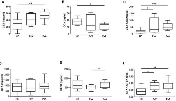
(r = -0.711,P= 0.014) indicating the reciprocal relationship between both parameters (Table 1).
Serum levels of 1,25(OH)
2D
3, calcium and calcitonin
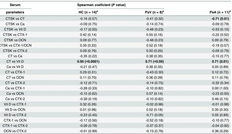
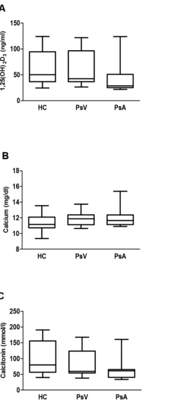
Next, 1,25(OH)2D3, calcium and CT levels in serum from patients with PsV and PsA, and con-trols were analysed (Fig 2). No significant differences between these groups were observed as expected. However, we found a significant positive Spearman’s coefficient correlation between CT and 1,25(OH)2D3in patients and controls, indicating a direct and very strong relationship between increase or decrease of both variables in controls and a less strong relationship in case of psoriasis patients (HC, r = 0.947,P<0.0001; PsV, r = 0.714,P= 0.047; PsA, r = 0.711,
P= 0.014;Table 1).
Comparison of TRAP enzymatic activity between cultured PBMCs
PBMCs from controls and patients with PsV and PsA were incubated with or without RANKL/M-CSF (RM) in the presence or absence of 1,25(OH)2D3(Vit D). Thereafter, TRAP enzymatic activity was measured in order to assess osteoclast activity. No differences were observed between PBMCs from patients and controls under culture control conditions, but in the presence of RM, PBMCs of PsA showed higher TRAP enzymatic activity compared with PBMCs from PsV and controls (P<0.05,P= 0.041, respectively;Fig 3A–3C). However, a sig-nificant higher number of PsA multinucleated TRAP+ cells per field was observed in the absence or presence of RM compared with PBMCs of PsV and controls (P<0.001,Fig 4A–4C). Next the effect of 1,25(OH)2D3on TRAP activity was analysed in PBMCs of HC, PsV and PsA. Fig 1. Bone turnover markers in serum from psoriasis patients and controls.Box and whisker plots show serum concentrations of A) CTX-1, B) OCN, C) CTX-1/OCN ratio, D) CTX-2, E) CTSK, and F) CTX-1/CTSK ratio in healthy controls (HC, n = 14), and patients with psoriasis vulgaris (PsV, n = 8) and psoriatic arthritis (PsA, n = 11) measured by ELISA kits. Box and whisker plots represent median with minimum to maximum values.*P<0.05,**P<0.01 and
***P<0.001 indicate statistically significant differences obtained by one-way ANOVA with Dunnetts’s multiple comparison test; and #P<0.05 only by t-test.
doi:10.1371/journal.pone.0153094.g001
We observed no effect of 1,25(OH)2D3in the case of control PBMCs while 1,25(OH)2D3 slightly inhibited TRAP activity from PBMCs of PsV compared with culture control conditions (P<0.05). Furthermore, 1,25(OH)2D3diminished TRAP activity in PBMCs of PsA, even in the presence of RM compared with culture control conditions (P= 0.008,P= 0.040, respectively) and with RM alone (P<0.05) (Fig 3A–3C). However, 1,25(OH)2D3diminished the number of multinucleated TRAP+ cells from all groups in the presence of RM, but in a greater proportion and even in the absence of RM in PBMCs of PsA (P<0.001,Fig 4A–4C).
Differential cytokine secretion during osteoclastogenesis
in vitro
Vitamin D plays an important role in the modulation of the immune system [19–24]. There-fore, we assessed the effect of vitamin D on cytokine secretion profile from psoriatic PBMCs upon osteoclastogenesis. Data was normalized due to baseline secretion differences between individual samples from the same group and treatment, as follows: For each sample, culture condition and cytokine, a fold change was calculated; equal to the cytokine concentration in the stimulated (RM, Vit D and RM+VitD) divided by the culture control condition (without RM, Vit D and RM+VitD) (Fig 5).
Table 1. Spearman coefficient correlation analysis of serum parameters in healthy controls and patients with psoriasis vulgaris and psoriatic arthritis.
Serum Spearman coeficient (Pvalue)
parameters HC (n = 14)a PsV (n = 8)a PsA (n = 11)a
CTSK vs CT -0.16 (0.57) -0.41 (0.32) -0.71 (0.01)
CTSK vs Ca -0.09 (0.75) -0.14 (0.74) -0.09 (0.79)
CTSK vs Vit D -0.17 (0.55) -0.48 (0.23) -0.53 (0.10)
CTSK vs CTX-1 0.42 (0.14) 0.55 (0.16) -0.22 (0.52)
CTSK vs OCN 0.09 (0.77) -0.48 (0.23) 0.09 (0.79)
CTSK vs CTX-1/OCN 0.35 (0.22) 0.52 (0.18) -0.19 (0.57)
CTSK vs CTX-2 0.09 (0.76) 0.50 (0.20) -0.09 (0.79)
CT vs Ca -0.35 (0.22) 0.38 (0.35) -0.10 (0.77)
CT vs Vit D 0.95 (<0.0001) 0.71 (<0.05) 0.71 (0.01)
Ca vs Vit D -0.21 (0.47) 0.38 (0.35) 0.05 (0.89)
CT vs CTX-1 0.29 (0.31) -0.43 (0.30) 0.12 (0.72)
CT vs OCN 0.11 (0.70) 0.36 (0.39) 0.11 (0.76)
CT vs CTX-2 -0.12 (0.71) -0.14 (0.75) -0.32 (0.34)
Ca vs CTX-1 -0.28 (0.33) -0.10 (0.82) 0.00 (1.00)
Ca vs OCN -0.15 (0.62) 0.57 (0.14) -0.23 (0.50)
Ca vs CTX-2 -0.39 (0.19) -0.10 (0.82) 0.46 (0.15)
Vit D vs CTX-1 0.32 (0.26) -0.02 (0.96) -0.01 (0.98)
Vit D vs OCN 0.01 (0.98) 0.52 (0.18) 0.35 (0.30)
Vit D vs CTX-2 -0.23 (0.45) -0.71 (0.05) 0.05 (0.89)
CTX-1 vs OCN -0.17 (0.56) -0.52 (0.18) -0.10 (0.77)
CTX-1 vs CTX-2 -0.09 (0.78) -0.37 (0.37) -0.04 (0.92)
OCN vs CTX-2 -0.01 (0.99) -0.13 (0.76) 0.36 (0.29)
Abbreviations: HC, healthy controls; PsV, psoriasis vulgaris; PsA, psoriatic arthritis; CTSK, cathepsin K; CT, calcitonin; Ca, calcium; Vit D, 1,25(OH)2D3; CTX-1, C-telopeptide of type I collagen; OCN, osteocalcin; CTX-2, C-telopeptide of type II collagen.
an is the number of participants.P<0.05.
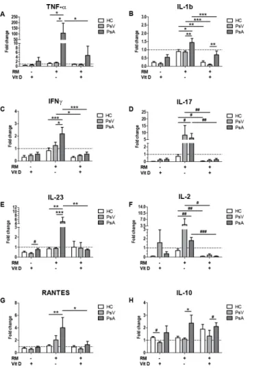
PBMCs of PsA in the presence of RM showed an increased secretion of cytokines such as TNF-α, IL-1b, IFN-γ, IL-17, IL-23, IL-2 and RANTES (91.83 fold,P<0.05; 1.62 fold,P<0.05; 2.64 fold,P<0.001; 8.59 fold,P= 0.032; 5.89 fold,P<0.01; 2.52 fold,P= 0.008; 3.63 fold,
Fig 2. Levels of 1,25(OH)2D3, calcium and calcitonin in serum from psoriasis patients and controls. Box and whisker plots show serum concentrations of A) 1,25(OH)2D3, B) calcium and C) calcitonin, in healthy controls (HC, n = 14), and patients with psoriasis vulgaris (PsV, n = 8) and psoriatic arthritis (PsA, n = 11) measured by ELISA kits. Box and whisker plots represent median with minimum to maximum values.
doi:10.1371/journal.pone.0153094.g002
Fig 3. Effect of 1,25(OH)2D3on TRAP activity from psoriasis and control PBMCs during
osteoclastogenesis.PBMCs from A) healthy controls (HC, n = 14), and patients with B) psoriasis vulgaris (PsV, n = 9) and C) psoriatic arthritis (PsA, n = 11) were cultured with or without RANKL/M-CSF (RM) in the presence or in the absence of 1,25(OH)2D3(Vit D); where n is the number of participants. TRAP enzymatic activity was measured in culture supernatants after 14 days. Box and whisker plots represent median with minimum to maximum values.*P<0.05 indicate statistically significant differences obtained by one-way ANOVA with Dunnetts’s multiple comparison test, and, #P<0.05 and ##P<0.01 only by Mann-Whitney or t-test. aP<0.05 indicate statistically significant differences between PsA and PsV cultured with RM obtained by one-way ANOVA with Dunnetts’s multiple comparison test, and, bP<0.05 between PsA and HC cultured with RM only by Mann-Whitney or t-test.
Fig 4. Effect of 1,25(OH)2D3on TRAP+ osteoclast number from psoriasis and control PBMCs during
osteoclastogenesis.PBMCs from A) healthy controls (HC, n = 4), and patients with B) psoriasis vulgaris (PsV, n = 4) and C) psoriatic arthritis (PsA, n = 4) were cultured with or without RANKL/M-CSF (RM) in the presence or in the absence of 1,25(OH)2D3(Vit D) where n represents the number of samples. TRAP + multinucleated cells (3 nuclei) per visual field, identified as mature osteoclasts, were counted after 14 days. Columns represent mean±SD.*P<0.05,**P<0.01 and***P<0.001 indicate statistically significant differences obtained by one-way ANOVA with Dunnetts’s multiple comparison test. aP<0.001 indicate statistically significant differences between PsA and PsV, and, PsA and HC cultured with and without RM, obtained by one-way ANOVA with Dunnetts’s multiple comparison test.
doi:10.1371/journal.pone.0153094.g004
P<0.01, respectively) compared with PBMCs of HC (Fig 5A–5G). Furthermore, PBMCs of PsA showed higher secretion of TNF-α, IL-1b, IFN-γ, IL-23, IL-10 (71.49 fold,P<0.05; 1.69 fold,P<0.01; 1.76 fold,P<0.05; 5.68 fold,P<0.001; 2.16 fold,P<0.05, respectively) compared with PBMCs of PsV (Fig 5A–5C, 5E and 5H).
Addition of 1,25(OH)2D3decreased levels of IL-1b (2.09 fold,P<0.0001; 9.85 fold, P<0.0001; 3.74 fold,P<0.01, respectively), IL-17 (22.77 fold,P= 0.008; 51.54 fold,P= 0.007; 11.93 fold,P= 0.02, respectively) and IL-2 (20.0 fold,P<0.0001; 18.51 fold,P= 0.035; 8.11 fold,P= 0.008, respectively) by PBMCs of PsA, PsV and HC; IFN-γby PsA and PsV (3.87 fold, P<0.0001; 2.79 fold,P<0.05, respectively); and TNF-α, IL-23 and RANTES by PsA (22.83 fold,P<0.05; 6.69 fold,P<0.01; 3.12 fold,P<0.05, respectively) compared with RM alone (Fig
5A–5G). On the other hand, PBMCs of PsA secreted higher levels of IL-1b and IL-10 in the Fig 5. Effect of 1,25(OH)2D3on cytokine secretion from PBMCs of psoriasis patients and controls. PBMCs from healthy controls (HC, white), and patients with psoriasis vulgaris (PsV, light grey) and psoriatic arthritis (PsA, dark grey) were cultured with or without RANKL/M-CSF (RM), and in the presence or in the absence of 1,25(OH)2D3(Vit D). After 14 days supernatant concentrations of A) TNF-α, B) IL-1b, C) IFN-γ, D) IL-17, E) IL-23, F) IL-2, G) RANTES and H) IL-10 were measured by Q-Plex multiplex arrays. Columns represent mean±SD of fold change of cytokine secretion compared to cytokine levels under culture conditions in the absence of RM and/or Vit D represented by dotted lines. HC, n = 5; PsV, n = 7; and PsA, n = 5; where n is the number of participants. Significant differences were obtained by two way ANOVA followed by Bonferroni post hoc test.*P<0.05,**P<0.01 and***P<0.001 are statistical significant
differences obtained by the Bonferroni post hoc test. #P<0.05, ##P<0.01 and ###P<0.001 indicate statistical significant differences only obtained by Mann-Whitney test.
presence of RM+Vit D (7.95 fold,P<0.01 and 1.58 fold,P= 0.048, respectively) compared with PsV (Fig 5B and 5H). However, the effect of 1,25(OH)2D3in the presence of RM showed no differences in any of the cytokines compared with the effect of 1,25(OH)2D3alone (Fig 5).
Discussion
For decades the beneficial effects of sunlight in the treatment of skin diseases such as psoriasis vulgaris, and immune system modulation are known. After sunlight exposure of the skin, 7-dehydrocholesterol converts to pre-vitamin D3[33], which is hydroxylated in the liver into 25-hydroxyvitamin D3[25-(OH)D3] [34] and in the kidney into 1,25-dihydroxyvitamin D3 [1,25(OH)2D3], the active vitamin D metabolite involved in extracellular calcium homeostasis and bone metabolism. Furthermore, lack of sun exposure and adequate vitamin D supply results in vitamin D deficiency and musculoskeletal pathologies [35,36]. Epidemiological stud-ies show that vitamin D deficiency is frequent in patients with psoriasis vulgaris and psoriatic arthritis [37–40]. Although phototherapy and topical application of vitamin D analogs have been used as first-line treatment with satisfactory results in the treatment of psoriasis vulgaris [1,2], therapies including vitamin D in patients with psoriatic arthritis are currently not avail-able. Pilot studies with oral vitamin D analogs supplementation have demonstrated efficacy and safety in the treatment of psoriatic arthritis [41,42] and psoriasis vulgaris [43,44]. How-ever, there have been only few randomized controlled trials on vitamin D supplementation, which include patients with psoriasis vulgaris but none with psoriatic arthritis [45–48]. More-over, the only finished randomized controlled trial did not show any vitamin D benefit [45] while the other three trials have not yet been completed [46–48]. More studies are needed to confirm if correction of deficiency would result in a statistically significant clinical improve-ment. On the other hand, to date, many studies have focused on the evaluation of the effects of cytokines and immune cells on osteoclast differentiation, but few on the reciprocal effects of osteoclasts on immune cells under physiological and pathological conditions. We aimed to study the osteoclastogenic potential and cytokine profile of PBMCs from patients with psoria-sis vulgaris and psoriatic arthritis in response to 1,25(OH)2D3.
Osteoclasts are important players in calcium homeostasis, and calcitonin is a hormone which reduces blood calcium inhibiting bone resorption as well as osteoclast formation [12]. We assessed the calcium and bone resorption marker levels in patients with psoriasis vulgaris and psoriatic arthritis. No differences in serum levels of 1,25(OH)2D3, calcium and CT between patients and controls were observed. However, our results showed a strong direct positive Spearman’s correlation between CT and 1,25(OH)2D3in HC, and a less strong Spearman’s coeficient of about 0.7 in all psoriasis patients.
CTSK, highly expressed in osteoclasts, is involved in the degradation of type I collagen [11], and OCN secreted by osteoblasts, is involved in bone formation [49]. Besides CTX-1/OCN ratios, we determined also the CTX-1/CTSK ratios in psoriasis patients and controls as CTX-1 is a product of collagen type I degradation by CTSK [32]. Such a ratio has not been reported before. We confirmed an altered bone remodelling in PsA patients demonstrated by higher CTX-1, lower OCN serum levels, and also higher CTX-1/OCN and CTX-1/CTSK ratios com-pared with controls. As CTX-1 is present in scar tissue as well as in dermis, tendons and liga-ments, it was not surprising that also PsV patients with an impaired skin barrier and altered wound healing had slightly higher serum levels of CTX-1/OCN and CTX-1/CTSK ratios as controls. However, values were still lower compared with those found in patients with PsA. Although, recently CTSK has been found highly expressed in psoriasis patient skin and involved in development of psoriasis-like skin lesions, inflammation and bone erosion in mouse models [50,51], we found only significant higher CTSK levels in patients with PsA
compared with PsV. Furthermore, our results showed a negative high coefficient correlation between CT and CTSK serum levels in patients with PsA but no correlation in case of PsV and HC. It is known that CT inhibits bone resorption in response to high serum calcium levels [52]. Our results suggest an altered osteoclast resorption response to hypercalcemic serum con-ditions which might explain partially an increase in the degradation of type 1 collagen by CTSK in the case of patients with PsA.
Psoriatic arthritis has been associated with high numbers of circulating osteoclast precur-sors; and osteoclasts, differentiated from those precursors have increased resorptive activity [29]. 1,25(OH)2D3promotes osteoclast differentiation by increasing expression of mature oste-oclast-associated genes [53] and stimulating bone resorption [54,55], but also it decreases resorptive activity in osteoclasts matured and maintained in the presence of 1,25(OH)2D3[56]. Moreover, 1,25(OH)2D3inhibits RANKL-induced osteoclastic differentiation in the absence of osteoblastsin vitro[57]. Our results showed high numbers per field of multinucleated TRAP + cells in the presence of RM by PBMCs of PsA, as already reported [58]. In addition, we observed high TRAP activity in the presence of RM by PBMCs of PsA, and also high multinu-cleated TRAP+ cell numbers in the absence of RM. Moreover, to our knowledge, it has not been reported before that 1,25(OH)2D3inhibited TRAP activity by PBMCs of PsV and PsA, and reduced TRAP+ cell numbers in a greater extent by PsA. No increases in TRAP activity in the presence of RM, but high multinucleated TRAP+ cell numbers by all PBMCs were observed. These discrepancies could be explained by the heterogeneity of the PBMCs popula-tion [59] with osteoclast precursors which in turn comprise several subpopulations, even with different proliferative capacities [60,61]. In addition, TRAP is expressed and secreted also by other cells than mature osteoclasts such as macrophages and dendritic cells [62].
Immune cells play an important role in the pathology of psoriasis, and together with osteo-clasts in the pathology of joint inflammation in patients with PsA. Osteoosteo-clasts can present anti-gens to T cells [63], secrete chemokines such as IL-8 and RANTES [64] and recruit T cellsin vitro[65]. Osteoclasts may express IL-6, TNF-α, IL-1α, IL-1βand macrophage inflammatory protein-1αin vivo[66–68]. Anti-CD3-stimulated PBMCs of PsA secreted higher IL-2 IFN-γ
and IL-10, lower IL-17, and showed no differences in TNF-αsecretion [69]. Our results showed that RM-stimulated PBMCs of PsA secreted higher levels of proinflammatory cytokines such as TNF-α, IL-1b, IFN-γ, IL-17, IL-23, IL-2 and RANTES compared with control PBMCs, and additionally higher TNF-α, IL-1b, IFN-γ, IL-23 and IL-10 compared with PBMCs of PsV, sug-gesting that these cytokines may contribute to the joint inflammation scenery in patients with PsA. No differences in any cytokine secretion were observed between RM-stimulated PBMCs of PsV and HC; which correspond to those authors who reported no differences in IL-10, IL-1β
and IL-23 levels of LPS-stimulated PBMCs from cutaneous psoriasis [70]. Here we show that PBMCs from patients with PsV and PsA have differential cytokine secretion in response to 1,25(OH)2D3. In the presence of RM, 1,25(OH)2D3decreased levels of IL-1b, IL-17 and IL-2 by PsA, PsV and HC; IFN-γby PsA and PsV; and TNF-α, IL-23 and RANTES by PsA after 14 days compared with RM-stimulated PBMCs. However, 1,25(OH)2D3inhibited secretion of any tested cytokine regardless of the presence or absence of RM.
IL-23 and the anti-inflammatory cytokine IL-10 compared with PBMCs of PsV; with differ-ences in response to 1,25(OH)2D3.
Our data provides new insight into the different cytokine secretion profiles of PBMCs in the circulation of patients with PsA and PsV, and differences in their capacity to differentiate into osteoclasts and respond to 1,25(OH)2D3. Therefore, these data also suggest the development of therapeutic strategies including vitamin D for patients with psoriatic arthritis.
Supporting Information
S1 Table. Subjects demographics from patients with PsV and PsA, and healthy controls.
Data from age variable are shown as mean ± SD.One-way ANOVA with Bonferroni ’s multi-ple comparison test.
Two-way ANOVA with Bonferroni post test analysis. (DOC)
Acknowledgments
We would like to thank the Department of Dermatology from the Jena University Hospital for their help in collecting blood samples and data from psoriasis patients specially the laboratory of Experimental Dermatology III.
Author Contributions
Conceived and designed the experiments: SC JN. Performed the experiments: SC. Analyzed the data: SC JN. Contributed reagents/materials/analysis tools: SC NK. Wrote the paper: SC JN.
References
1. Soleymani T, Hung T, Soung J. The role of vitamin D in psoriasis: a review. Int J Dermatol. 2015; 54: 383–392. doi:10.1111/ijd.12790PMID:25601579
2. Lim HW, Silpa-archa N, Amadi U, Menter A, Van Voorhees AS, Lebwohl M. Phototherapy in dermatol-ogy: A call for action. J Am Acad Dermatol. 2015; 72: 1078–1080. doi:10.1016/j.jaad.2015.03.017
PMID:25981004
3. Ash P, Loutit JF, Townsend KM. Osteoclasts derived from haematopoietic stem cells. Nature 1980; 283: 669–670. PMID:7354855
4. Takahashi N, Akatsu T, Udagawa N, Sasaki T, Yamaguchi A, Moseley JM, et al. Osteoblastic cells are involved in osteoclast formation. Endocrinology 1988; 123: 2600–2602. PMID:2844518
5. Fuller K, Wong B, Fox S, Choi Y, Chambers TJ. TRANCE is necessary and sufficient for osteoblast-mediated activation of bone resorption in osteoclasts. J Exp Med. 1998; 188: 997–1001. PMID:
9730902
6. Tanaka S, Takahashi N, Udagawa N, Tamura T, Akatsu T, Stanley ER, et al. Macrophage colony-stim-ulating factor is indispensable for both proliferation and differentiation of osteoclast progenitors. J Clin Invest. 1993; 91: 257–263. PMID:8423223
7. Ishida N, Hayashi K, Hoshijima M, Ogawa T, Koga S, Miyatake Y, et al. Large scale gene expression analysis of osteoclastogenesis in vitro and elucidation of NFAT2 as a key regulator. J Biol Chem. 2002; 277: 41147–41156. PMID:12171919
8. Takayanagi H, Kim S, Koga T. Induction and activation of the transcription factor NFATc1 (NFAT2) inte-grate RANKL signalling in terminal differentiation of osteoclasts. Dev Cell. 2002; 3: 889–901. PMID:
12479813
9. Saftig P, Hunziker E, Wehmeyer O, Jones S, Boyde A, Rommerskirch W, et al. Impaired osteoclastic bone resorption leads to osteopetrosis in cathepsin-K-deficient mice. Proc Natl Acad Sci USA. 1998; 95: 13453–13458. PMID:9811821
10. Ek-Rylander B, Flores M, Wendel M, Heinegård D, Andersson G. Dephosphorylation of osteopontin and bone sialoprotein by osteoclastic tartrate-resistant acid phosphatase. Modulation of osteoclast adhesion in vitro. J Biol Chem. 1994; 269: 14853–14856. PMID:8195113
11. Nicholson GC, Moseley JM, Sexton PM, Mendelsohn FA, Martin TJ. Abundant calcitonin receptors in isolated rat osteoclasts. Biochemical and autoradiographic characterization. J Clin Invest. 1986; 78: 355–360. PMID:3016026
12. Wallach S, Rousseau G, Martin L, Azria M. Effects of calcitonin on animal and in vitro models of skeletal metabolism. Bone 1999; 25: 509–516. PMID:10574570
13. Shinki T, Ueno Y, DeLuca HF, Suda T. Calcitonin is a major regulator for the expression of renal 25-hydroxyvitamin D3-1alpha-hydroxylase gene in normocalcemic rats. Proc Natl Acad Sci USA. 1999; 96: 8253–8258. PMID:10393981
14. Atkins GJ, Anderson PH, Findlay DM, Welldon KJ, Vincent C, Zannettino AC, et al. Metabolism of vita-min D(3) in human osteoblasts: evidence for autocrine and paracrine activities of 1α ,25-dihydroxyvita-min D(3). Bone 2007; 40: 1517–1528. PMID:17395559
15. Horwood NJ, Elliott J, Martin TJ, Gillespie MT. Osteotropic agents regulate the expression of osteoclast differentiation factor and osteoprotegerin in osteoblastic stromal cells. Endocrinology 1998; 139: 4743–4746. PMID:9794488
16. Adams JS, Beeker TG, Hongo T, Clemens TL. Constitutive expression of a vitamin D 1-hydroxylase in a myelomonocytic cell line: a model for studying 1,25-dihydroxyvitamin D production in vitro. J Bone Miner Res. 1990; 5: 1265–1269. PMID:1963733
17. Reichel H, Koeffler HP, Norman AW. Synthesis in vitro of 1,25-dihydroxyvitamin D3 and 24,25-dihy-droxyvitamin D3 by interferon-γ-stimulated normal human bone marrow and alveolar macrophages. J Biol Chem. 1987; 262: 10931–10937. PMID:3112152
18. Kogawa M, Anderson PH, Findlay DM, Morris HA, Atkins GJ. The metabolism of 25-(OH)-vitamin D(3) by osteoclasts and their precursors regulates the differentiation of osteoclasts. J Steroid Biochem Mol Biol. 2010; 121: 277–280. doi:10.1016/j.jsbmb.2010.03.048PMID:20304055
19. D’Ambrosio D, Cippitelli M, Cocciolo MG, Mazzeo D, Di Lucia P, Lang R, et al. Inhibition of IL-12 pro-duction by 1,25-dihydroxy-vitamin D3. Involvement of NF-kappa B downregulation in transcriptional repression of the p40 gene. J Clin Invest. 1998; 101: 252–262. PMID:9421488
20. Lemire JM, Archer DC, Beck J, Spiegelberg HL. Immunosuppressive actions of 1,25-dihydroxyvitamin D3: preferential inhibition of Th 1 functions. J Nutr. 1995; 125: 1704S–1708S. PMID:7782931 21. Muller K, Diamant M, Bendtzen K. Inhibition of production and function of interleukin-6 by
1,25-dihy-droxyvitamin D3. Immunol Lett. 1991; 28: 115–120. PMID:1885209
22. Raychaudhuri S, Mitra A, Datta-Mitra A. Immunomodulatory mechanisms of action of calcitriol in Psori-asis. Indian J Dermatol. 2014; 59: 116–122. doi:10.4103/0019-5154.127668PMID:24700927 23. Heine G, Niesner U, Chang HD, Steinmeyer A, Zügel U, Zuberbier T, et al. 1,25-Dihydroxyvitamin D(3)
promotes IL-10 production in human B cells. Eur J Immunol. 2008; 38: 2210–2218. doi:10.1002/eji. 200838216PMID:18651709
24. Xystrakis E, Kusumakar S, Boswell S, Peek E, Urry Z, Richards DF, et al. Reversing the defective induction of IL-10-secreting regulatory T cells in glucocorticoid-resistant asthma patients. J Clin Invest. 2006; 116: 146–155. PMID:16341266
25. Choe YB, Hwang YJ, Hahn HJ, Jung JW, Jung HJ, Lee YW, et al. A comparison of serum inflammatory cytokines according to phenotype in patients with psoriasis. Br J Dermatol. 2012; 167: 762–767. doi:
10.1111/j.1365-2133.2012.11038.xPMID:22564054
26. Nakajima H, Nakajima K, Tarutani M, Morishige R, Sano S. Kinetics of circulating Th17 cytokines and adipokines in psoriasis patients. Arch Dermatol Res. 2011; 303: 451–455. doi: 10.1007/s00403-011-1159-3PMID:21681565
27. Dalbeth N, Pool B, Smith T, Callon KE, Lobo M, Taylor WJ, et al. Circulating mediators of bone remod-eling in psoriatic arthritis: implications for disordered osteoclastogenesis and bone erosion. Arthritis Res Ther. 2010; 12: R164. doi:10.1186/ar3123PMID:20796300
28. Szentpetery A, McKenna MJ, Murray BF, Ng CT, Brady JJ, Morrin M, et al. Periarticular bone gain at proximal interphalangeal joints and changes in bone turnover markers in response to tumor necrosis factor inhibitors in rheumatoid and psoriatic arthritis. J Rheumatol. 2013; 40: 653–662. doi:10.3899/ jrheum.120397PMID:23457381
29. Ritchlin CT, Haas-Smith SA, Li P, Hicks DG, Schwarz EM. Mechanisms of TNF-alpha- and RANKL-mediated osteoclastogenesis and bone resorption in psoriatic arthritis. J Clin Invest. 2003; 111: 821–
831. PMID:12639988
31. MagaròM, Altomonte L, Mirone L, Zoli A, Tricerri A. Serum osteocalcin as an index of bone turnover in active rheumatoid arthritis and in active psoriatic arthritis. Clin Rheumatol. 1989; 8: 494–498. PMID:
2612118
32. Garnero P, Ferreras M, Karsdal MA, Nicamhlaoibh R, Risteli J, Borel O, et al. The type I collagen frag-ments ICTP and CTX reveal distinct enzymatic pathways of bone collagen degradation. J Bone Miner Res. 2003; 18: 859–867. PMID:12733725
33. DeLuca HF, Schnoes HK. Metabolism and mechanism of action of vitamin D. Ann Rev Biochem. 1976; 45: 631–666. PMID:183601
34. Cheng JB, Levine MA, Bell NH, Mangelsdorf DJ, Russell DW. Genetic evidence that the human CYP2R1 enzyme is a key vitamin D 25-hydroxylase. Proc Natl Acad Sci USA. 2004; 101: 7711–7715. PMID:15128933
35. Chen TC, Chimeh F, Lu Z, Mathieu J, Person KS, Zhang A, et al. Factors that influence the cutaneous synthesis and dietary sources of vitamin D. Arch Biochem Biophys. 2007; 460: 213–217. PMID:
17254541
36. Holick MF, Chen TC. Vitamin D deficiency: a worldwide problem with health consequences. Am J Clin Nutr. 2008; 87: 1080S–1086S. PMID:18400738
37. Gisondi P, Rossini M, Di Cesare A, Idolazzi L, Farina S, Beltrami G, et al. Vitamin D status in patients with chronic plaque psoriasis. Br J Dermatol. 2012; 166: 505–510. doi:10.1111/j.1365-2133.2011. 10699.xPMID:22013980
38. Grazio S, NaglićĐB, AnićB, GrubišićF, Bobek D, Bakula M, et al. Vitamin D serum level, disease activ-ity and functional abilactiv-ity in different rheumatic patients. Am J Med Sci. 2015; 349: 46–49. doi:10.1097/ MAJ.0000000000000340PMID:25310509
39. Suárez-Varela MM, Reguera-Leal P, Grant WB, Rubio-López N, Llopis-González A. Vitamin D and psoriasis pathology in the Mediterranean region, Valencia (Spain). Int J Environ Res Public Health. 2014; 11: 12108–12117. doi:10.3390/ijerph111212108PMID:25429679
40. Touma Z, Eder L, Zisman D, Feld J, Chandran V, Rosen CF, et al. Seasonal variation in vitamin D levels in psoriatic arthritis patients from different latitudes and its association with clinical outcomes. Arthritis Care Res (Hoboken). 2011; 63: 1440–1447.
41. Gaál J, Lakos G, Szodoray P, Kiss J, Horváth I, Horkay E, et al. Immunological and clinical effects of alphacalcidol in patients with psoriatic arthropathy: results of an open, follow-up pilot study. Acta Derm Venereol. 2009; 89: 140–144. doi:10.2340/00015555-0555PMID:19325997
42. Huckins D, Felson DT, Holick M. Treatment of psoriatic arthritis with oral 1,25-dihydroxyvitamin D3: a pilot study. Arthritis Rheum. 1990; 33: 1723–1727. PMID:2242069
43. Finamor DC, Sinigaglia-Coimbra R, Neves LC, Gutierrez M, Silva JJ, Torres LD, et al. A pilot study assessing the effect of prolonged administration of high daily doses of vitamin D on the clinical course of vitiligo and psoriasis. Dermatoendocrinol. 2013; 5: 222–234. doi:10.4161/derm.24808PMID:
24494059
44. Takamoto S, Onishi T, Morimoto S, Imanaka S, Yukawa S, Kozuka T, et al. Effect of 1 alpha-hydroxy-cholecalciferol on psoriasis vulgaris: a pilot study. Calcif Tissue Int. 1986; 39: 360–364. PMID:
3100000
45. Hata TR, Audish D, Kotol P, Coda A, Kabigting F, Miller J, et al. A randomized controlled double-blind investigation of the effects of vitamin D dietary supplementation in subjects with atopic dermatitis. J Eur Acad Dermatol Venereol. 2014; 28: 781–789. doi:10.1111/jdv.12176PMID:23638978
46. Scragg R, Waayer D, Stewart AW, Lawes CM, Toop L, Murphy J, et al. The Vitamin D Assessment (ViDA) Study: design of a randomized controlled trial of vitamin D supplementation for the prevention of cardiovascular disease, acute respiratory infection, falls and non-vertebral fractures. J Steroid Biochem Mol Biol. 2015.http://dx.doi.org/10.1016/j.jsbmb.2015.09.010.
47. Chulalongkorn University (Sponsor). The efficacy of vitamin D3 for the treatment of chronic plaque type psoriatic patients with vitamin D deficiency and insufficiency: a randomized controlled trial. Available:
https://clinicaltrials.gov/ct2/show/NCT01339741
48. Pontificia Universidad Catolica de Chile (Sponsor). Effect of vitamin D supplementation on metabolic parameters of patients with moderate to severe psoriasis. Available:https://clinicaltrials.gov/ct2/show/ NCT02271971
49. Hauschka PV, Lian JB, Cole DE, Gundberg CM. Osteocalcin and matrix Gla protein: vitamin K-depen-dent proteins in bone. Physiol Rev. 1989; 69: 990–1047. PMID:2664828
50. Hao L, Zhu G, Lu Y, Wang M, Jules J, Zhou X, et al. Deficiency of cathepsin K prevents inflammation and bone erosion in rheumatoid arthritis and periodontitis and reveals its shared osteoimmune role. FEBS Lett. 2015; 589: 1331–1339. doi:10.1016/j.febslet.2015.04.008PMID:25896020
51. Hirai T, Kanda T, Sato K, Takaishi M, Nakajima K, Yamamoto M, et al. Cathepsin K is involved in devel-opment of psoriasis-like skin lesions through TLR-dependent Th17 activation. J Immunol. 2013; 190: 4805–4811. doi:10.4049/jimmunol.1200901PMID:23543761
52. Friedman J, Raisz LG. Thyrocalcitonin: inhibitor of bone resorption in tissue culture. Science 1965; 150: 1465–1467. PMID:5892553
53. Gu J, Tong XS, Chen GH, Wang D, Chen Y, Yuan Y, et al. Effects of 1α,25-(OH)2D3on the formation and activity of osteoclasts in RAW264.7 cells. J Steroid Biochem Mol Biol. 2015; 152: 25–33. doi:10. 1016/j.jsbmb.2015.04.003PMID:25864627
54. Holtrop ME, Cox KA, Clark MB, Holick MF, Anast CS. 1,25-dihydroxycholecalciferol stimulates osteo-clasts in rat bones in the absence of parathyroid hormone. Endocrinology 1981; 108: 2293–2301. PMID:6894424
55. Raisz LG, Trummel CL, Holick MF, DeLuca HF. 1,25-dihydroxycholecalciferol: a potent stimulator of bone resorption in tissue culture. Science 1972; 175: 768–769. PMID:4333399
56. Kogawa M, Findlay DM, Anderson H, Ormsby R, Vincent C, Morris HA, et al. Osteoclastic metabolism of 25(OH)-vitamin D3: A potential mechanism for optimization of bone resorption. Endocrinology 2010; 151: 4613–4625. doi:10.1210/en.2010-0334PMID:20739402
57. Lee SK, Kalinowski J, Jastrzebski S, Lorenzo JA. 1,25(OH)2 vitamin D3-stimulated osteoclast forma-tion in spleen-osteoblast cocultures is mediated in part by enhanced IL-1 alpha and receptor activator of NF-kappa B ligand production in osteoblasts. J Immunol. 2002; 169: 2374–2380. PMID:12193704 58. IkićM, JajićZ, LazićE, IvčevićS, GrubišićF, MarušićA, et al. Association of systemic and intra-articular
osteoclastogenic potential, pro-inflammatory mediators and disease activity with the form of inflamma-tory arthritis. Int Orthop. 2014; 38: 183–192. doi:10.1007/s00264-013-2121-0PMID:24100919 59. Grage-Griebenow E, Flad HD, Ernst M. Heterogeneity of human peripheral blood monocyte subsets. J
Leukoc Biol. 2001; 69: 11–20. PMID:11200054
60. Komano Y, Nanki T, Hayashida K, Taniguchi K, Miyasaka N. Identification of a human peripheral blood monocyte subset that differentiates into osteoclasts. Arthritis Res Ther. 2006; 8: R152. PMID:
16987426
61. Lari R, Kitchener PD, Hamilton JA. The proliferative human monocyte subpopulation contains osteo-clast precursors. Arthritis Res Ther. 2009; 11: R23. doi:10.1186/ar2616PMID:19222861
62. Janckila AJ, Parthasarathy RN, Parthasarathy LK, Seelan RS, Hsueh YC, Rissanen J, et al. Properties and expression of human tartrate-resistant acid phosphatase isoform 5a by monocyte-derived cells. J Leukoc Biol. 2005; 77: 209–218. PMID:15542543
63. Li H, Hong S, Qian J, Zheng Y, Yang J, Yi Q. Cross talk between the bone and immune systems: osteo-clasts function as antigen-presenting cells and activate CD4+ and CD8+ T cells. Blood 2010; 116: 210–217. doi:10.1182/blood-2009-11-255026PMID:20304810
64. Pappalardo A, Thompson K. Novel immunostimulatory effects of osteoclasts and macrophages on humanγδT cells. Bone 2015; 71: 180–188. doi:10.1016/j.bone.2014.10.019PMID:25445456 65. Grassi F, Manferdini C, Cattini L, Piacentini A, Gabusi E, Facchini A, et al. T cell suppression by
osteo-clasts in vitro. J Cell Physiol. 2011; 226: 982–990. doi:10.1002/jcp.22411PMID:20857429
66. O’Keefe R, Teot L, Singh D, Puzas JE, Rosier RN, Hicks DG. Osteoclasts constitutively express regula-tors of bone resorption: an immunohistochemical and in situ hybridization study. Lab Invest. 1997; 76: 457–465. PMID:9111508
67. Rahimi P, Wang C, Stashenko P, Lee SK, Lorenzo JA, Graves DT. Monocyte chemoattractant protein-1 expression and monocyte recruitment in osseous inflammation in the mouse. Endocrinology protein-1995; 136: 2752–2759. PMID:7750500
68. Rothe L, Collin-Osdoby P, Chen Y, Sunyer T, Chaudhary L, Tsay A, et al. Human osteoclasts and oste-oclast-like cells synthesize and release high basal and inflammatory stimulated levels of the potent che-mokine interleukin-8. Endocrinology 1998; 139: 4353–4363. PMID:9751519
69. BosèF, Capsoni F, Molteni S, Raeli L, Diani M, Altomare A, et al. Differential expression of interleukin-2 by anti-CD3-stimulated peripheral blood mononuclear cells in patients with psoriatic arthritis and patients with cutaneous psoriasis. Clin Exp Med. 2014; 39: 385–390.