Influence of Sodium Hypochlorite-Based Irrigants
on the Susceptibility of Intracanal Microbiota to
Biomechanical Preparation
Janir Alves SOARES1 Donaldo Rosa PIRES JÚNIOR2
1Discipline of Endodontics, Department of Dentistry and 2Discipline of Microbiology,
Department of Science of the Heath, Federal University of Vales do Jequitinhonha e Mucuri, Diamantina, MG, Brazil
This study evaluated the microbiological conditions of root canals, using smears and culture from anterior teeth and premolars with necrotic pulps associated with chronic periapical pathologies, before and after biomechanical preparation (BMP). During double-flared instrumentation, 1, 2.5 and 5% sodium hypochlorite (NaOCl)-based irrigants were used in 3 groups: GI (n=39), GII (n=36) and GIII (n=36), respectively. Before BMP, all cultures were positive and the smears showed microbiologically diverse morphotypes, including fusiforms, pleomorphic, rods, cocci and filaments. Quantitetively, 20, 20 and 23 morphotypes were identified in GI, GII and GIII, respectively). After BMP, the percentages of negative cultures in GI, GII and GIII were 74.2%, 86.3% and 93.4% (p>0.05) and the number of morphotypes decreased to 14, 15 and 5, respectively. All teeth with 2 root canals and/or associated fistulas were microbiologically negative after BMP, regardless of irrigant concentration. Gram-negative morphotypes were more susceptible to the action of irrigants. After irrigation with 5% NaOCl, only structural arrangements consisting of Gram-positive cocci and bacilli persisted. Thus, BMP plus 5% NaOCl offered the best antiseptic potential because in the few positive cultures a significant reduction in the number of microbiological morphotypes was also shown (p<0.05).
Key Words: root canal therapy, biomechanical preparation, irrigating solutions, microorganisms, sodium hypochlorite.
Correspondence: Prof. Dr. Janir Alves Soares, Universidade Federal dos Vales do Jequitinhonha e Mucuri, Campi I, Rua da Glória, 187, 39100-00 Diamantina, MG, Brasil. Tel: +55-38-3531-2901. Fax: +55-38-3531-1811. email: [email protected]
INTRODUCTION
The root canals of teeth with necrotic pulps associated with periapical pathology contain decom-posed pulp and a diverse reservoir of microorganisms as well as byproducts from pulp necrosis and bacterial metabolism (1). These microorganisms are mainly those that inhabit the oral cavity, the upper respiratory tract or the gastrointestinal system. The number of species identified in the mouth can reach over 530. Of this group, more than one third has already been isolated from root canals (2). Endodontic infection is endog-enous and predominantly polymicrobial and a possible correlation has been proposed between the magnitude of radiographic alteration and both the quantity and diversity of the endodontic microbiota (3).
With respect to morphotypes and their staining reactions, most are positive cocci and
in amorphous exopolysaccharide structures, adhering to dentin surfaces (biofilms). A scanning electron mi-croscopic study (5) showed a high incidence of straight and curved bacilli, cocci, spirochetes, coco-bacilli rods and filaments, as well as microcolonies composed of cocci, bacilli or yeasts in the apical third of root canals associated with periapical lesions. Sen et al. (6) have reported structural arrangements of a corn-cob struc-ture and Nair (7) identified cellular arrangements in hyphal-like or palisade-like structures.
Classic studies have shown the role of microor-ganisms in formation of chronic abscesses, cysts and periapical granulomas. Endodontics has sought an ef-fective method to control intracanal infection (1,8). Initially, control of root canal microbiota was predomi-nantly based on locally applied medication. Only after a long time and innumerable changes of balls of cotton wool or paper points soaked in a variety of antiseptic substances, biomechanical preparation (BMP) was in-troduced to endodontic therapy, representing a signifi-cant advance in the control of microorganisms that are the focus of infection (4).
In an effort to remove organic material, microor-ganisms and their toxins, numerous types of irrigating solutions have been proposed, which in conjunction with the mechanical action of files and reamers, have achieved a satisfactory debridement and antisepsis of root canals (1). Sodium hypochlorite (NaOCl)-based solutions have been the most commonly recommended since the 1950s, although there has been no consensus about the best concentration to use (8). Considering that most of its properties are influenced by free chlorine concentration, the purpose of this study was to evaluate the antiseptic effect of various NaOCl-based irrigants used for BMP of root canals associated with chronic periapical pathologies in asymptomatic patients.
MATERIAL AND METHODS
Patients treated at the Clinic of Endodontics of Federal University of Vales do Jequitinhonha e Mucuri (Brazil) were enrolled in this study. The research project was approved by the Ethics in Research Committee and the patients signed an informed consent form.
The sample comprised 93 anterior teeth and premolars with advanced carious lesions, necrotic pulp and periapical radiolucences, from 83 asymptomatic patients aged 14-53 years, making a total of 111 root
canals randomly assigned to 3 groups: I (n=39), II (n=36) and III (n=36), with 31, 32 and 30 teeth, respectively.
smears were prepared and Gram stained. Using an immersion optical microscope, the morphotypes were recorded (approximately 20 fields per slide). The num-ber of microbial morphotypes in the root canals before and after BMP, as well as the incidence of negative microbiological culture after BMP, were analyzed sta-tistically by the chi-square test at 5% significance level.
RESULTS
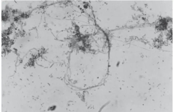
The results are presented on Tables 1, 2 and 3. In the first microbiological evaluation all samples had microorganisms, but a larger number of morphotypes were identified in the primary root canal smears. In contrast, in the second microbiological evaluation, a larger number of morphotypes were identified in the cultures. Before BMP, 24 microbiological morphotypes
(see examples on Figs. 1-3) were identified from 111 root canals (Tables 1 and 3). The number of morphotypes ranged from 2 to 16 per root canal, median of 10 (Table 3). After BMP, root canals in groups I, II and III were 74.2%; 86.3% and 93.4% microbiologically negative respectively, but without significant difference (χ2=4.0;
p=0.1351) (Table 2). After BMP, 92.6% of the cultures of the 15 microbiologically positive root canals re-mained polymicrobial. The cultures obtained after BMP revealed 16 microbiological forms, 14, 15 and 5 morphotypes being recovered in groups I, II and III, respectively (Table 2). The 18 teeth with two root canals had negative microbiological culture after BMP irrespective of the irrigant. Similar result was found for the 18 teeth with sinus tract (Table 3). After BMP, the predominant microorganisms in Group I were: Gram-positive rods, staphylococci, Gram-Gram-positive strepto-cocci and diplostrepto-cocci. Group II revealed Gram-positive cocci in isolation or in chains, Gram-positive fusiforms, Gram-positive filaments and Gram-positive diplobacilli and streptococci. Group III revealed Gram-positive V-shaped rods, Gram-positive cocci in isolation or in chains, Gram-positive diplococci and staphylococci occurred at the same frequency (Table 1).
DISCUSSION
All teeth with necrotic pulp associated with periapical radiolucent areas had a variable number of microbiological morphotypes in the root canal, mainly rods, cocci, fusiforms and filaments, Gram-positive
Figure 2. Uncountable cocci, bacilli, filamentous microorganisms, pseudo-hyphae and some neutrophils.
Figure 1. Cocci and bacilli within extensive interlaced filaments.
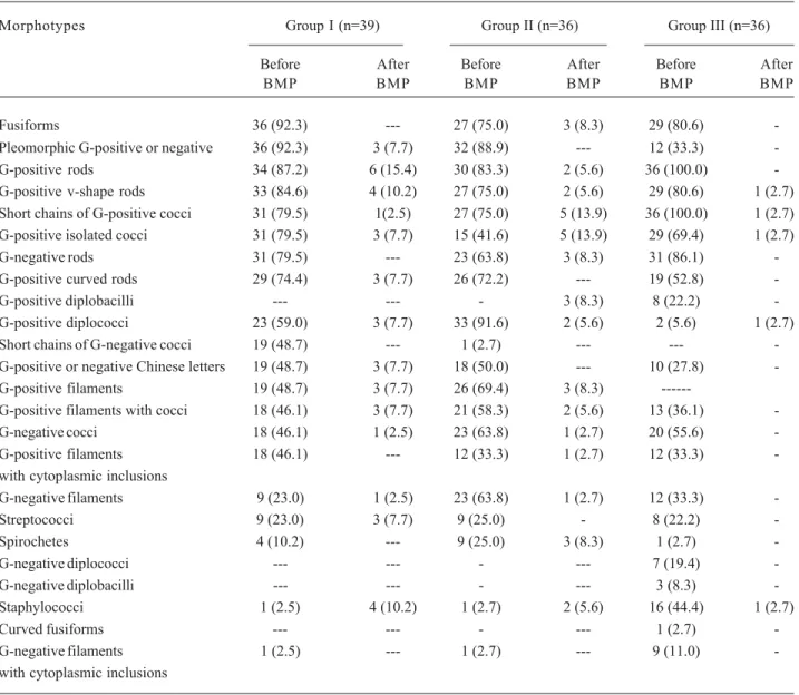
Table 1. Relative and absolute incidence of microbial morphotypes, in root canals before and after biomechanical preparation in the 3 experimental groups.
Morphotypes Group I (n=39) Group II (n=36) Group III (n=36)
Before After Before After Before After
BMP BMP BMP BMP BMP BMP
Fusiforms 36 (92.3) --- 27 (75.0) 3 (8.3) 29 (80.6) -Pleomorphic G-positive or negative 36 (92.3) 3 (7.7) 32 (88.9) --- 12 (33.3) -G-positive rods 34 (87.2) 6 (15.4) 30 (83.3) 2 (5.6) 36 (100.0) -G-positive v-shape rods 33 (84.6) 4 (10.2) 27 (75.0) 2 (5.6) 29 (80.6) 1 (2.7) Short chains of G-positive cocci 31 (79.5) 1(2.5) 27 (75.0) 5 (13.9) 36 (100.0) 1 (2.7) G-positive isolated cocci 31 (79.5) 3 (7.7) 15 (41.6) 5 (13.9) 29 (69.4) 1 (2.7) G-negative rods 31 (79.5) --- 23 (63.8) 3 (8.3) 31 (86.1) -G-positive curved rods 29 (74.4) 3 (7.7) 26 (72.2) --- 19 (52.8) -G-positive diplobacilli --- --- - 3 (8.3) 8 (22.2) -G-positive diplococci 23 (59.0) 3 (7.7) 33 (91.6) 2 (5.6) 2 (5.6) 1 (2.7) Short chains of G-negative cocci 19 (48.7) --- 1 (2.7) --- --- -G-positive or negative Chinese letters 19 (48.7) 3 (7.7) 18 (50.0) --- 10 (27.8) -G-positive filaments 19 (48.7) 3 (7.7) 26 (69.4) 3 (8.3)
---G-positive filaments with cocci 18 (46.1) 3 (7.7) 21 (58.3) 2 (5.6) 13 (36.1) -G-negative cocci 18 (46.1) 1 (2.5) 23 (63.8) 1 (2.7) 20 (55.6) -G-positive filaments 18 (46.1) --- 12 (33.3) 1 (2.7) 12 (33.3) -with cytoplasmic inclusions
G-negative filaments 9 (23.0) 1 (2.5) 23 (63.8) 1 (2.7) 12 (33.3) -Streptococci 9 (23.0) 3 (7.7) 9 (25.0) - 8 (22.2) -Spirochetes 4 (10.2) --- 9 (25.0) 3 (8.3) 1 (2.7) -G-negative diplococci --- --- - --- 7 (19.4) -G-negative diplobacilli --- --- - --- 3 (8.3) -Staphylococci 1 (2.5) 4 (10.2) 1 (2.7) 2 (5.6) 16 (44.4) 1 (2.7)
Curved fusiforms --- --- - --- 1 (2.7)
-G-negative filaments 1 (2.5) --- 1 (2.7) --- 9 (11.0) -with cytoplasmic inclusions
BMP = Biomechanical preparation; (--- ) = Absent; Outside brackets = Relative frequency; Inside brackets = Absolute frequency.
Table 2. Incidence of positive microbiological cultures and microbial morphotypes before and after biomechanical preparation.
Groups Before biomechanicalpreparation After biomechanical preparation
Positive cultures Number of morphotypes Positive cultures Number of morphotypes
I (n=39) 39 (100%) 20 10 (25.8%) 14
II (n=36) 36 (100%) 20 5 (13.7%) 15
III (n=36) 36 (100%) 23 2 (6.6%) 5*
and Gram-negative bacteria. This microbiological di-versity agrees with previous histobacteriological (1,4) and microbial culture studies (8). The role of microor-ganisms in periapical pathogenesis has been well estab-lished and several methodologies have been used to characterize the microbiota in root canals associated with chronic periapical pathology and evaluate the effectiveness of endodontic techniques to eliminate microorganisms from root canals.
It is known that 4 to 7 microbial species on average (and a maximum of 16) can be retrieved per
canal using current techniques (4). However, in the present study, the incidence of morphotypes varied from 2 to16 (9.6 per root canal on average). It is difficult to establish a correlation between the number of micro-biological morphotypes and bacterial species. Never-theless, some authors (9,10) emphasize that to evaluate the intracanal microbiota, Gram stained smears are easy and economical and state that there is a significant correlation with the results of microbiological culture. It must be emphasized that these methodologies show temporal variation. Thus, before BMP, a larger density and diversity of morphotypes in smears, obtained di-rectly from root canals was observed compared to the respective cultures. In contrast, after BMP, the cultures provided smears with a greater density of microorgan-isms. Such a pattern was possibly be due to the number of microorganisms in the root canals and/or the sensi-tivity of the methodology used. Therefore, with a large number of microorganisms, the smears tend to be more precise; in contrast, with a small number, preliminary culture improves the observation of viable microorgan-isms. In this way, the microbiological aspects of the initial smears obtained from root canals were compared to the smears resulting from culture after BMP.
After BMP, an increasing incidence of negative culture can be observed using irrigants of increasing concentration. This is consistent with the findings of a previous in vitro study (11), which showed that an antiseptic solution acts according to the biochemical principle that depends on the concentration of the active element. In this case, it is directly proportional to the concentration of free chlorine, which, in turn, depends on the concentration of hypochloric acid in the solution. This causes a bactericidal effect through oxidation and hydrolysis of sulphide groups of enzymes located on bacterial membranes and cytoplasm (12). Gutiérrez et al. (13) attributed the bactericidal action to plasmosis, in which cuboid crystals of sodium chlorate, the source of NaOCl (12), close precipitate close to bacteria and is found in dentinal tubules at a depth of up to 220 μm.
No significant differences were found between the negative microbiological cultures and the different NaOCl concentrations. Perhaps, less concentrated so-lutions used copiously and frequently have their antimi-crobial effect increased, becoming as clinically effec-tive as 5% NaOCl. Because all solutions were used in Table 3. Incidence of microbial morphotypes before BMP,
numbers of root canals, sinus tract and positive microbiological cultures after BMP.
Tooth Group I Group II Group III (n=31) (n=32) (n=30)
1 4# 11 3
2 5+ 11 11+
3 3+ 9# 7#
4 5 9+ 9*
5 6* 8 6
6 9+ 9# 9
7 6 10 12
8 10* 12* 14
9 9+ 11# 10*
10 10# 13 11
11 10 9+ 13*
12 11# 9* 10
13 9+ 9 13
14 9+ 10# 12*
15 8# 5 8*
16 10+ 13 10*
17 11+ 4+ 9
18 10 13 12
19 8 11 11#
20 11# 13+ 10*
21 12 11 4#
22 16 2* 11*
23 15# 7* 13*
24 16 6 12#*
25 10 13+ 14+
26 9+ 9 12
27 8# 10 6*
28 6 7 11*#
29 4 11 3
30 6# 7 5#
31 9+ 5
--32 -- 11
the same way and under similar clinical conditions, 5% NaOCl would have a better antiseptic performance (i.e., produce 100% negative microbiological culture), which did not occur, suggesting that variable concentration, frequency and volume have a limited enhancing effect on the antiseptic efficacy of irrigants. This limit corre-sponds to its maximum clinical efficacy, which, once reached, cannot be increased significantly by either the amount applied after each file, the active ingredient concentration or the increase of irrigation frequency.
After BMP, the incidence of negative microbio-logical culture was not significantly influenced by the concentration of irrigants. The solutions presented a similar antisepsis pattern against Gram-negative and positive microorganisms. However, 5% NaOCl was significantly more effective against Gram-positives. Sydney and Estrela (14) showed that Gram-negative bacteria were more sensitive to BMP, while the Gram-positive bacteria were more resistant, with the highest incidence of Peptostreptococcus prevoti and Clostridium
histolyticum. After BMP and placement of calcium
hydroxide-based intracanal dressings in dogs’ teeth with induced chronic periapical lesions, Soares et al. (1) observed a predominantly Gram-positive microbiota in the root canal system. The different susceptibilities of microorganisms may be partially attributed to the com-position and structure of their cell walls. Thus, although the cell wall of Gram-negative bacteria is more complex, containing glycopeptides, lipoproteins, an external mem-brane and lipopolysaccharides, apparently the cell struc-tures of Gram-positive microorganisms offer greater resistance to endodontic procedures, possibly deter-mined by the structure of glycopeptides in the cell wall, which is thicker in Gram-positive microorganisms.
The incidence of morphotypes in the canals of teeth with fistulas varied from 3 to 16 (9.8 on average)
versus 2 to 15 (9.5 on average) for teeth without fistulas. This raises the possibility that the occurrence of a fistula is independent on the number of morphotypes in the root canal and may be dependant on the interaction between certain bacterial species and the host’s immunological system (4,8). All 18 teeth with associated sinus tracts were microbiologically negative after BMP. In part, it may be attributed to the fact that 12 teeth (66.6%) had their canals irrigated with 5% NaOCl, which increased the chances of eliminating the microbiota.
Perhaps the antiseptic effect of irrigants also correlates with the ability to remove debris from the root
canals, which, in turn, is influenced, not only by the concentration of the active ingredient, but also by the quantity and frequency of irrigation as well as the amount of root canal enlargement. Nery et al. (15) demonstrated that the ability to remove debris lodged in the apical third depends much more on the frequency of using an irrigating solution than on the technique of irrigation or the type of irrigating solution used, while Orstavik et al. (16) highlighted the importance of wid-ening the root canal. The influence of the time that the irrigating solution remained in the root canal may explain why all teeth with 2 roots produced negative cultures irrespective of the irrigants solution used because the time for irrigation and instrumentation was practically twice as long as that spent for a single-rooted tooth.
The irrigants showed a varying effect on the microbial forms. Thus, some morphotypes were re-duced or even eliminated, a result that increased with higher concentrations of free chlorine but not always. This uneven pattern of endodontic microbiota suscep-tibility to BMP has been shown.It has been reported (8) that microbiological samples obtained after BMP plus 2.5% NaOCl revealed that, numerically, some species were reduced, some increased and others remained the same. However, in our study, 5% NaOCl had a more specific antiseptic effect because it eliminated 77.3% of the identified morphotypes. Therefore, the root canal microbiota can be significantly altered by the prepara-tion of teeth with necrotic pulp associated with radiolu-cent periapical areas using 5% NaOCl as irrigant. In contrast, the use of chlorine solutions in lower concen-trations immediately followed by root canal obturation often leads to failure because of the persistence of radiolucent areas is associated with a high incidence of Gram-positive cocci and bacilli, distributed in the root canal and apical cementum lacunae, which are respon-sible for marked periapical tissue disorganization (17). In addition to giving the main root canal a conical shape, BMP has other advantages. Each milligram of necrotic tissue removed contains 107.7± 0.6
the previous pattern of colonization (1,14,19). From a microbiological standpoint, the main root canal and its ramifications should receive the best disinfection protocol possible. Several calcium hydrox-ide-base dressings have been recommended with this objective (1,4,20). Although the gas requirements for obligatory anaerobes were not used, the results of this study suggest that, in terms of negative microbiological culture, the antiseptic efficacy of irrigants was not influenced by their concentration. Regarding the elimi-nation of microbiological morphotypes, only 5% NaOCl had a strong impact on root canal microbiota because in the few positive microbiological cultures a significant reduction was shown in the microbiological morphotypes. Therefore, BMP with use of 5% NaOCl will reduce the number of microorganisms in the main root canal for the intracanal dressing to eliminate.
RESUMO
Este estudo avaliou as condições microbiológicas dos canais radiculares, por meio de esfregaços e culturas de dentes anteriores e pré-molares com necrose pulpar associada à radiolucidezes periapicais, antes e após o preparo biomecânico (PBM). Utilizou-se a técnica de instrumentação biescalonada coadjuvada por soluções de hipoclorito de sódio (NaOCl) a 1, 2,5 ou 5% nos grupos I (n=39), II (n=36) e III (n=36), respectivamente. Antes do PBM havia 100% de culturas positivas e os esfregaços proveram diversificados morfotipos microbiológicos, sendo 20, 20 e 23 nos grupos I, II e III, respectivamente. Após o PBM, o percentual de culturas negativas nos grupos I, II e III foi 74,2%, 86,3 e 93,4% (p>0,05) e a incidência de morfotipos declinou para 14, 15 e 5, respectivamente. Todos os dentes birradiculados e/ou portadores de fístulas apresentaram-se microbiologicamente negativos após o PBM, independentemente do irrigante utilizado. Os morfotipos Gram-negativos foram mais suscetíveis à ação do PBM. Após o PBM persistiram apenas cocos e bacilos Gram-positivos no grupo III. Portanto, o PBM coadjuvado por solução de NaOCl a 5%, proporcionou o melhor desempenho anti-séptico, pois, nas poucas culturas positivas, houve também significativa redução do número de morfotipos microbiológicos (p<0.05).
REFERENCES
1 . Soares JA, Leonardo MR, Silva LAB, Tanomaru Filho M, Ito IY. Effect of biomechanical preparation and calcium hydrox-ide pastes on the anti-sepsis of root canal systems in dogs. J App Oral Sci 2005;13:93-100.
2 . Debelian GJ, Olsen I, Tronstad L. Bacteremia in conjunction with endodontic therapy. Endod Dent Traumatol 1995;11:142-149.
3 . Sundqvist G. Ecology of the root canal flora. J Endod 1992;18:427-429.
4 . Soares JA, Leonardo MR, Silva LAB, Tanomaru Filho M, Ito
IY. Effect of rotary instrumentation and of the association of calcium hydroxide and chlorhexidine on the anti-sepsis of root canal systems in dogs. Braz Oral Res 2006;20:120-126. 5 . Molven O, Olsen I, Kerekes K. Scanning electron microscopy of bacteria in the apical part of root canals in permanent teeth with periapical lesions. Endod Dent Traumatol 1991;7:226-229.
6 . Sen BH, Piskin B, Dermirci T. Observation of bacteria and fungi in infected root canals and dentinal tubules by SEM. Endod Dent Traumatol 1995;11:6-9.
7 . Nair PNR. Light and electron microscopic studies of root canal flora and periapical lesions. J Endod 1987;13:29-39. 8 . Gomes BPFA, Lilley JD, Drucker DB. Variations in the
sus-ceptibilities of components of the endodontic microflora to biomechanical procedures. Int Endod J 1996;29:235-241. 9 . Pajari U, Ahola R, Backman T, Hietala EL, Tjäderhane L,
Larmas M. Evaluation of Gram’s method of staining for the prognosis of root canal treatment in nonvital dental pulps. Oral Surg Oral Med Oral Pathol 1993;76:91-99.
10. Kuruvilla JR, Kamath P. Antimicrobial activity of 2.5% sodium hypochlorite and 0.2% chlorhexidine gluconate sepa-rately and combined, as endodontic irrigants. J Endod 1998;24:472-475.
11. Yesilsoy C, Whitaker E, Cleveland D, Phillips E, Trope M. Antimicrobial and toxic effects of established and potential root canal irrigants. J Endod 1995;21:513-515.
12. Siqueira Jr JF, Batista MMD, Fraga RC, Uzeda M Antibacterial effects of endodontic irrigants on Black-pigmented Gram-negative anaerobes and facultative bacteria. J Endod 1998;24:414-416.
13. Gutiérrez JH, Jofré A, Villena F. Scanning electron micro-scope study on the action of endodontic irrigants on bacteria invading the dentinal tubules. Oral Surg Oral Med Oral Pathol 1990;69:491-501.
14. Sydney GB, Estrela C. Influence of root canal preparation on anaerobic bacteria in teeth with asymptomatic apical peri-odontitis. Braz Endod J 1996;1:7-10.
15. Nery MJ, Holland R, Souza V. Efficacy of different irrigating techniques and irrigants for removal of debris from root canals. Rev Odontol Araçatuba 1982;3:21-27.
16. Orstavik D, Kerekes K, Molven O. Effects of extensive apical reaming and calcium hydroxide dressing on bacterial infection during treatment of apical periodontitis: a pilot study. Int Endod J 1991;24:1-7.
17. Leonardo MR, Almeida WA, Ito IY, Silva LAB. Radiographic and microbiologic evaluation of postreatment apical and periapical repair of root canals of dog’s teeth with experi-mentally induced chronic lesion. Oral Surg Oral Med Oral Pathol 1994;78:23-28.
18. Zavistoski J, Dzink J, Onderdonk A, Bartlett J. Quantitative bacteriology of endodontic infections. Oral Surg Oral Med Oral Pathol 1980;49:171-174.
19. Sjögren U, Fidgor D, Persson S, Sundqvist G. Influence of infection at the time of root filling on the outcome of endodontic treatment of teeth with apical periodontitis. Int Endod J 1997;30:297-306.
20. Soares JA, Leonardo MR, Silva LAB, Tanomaru Filho M, Ito IY. Elimination of intracanal infection in dogs‘ teeth with induced periapical lesions after rotary instrumentation: Influ-ence of different calcium hydroxide pastes. J Appl Oral Sci. 2006;14:172-177.