Patrícia Daniela Melchiors Angst(a) Flávia Benetti Piccinin(a)
Rui Vicente Oppermann(a) Rosemary Adriana Chiérici Marcantonio(b)
Sabrina Carvalho Gomes(a)
(a) Department of Periodontology, Dental School, Univ Federal do Rio Grande do Sul - UFRGS, Porto Alegre, RS, Brazil.
(b) Department of Periodontology, Univ Estadual Paulista – Unesp, Araraquara, SP, Brazil.
Corresponding Author: Sabrina Carvalho Gomes
E-mail: [email protected]
Response of molars and non-molars
to a strict supragingival control in
periodontal patients
Abstract: The posterior position in the arches is one of the factors that underlies the poor prognosis of molar teeth (M). It is speculated that M do not beneit from the oral hygiene routine as well as non-molars (NM) do. This study evaluated the response of M and NM to supragingival control during a 6-month period in 25 smokers (S) and 25 never-smokers (NS) with moderate-to-severe periodontitis. One calibrated examiner as-sessed visible plaque (VPI) and gingival bleeding (GBI) indexes, periodon-tal probing depth (PPD), bleeding on probing (BOP), and clinical attach-ment loss (CAL) at days 0 (baseline), 30 and 180. At baseline, M showed signiicantly higher mean values of VPI (p = 0.017) and PPD (p < 0.001) compared with NM; CAL was also greater in M (p < 0.001) and was af-fected by smoking (p = 0.007). The reductions obtained for periodontal indicators at day 180 showed similar responses between M and NM. For CAL, M (NS 0.57 ± 0.50; S 0.67 ± 0.64) and NM (NS 0.38 ± 0.23; S 0.50 ± 0.33) reached an almost signiicant difference (p = 0.05). Smoking did not inluence the response to treatment. Multilevel analysis revealed that, only for PDD reductions, the interaction between sites, teeth and patient was signiicant (p < 0.001). It was concluded that M beneit from an adequate regimen of supragingival bioilm control; therefore, suprag-ingival condition should be considered in the prognosis of molar teeth.
Descriptors: Tooth; Smoking; Periodontitis; Dental Scaling.
Introduction
Supragingival control has been considered a sine qua non condition to the establishment and maintenance of periodontal health.1,2 Although this
issue is not recent, the evidence that reinforces that supragingival control inluences the subgingival environment to a large extent, both from a clinical and a microbiological standpoint, is recent.3-5 Thus, considering
this relationship, no subgingival therapy is successful when an adequate control of supragingival bioilm is not ensured. However, an adequate supragingival plaque control depends on various factors, including the recognized dificulty to obtaining patient compliance regarding to the daily routine, as well as the intraoral condition, such as tooth position on the arches.
Molars (M) are considered those with more doubtful prognosis.6 The
presence of furcation defects, divergence of roots and dimensions of the furcation entrances; concavities on the root surfaces; cervical enamel
Declaration of Interests: The authors certify that they have no commercial or associative interest that represents a conflict of interest in connection with the manuscript.
Submitted: Aug 24, 2012
projections; and even the more posterior position in the arches are considered some of the factors that inluence the establishment and progression of peri-odontal destruction of those teeth.7 For example,
increased periodontal probing depth (PPD), clinical attachment loss (CAL) and a higher percentage of missing teeth are observed for M.8-11 In this
scenar-io, the initial observations of Nordland et al.12 and
Loos et al.13 are particularly important. They
com-pared free surfaces of M and non-molars (NM) with those having furcation involvement and observed that the worst condition was always associated with M. Nevertheless, most studies have not evalu-ated the response of different teeth to systematic periodontal intervention. Prasad et al.14 observed
that, regardless of the arch, gender or experiment period, M harbored a signiicantly higher frequency of plaque as compared to NM or to the whole arch values. Similar behavior was reported by Sreeniva-san et al.15. They observed higher bioilm and
gingi-vitis scores, as well as higher bacterial counts, in M. Furthermore, de novoplaque formation may differ between the teeth groups. Furuichi et al.16 reported
that M, especially the mandibular ones, had higher plaque scores during a shorter time period.
Studies investigating the eficacy of various treat-ments to remove the subgingival bioilm also high-lighted the possible effects of differences between the teeth with respect to their anatomy and posi-tions in the arch. Starting with the results of Pihl-strom et al.8 in the 1980s to those of Rosling et al.
in 2001,10 it was observed, in patients undergoing
maintenance, that M, compared with NM, showed higher PPDs and CAL means.
On the other hand, periodontal condition is also affected by risk factors, particularly tobacco and diabetes. Smokers have been reported to have the worst periodontal conditions. However, although smoking is clearly related to the establishment and progression of periodontitis,17 its association with
local factors is still unknown.
So, the aim of the present secondary analysis was to determine to which extent M and NM teeth ben-eit from a strictly supragingival bioilm control in both smoker and never-smoker periodontal patients.
Methodology
Ethical considerations
This investigation was conducted in accordance with the Declaration of Helsinki. The protocol was approved by the ethics committee of the Fed-eral University of Rio Grande do Sul (UFRGS).
Study design and sample
This is a secondary analysis of a previous lon-gitudinal, single-arm clinical trial with supragingi-val bioilm control as the intervention and smok-ing as a risk factor.4
Sample
Sample size was calculated based on PPD, wherein it was estimated that to achieve 80% power to detect differences in PPD of 0.8–1.0 mm among smokers (S) and never-smokers (NS) with
α < 0.05, 25 individuals in each experimental group were needed.
A consecutive sample was selected from those patients seeking periodontal treatment at the School of Dentistry of UFRGS (July 2003 – August 2004)
The volunteers underwent a clinical exam if they initially met the following criteria:
• signing an informed consent;
• absence of an adverse systemic condition; • non-pregnant woman;
• no periodontal treatment in the previous 12 months;
• no intake of antibiotic or anti-inlammatory in the past 6 and 3 months, respectively;
• absence of prescription to a regular chemical plaque control.
Statistical analysis
Data analysis was performed using the comput-er program SPSS v.17.0 (SPSS Inc., Chicago, USA). Level of signiicance was set at 5%, and the indi-vidual was considered as the unit of analysis.
The analysis included 50 individuals with NM (25 S; 25 NS) and 48 individuals with M (25 S; 23 NS) since two patients had no M. For the descrip-tive analysis, NM (incisors, canines and premolars) and M (molars) teeth were grouped within an indi-vidual. In sequence, the average percentage of sites positive to VPI, GBI and BOP was calculated. Mean values were calculated for PPD and CAL. Normal distribution of data was tested by the Kolmogo-rov-Smirnov test. Linear models for two factors (smoking and dental group) were used to calculate estimates (mean ± SD) for comparison at baseline. Reductions in periodontal indicators at the end of the experimental period were also calculated and analyzed. The multilevel analysis considered sites, nested in teeth, and nested in patients, by means of linear mixed models.
Results
Baseline data are expressed in Table 2. It can be observed that VPI (p = 0.017), PPD (p < 0.001) and CAL (p < 0.001) were signiicantly higher in M than in NM. When exposure to tobacco was considered, smokers showed more pronounced CAL for both M and NM (p = 0.007).
The percentage reduction in periodontal indica-tors throughout the treatment is shown in Table 3. Signiicant and similar reductions were observed for all indicators during the study. The greatest reduc-tions were observed within the irst 30 days (data not shown). Smoking did not affect the response to therapy. However, the reductions observed in CAL for M (NS 0.57 ± 0.50; S 0.67 ± 0.64) compared with NM (NS 0.38 ± 0.23; S 0.51 ± 0.33) were close to being statistically signiicant (p = 0.05).
Multilevel analysis revealed that only for PPD reduction was the interaction between the three lev-els (sites, teeth and patient) signiicant (p < 0.001). Reductions were higher for approximal sur-faces (NS: NM = 1.22 ± 1.33; M = 1.76 ± 1.81; S: NM = 1.35 ± 1.33; M = 1.37 ± 1.57) com-Reproducibility measures
One trained and calibrated examiner (F.B.P.) reached a weighted Kappa value (± 1 mm) for PPD of 0.98 and 0.95, respectively, before and during the study.
Clinical Procedures
Periodontal examination
A full-mouth exam (periodontal probe, Neumar Inc., São Paulo, Brazil) was conducted at six sites per tooth (mesiobuccal, buccal, distobuccal, mesio-lingual, mesio-lingual, distolingual). Visible plaque (VPI)18
and Gingival bleeding (GBI)18 indexes, PPD,
bleed-ing on probbleed-ing (BOP) and CAL were assessed. PPD and CAL measurements were taken in millimeters and rounded to the nearest whole millimeter.
Clinical data were collected at day 0 (baseline), 30 and 180.
Experimental interventions
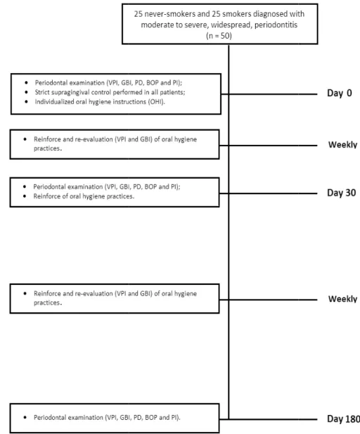
Strict supragingival control was established from days 0 to 180 (Figure 1). Initially the sub-jects received full-mouth supragingival debride-ment (Nos. 11-12; 13-14 Gracey curettes; Neumar) and oral hygiene instructions. Briely, subjects were trained in the Bass technique. A soft-bristle tooth-brush and a luoride toothpaste (CloseUp, Unilever, Valinhos, Brazil) were provided along 180 days.4
The patients were supervised weekly, and their ad-herence to the clinical instructions, as well as to supragingival control (VPI and GBI), was checked. Reinforcement regarding oral hygiene was provid-ed on an individual basis by a periodontist (MSc), not involved with the clinical exams (S.C.G).
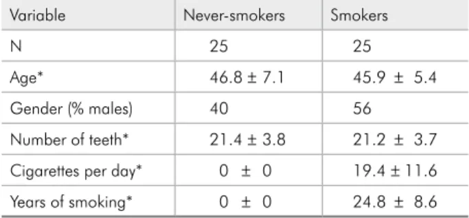
Table 1 - Baseline characteristics of patients according to experimental group.
Variable Never-smokers Smokers
N 25 25
Age* 46.8 ± 7.1 45.9 ± 5.4
Gender (% males) 40 56
Figure 1 - Flowchart of experimental procedures.
pared with free surfaces (NS: NM = 0.62 ± 1.10, M = 0.67 ± 1.24; S: NM = 0.71 ± 1.08, M = 0.93 ± 1.39) (Table 4).
Discussion
This investigation showed that both supra- and subgingival periodontal indicators were reduced for non-molar and molar teeth after a regimen of strict supragingival plaque control. Reductions observed for PPD were greater for M sites, while CAL did not differ between M and NM. Smoking did not inlu-ence this response.
The present study was a secondary analysis from a study reported earlier, performed as a single-arm clinical trial,4 that is, a single treatment distributed
among different groups. Thus, randomization of patients or treatment was not performed. Further-more, because of the longitudinal nature of the in-vestigation and also to the intra- and extraoral char-acteristics related to tobacco use, it was not possible to ensure that the examiner remained blinded to the study group. However, along the study subjects were identiied by number only and the examiner did not have access to the patients’ general health records.
Previous studies4,5 have shown that
Table 2 - Baseline periodontal indicators for patients sub-jected to supragingival control.
NM M
VPI (%)*
NS 89.9 ± 08.9 95.7 ± 06.8
p = 0.175 S 87.1 ± 14.6 92.3 ± 12.4
p = 0.017
GBI (%)*
NS 82.5 ± 13.7 83.5 ± 20.3
p = 0.064 S 74.7 ± 19.6 77.4 ± 19.2
p = 0.621
BOP (%)*
NS 14.3 ± 10.5 17.7 ± 18.6
p = 0.109 S 22.1 ± 16.4 21.5 ± 22.9
p = 0.688
PPD (mm)*
NS 3.49 ± 0.50 4.54 ± 0.91
p = 0.356 S 3.78 ± 0.65 4.53 ± 0.75
p = 0.000
CAL (mm)*
NS 3.23 ± 0.94 4.39 ± 1.38
p = 0.007 S 4.05 ± 1.12 4.88 ± 1.23
p = 0.000
NS: never-smokers; S: smokers; NM: non-molar; M: molar; VPI: visible plaque index; GBI: gingival bleeding index; BOP: bleeding on prob-ing; PPD: periodontal probing depth; CAL: clinical attachment loss. *mean ± standard deviation. Bold letters: Comparison between M and NM. Italic letters: Comparison between S and NS.
Table 3 - Mean reduction in periodontal indicators at the end of the experimental period.
NM M
VPI (%)*π
NS 82.9 ± 10.7 79.9 ± 16.0 p = 0.772 S 82.4 ± 13.7 78.6 ± 19.4
p = 0.266
GBI (%)*π
NS 80.2 ± 12.7 82.0 ± 19.6 p = 0.131 S 74.4 ± 19.6 76.6 ± 18.7
p = 0.581
BOP (%)*π
NS 74.5 ± 13.7 65.6 ± 19.1 p = 0.782 S 70.8 ± 19.8 66.9 ± 29.6
p = 0.142
PPD (mm)*π
NS 1.01 ± 0.41 1.36 ± 0.75 p = 0.963 S 1.13 ± 0.53 1.25 ± 0.66
p = 0.059
CAL (mm)*π
NS 0.38 ± 0.23 0.57 ± 0.50 p = 0.226 S 0.50 ± 0.33 0.67 ± 0.64
p = 0.050
NS: never-smokers; S: smokers; NM: non-molar; M: molar; VPI: visible plaque index; GBI: gingival bleeding index; BOP: bleeding on prob-ing; PPD: periodontal probing depth; CAL: clinical attachment loss. *mean ± standard deviation. π Significant reduction for all indicators:
p = 0.000. Bold letters: Comparison between M and NM. Italic letters: Comparison between S and NS.
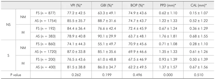
Table 4 - Multilevel analysis of mean reduction in periodontal indicators at the end of the experimental period.
VPI (%)* GBI (%)* BOP (%)* PPD (mm)* CAL (mm)*
NS
NM FS (n = 877) 77.2 ± 42.5 63.3 ± 49.1 74.9 ± 43.6 0.62 ± 1.10 0.15 ± 1.07 AS (n = 1754) 85.5 ± 35.7 88.7 ± 31.6 74.7 ± 43.7 1.22 ± 1.33 0.52 ± 1.22
M FS (n = 192) 84.4 ± 36.4 76.6 ± 42.4 72.4 ± 45.9 0.67 ± 1.24 0.36 ± 1.29 AS (n = 383) 78.9 ± 40.8 90.1 ± 29.9 63.7 ± 48.1 1.76 ± 1.81 0.68 ± 1.55
S
NM FS (n = 860) 74.1 ± 44.3 55.1 ± 49.7 70.9 ± 45.6 0.71 ± 1.08 0.28 ± 1.10 AS (n = 1720) 87.0 ± 33.8 85.1 ± 35.6 69.9 ± 46.6 1.35 ± 1.33 0.61 ± 1.26
M FS (n = 200) 76.5 ± 43.6 61.0 ± 48.8 67.5 ± 46.9 0.93 ± 1.39 0.50 ± 1.39 AS (n = 400) 81.5 ± 38.8 86.0 ± 34.7 62.2 ± 49.5 1.37 ± 1.57 0.67 ± 1.56
P value 0.262 0.199 0.496 0.000 0.510
NS: never-smokers; S: smokers; NM: non-molar; M: molar; FS: free sites; AS: approximal sites; VPI: visible plaque index; GBI: gingival bleeding index; BOP: bleeding on probing; PPD: periodontal probing depth; CAL: clinical attachment loss. *mean ± standard deviation.
This association may have been determined by ana-tomic features and by their positions in the arches, which supposedly inluence bioilm control during daily hygiene procedures.7 The latter could inluence
the subgingival environment and thus long-term periodontal stability.3
baseline. Sreenivasan et al.15 reported higher plaque
scores with de novo plaque formation being more pronounced in M. The present analysis showed that the percentage of visible bioilm declined signii-cantly during the irst 30 days for M and NM and remained the same between groups throughout the study (data not shown). Tobacco exposure did not affect the response.
On the other hand, the GBI was not affected by tooth type (p = 0.621) or by smoking (p = 0.064). During the experimental period, even though dif-ferences were observed between S and NS in the 30-to-80-day interval (data not shown), the reduc-tion achieved at day 180, compared with baseline, showed no differences. To the best of our knowl-edge, the relation between smoking and tooth type had never before been investigated. However, Sreenivasan et al.15 found more gingival
inlamma-tion in M than in NM.
The present investigation also demonstrated that, at baseline, PPD values were signiicantly greater in M (4.54 ± 0.91; 4.53 ± 0.75) than in NM (3.49 ± 0.50; 3.78 ± 0.65), respectively, for NS and S. Interestingly smoking did not inluence these re-sults. Our initial PPD values are higher than those observed by Rosling et al.,10 who studied normal
(NM 2.5 ± 0.4; M 2.8 ± 0.5) and highly susceptible (NM 2.9 ± 0.6; M 4.0 ± 0.8) individuals. This au-thor, as reported by the present study, also observed higher and signiicant PPD values for M. Miyamo-to et al.11 likewise reported similar results for NM
(3.50 mm) and M (4.36 mm).
It was also observed that CAL values were higher for M at baseline. Pihlstrom et al.8 observed
that M had greater CAL (M 4.14 mm and NM 3.40 mm, with a difference of 0.74 ± 0.1 mm be-tween them). The reported data are similar to ours: M (NS 4.39 ± 1.38; S 4.88 ± 1.23) and NM (NS 3.23 ± 0.94; S 4.05 ± 1.12).
Since there were differences between M and NM at baseline, an analysis solely expressed by means would not identify eventual differences in response to the supragingival control throughout the experi-mental period. Thus, the mean percentage of reduc-tion, for both supra- and subgingival indicators, was calculated. In general, it was observed that M and
NM responded similarly to treatment, except for the clinical attachment gain that was greater for M. However, it must be realized that, even with numer-ical differences for the mean reduction among the groups, the signiicance had a p value at the limit (p = 0.05) that underscores the importance of dis-cussing the clinical implications of such inding.
Our data showed that smoking did not inluence the response to therapy, which is in accordance with other studies. Some authors showed that, although S may exhibit a worse periodontal clinical condition, their response to periodontal therapy is somewhat similar to that obtained in NS.4,19
Finally, considering the interdependence between tooth sites, teeth and patients,20 a multilevel analysis
was performed using all six tooth sites. In sequence, the analysis took into consideration the approxi-mal and free surfaces as a whole. Interestingly, PPD reductions were greater at the approximal sites, in molar teeth, in smoker patients (Table 4). As this re-sult did not differ from those obtained on all sites individually, these data are not shown. This ind-ing seems to conirm the trend observed for PPD values expressed as means grouped by individuals (p = 0.059) (Table 3) and furthermore strengthens the effect of supragingival control even in areas hav-ing dificult access.
It is important to emphasize that, since the pres-ent study is a secondary analysis, a sample size was not calculated speciically to compare M and NM. However, because signiicant differences could be demonstrated, the number of individuals appears to be adequate. Nevertheless, to strengthen and con-irm the present results, clinical trials speciically designed to address whether M and NM respond similarly to periodontal treatment are needed.
Conclusions
Acknowledgements
National Research Council, Brasília, DF, Brazil (140428/2003-8); National Coordination of
Post-Graduate Education, Brasília, DF, Brazil (0550/04-3); and São Paulo Research Foundation, SP, Brazil (2003/09302-8).
References
1. Axelsson P, Nyström B, Lindhe J. The long-term effect of a plaque control program on tooth mortality, caries and peri-odontal disease in adults. Results after 30 years of mainte-nance. J Clin Periodontol. 2004 Sep;31(9):749-57.
2. Tezal M, Scannapieco FA, Wactawski-Wende J, Grossi SG, Genco RJ. Supragingival plaque may modify the effects of subgingival bacteria on attachment loss. J Periodontol. 2006 May;77(5):808-13.
3. Weidlich P, Souza MAL, Oppermann RV. Evaluation of the dentogingival area during early plaque formation. J Periodon-tol. 2001 Jul;72(7):901-10.
4. Gomes SC, Piccinin FB, Susin C, Oppermann RV, Marcan-tonio RAC. Effect of supragingival plaque control in smok-ers and nevsmok-ersmoksmok-ers: 6-month evaluation of patients with periodontitis. J Periodontol. 2007 Aug;78(8):1515-21. 5. Gomes SC, Piccinin FB, Oppermann RV, Susin C,
Marcan-tonio RA. The effect of smoking on gingival crevicular fluid volume during the treatment of gingivitis. Acta Odontol Lati-noam. 2009;22(3):201-6.
6. Heitz-Mayfield LJA, Trombelli L, Heitz F, Needleman I, Moles D. A systematic review of the effect of surgical debridement vs. non-surgical debridement for the treatment of chronic periodontitis. J Clin Periodontol. 2002;29 Suppl 3:92-102. 7. Matthews DC, Tabesh M. Detection of localized tooth-related
factors that predispose to periodontal infections. Periodontol 2000. 2004;34:136-50.
8. Pihlstrom BL, Oliphant TH, McHught RB. Molar and nonmo-lar teeth compared over 6 1/2 years following two methods of periodontal therapy. J Periodontol. 1984 Sep;55(9):499-504. 9. Svardström G, Weenström JL. Periodontal treatment decisions
for molars: an analysis of influencing factors and long-term outcome. J Periodontol. 2000 Apr;71(4):579-85.
10. Rosling B, Serino G, Hellström MK, Socransky SS, Lindhe J. Longitudinal periodontal tissue alterations during supportive therapy. Findings from subjects with normal and high
sus-ceptibility to periodontal disease. J Clin Periodontol. 2001 Mar;28(3):241-9.
11. Miyamoto T, Kumagai T, Lang MS, Nunn ME. Compliance as a prognostic indicator. II. Impact of patient’s compliance to the individual tooth survival. J Periodontol. 2010 Sep;81(9):1280-8.
12. Nordland P, Garrett S, Kiger R, Vanooteghem R, Hutchens LH, Egelberg J. The effect of plaque control and root debride-ment in molar teeth. J Clin Periodontol. 1987 Apr;14(4):231-6. 13. Loos B, Nylund K, Claffey N, Egelberg J. Clinical effects of
root debridement in molar and non-molar teeth. A 2-year follow-up. J Clin Periodontol. 1989 Sep;16(8):498-504. 14. Prasad KV, Sreenivasan PK, Patil S, Chhabra KG, Javali SB,
DeVizio W. Removal of dental plaque from different regions of the mouth after a 1-minute episode of mechanical oral hygiene. Am J Dent. 2011 Feb;24(1):60-4.
15. Sreenivasan PK, DeVizio W, Prasad KV, Patil S, Chhabra KG, Rajesh G, et al. Regional differences within the dentition for plaque, gingivitis, and anaerobic bacteria. J Clin Dent. 2010;21(1):13-9.
16. Furuichi Y, Lindhe J, Ramberg P, Volpe AR. Patterns of de novo plaque formation in the human dentition. J Clin Peri-odontol. 1992 Jul;19(6):423-33.
17. Armitage GC. Development of a classification system for periodontal diseases and conditions. Ann Periodontol. 1999 Dec;4(1):1-6
18. Ainamo J, Bay I. Problems and proposals for recording gin-givitis and plaque. Int Dent J. 1975 Dec;25(4):229-35. 19. Darby IB, Hodge PJ, Riggio MP, Kinane DF. Clinical and
microbiological effect of scaling and root planning in smoker and non-smoker chronic and aggressive periodontitis patients. J Clin Periodontol. 2005 Feb;32(2):200-6.