JOURNAL OF NANO- AND ELECTRONIC PHYSICS Р - Р
Vol. 5 No 1, 01007(4pp) (2013) Том5№ 1, 01007(4cc) (2013)
2077-6772/2013/5(1)01007(4) 01007-1 2013 Sumy State University
Luminescence Properties of Ce
3 +-Doped Terbium Aluminum Garnet Phosphor Prepared
with Use of Nanostructured Reagents
I.V. Berezovskaya
1,*, B.I. Zadneprovski
2, N.I. Poletaev
3, Yu.A. Doroshenko
3,
N.P. Efryushina
1, E.V. Zubar
1, V.P. Dotsenko
11 A.V. Bogatsky Physico-Chemical Institute, National Academy of Sciences of Ukraine, 86, Lustdorfskaya doroga,
65080 Odessa, Ukraine
2 Central Research and Development Institute of Chemistry and Mechanics, 115487 Moscow, Russia
3 Institute of Combustion and Advanced Technologies, Mechnikov Odessa National University, 2, Dvoryanskaya str., 65082 Odessa, Ukraine
(Received 07 November 2012; revised manuscript received 15 December 2012; published online 28 March 2013)
The paper describes the synthesis of Ce3 +-doped terbium aluminum garnet (TAG) phosphors with use
of nanostructured oxides of aluminum and rare earths. Aluminum oxide nanoparticles were obtained by gaseous-disperse synthesis and characterized by X-ray diffraction, differential thermal analysis and scan-ning electron microscopy. It was shown that the Ce3 + ions in TAG exhibit the intense broad band emission with a maximum at about 563 nm and the quantum efficiency of luminescence of the Тb3(1 –x)Ce3xAl5O12 ( = 0.03) phosphor was found as high as 0.83.
Keywords: Terbium Aluminum Garnet, Synthesis, Nanopowders, Luminescence.
PACS numbers: 78.47.jd, 81.07.Wx
*[email protected]
The article was reported at the 2ndInternational Conference «Nanomaterials: Applications Properties-2012»
1. INTRODUCTION
The white light-emitting diodes (LED’s) are one of the most promising alternatives to conventional electric light sources, i.e. incandescent lamps, Hg-containing fluorescent lamps. This concept has been commercially realized by using a combination of a blue LED emitting around 460 nm and yttrium aluminum garnet (Y3Al5O12, YAG) doped with Ce3 + ions as a yellow phos-phor. The restricting factor in using such white LED’s for general lighting is a low color rendering index (Ra < 80) that is caused by a deficit of red component in
the emission spectrum of YAG:Ce3 + [1, 2]. The short-comings of YAG:Ce3 +-based white LED’s stimulated the search for alternative compositions with the garnet structure [1, 2] and, in particularly, the attempts to improve the color characteristics of LED’s by substit u-tion of Y3 + with other rare earth (R) ions. Jang et al. [3] have studied the luminescent properties of Ce3 + ions in Tb3xY3(1 –x)Al5O12 (TYAG) solid solutions. It was found that the Tb3 + substitution induces larger crystal field splitting of the Ce3 + 5d configuration and shifts the Ce3 + emission band towards longer wavelengths. Since this emission is efficiently excited by photons in the 380-460 nm region, efficient white LED’s were fabrica t-ed by using a combination of (In, Ga)N chips emitting around 460 nm and TYAG:Ce3 + as a yellow-orange phosphor. Also, several groups of authors have studied the luminescent properties of Ce3 + ions in Тb
3Al5O12 (TAG) upon excitation in the 240-550 nm region [4-6]. It was shown that there is an efficient energy transfer between Tb3 + and Ce3 + in TAG and this material can be also of interest for development of new scintillators [5]. As a rule, Ce-containing garnets phosphors are pre-pared by solid state reactions between starting reagents at temperatures, which typically exceed 1500 C.
Even then, depending upon the preparation condi-tions some amounts of impurity phases such as RAlO3, CeO2 can be revealed in the final products [2, 7]. Be-sides, an insufficient mixing and low reactivity of raw materials often result in the significant difference in the Ce3 + concentrations within the grains and in the vicinity of grain boundaries [8]. In the present paper, we describe the synthesis of Тb3(1 –x)Ce3xAl5O12 (x = 0-0.03) luminescent materials by solid state reactions between aluminum oxide Al2O3 and mixed rare earth oxides. Our approach included the use of a nanosized metastable Al2O3, the use of oxalate precursor-derived rare earth oxides, the firing of reaction mixtures in the temperature region of a metastable alumina phase to α-Al2O3 transition. It was expected that an acceleration of the diffusion of Al3 + and O2- in temperature region of the Al2O3 phase transformation would stimulate the solid state reactions. The results of luminescent meas-urements on the obtained materials are also reported and discussed.
2. EXPERIMENTAL
I.V. BEREZOVSKAYA, B.I. ZADNEPROVSKI
,
N.I. POLETAEV, ET AL. J.NANO-ELECTRON.PHYS. 5, 01007 (2013)01007-2
3. RESULTS AND DISCUSSION
Nanosized Al2O3 was obtained by a gaseous-disperse synthesis. This method is based on the com-bustion of powdered metals due to exothermic oxidizing reactions between them and a gaseous oxidizer (typi-cally O2). The details of experimental setup used for the synthesis of nanosized Al2O3 can be found in the paper by Poletaev et al. [9]. The dispersed in N2 aluminum particles with an average size of 4.8 µm were injected through an inner tube into O2 stream. After ignition by an external source, a stable two-phase diffusion flame was observed. The resulting product was collected using a fabric filter. The XRD pattern of the as-prepared Al2O3, shown in Fig. 1, indicates that the powder contains , ,θ-phases of Al2O3, which are pre-sent in approximately the same quantities.
Fig. 1 – Powder X-ray diffraction pattern of as-prepared Al2O3
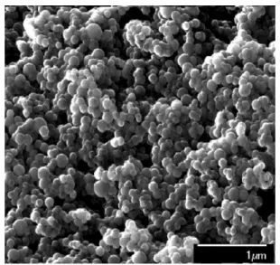
Fig. 2 – SEM photograph of as-prepared Al2O3
As can be seen from Fig. 2, its crystallites are of spherical in shape with 20-70 nm in diameter, and they exhibit a tendency to adhesion. Fig. 3 shows the particle size distribution of the as-prepared Al2O3. It is seen that the size of majority of the particles does not exceed 70 nm and the average size of crystallites was found to be ~ 49 nm. Probably, due to the size limitations of the analyzer used in this study this value somewhat exceeds the real one.
Fig. 3 – Particles size distribution of Al2O3
DTA curve of the as-prepared Al2O3 sample revealed a strong exothermic effect in the range 1200-1400 C with a maximum at 1296 C (see Fig. 4), which corre-sponds to the metastable alumina phase→-Al2O3 phase transition [10]. This phase transformation has been extensively studied, and at present, it is well known that -Al2O3 is formed through a nucleation and growth process, and depending upon chemical prehisto-ry of the precursor, degree of its cprehisto-rystallinity, the pre-sence of impurities etc. the transition temperature va-ries from 950 to 1350 C [10].
Fig. 4 – DTA curve of nanosized Al2O3
The second step in the preparation involved the for-mation of mixed rare earth oxides Tb4O7:Ce. To this end, the appropriate amounts of Tb4O7 (99.99 %) and CeO2 (99.99 %) were dissolved in a dilute HNO3 solution.
LUMINESCENCE PROPERTIES OF CE 3+-DOPED… J.NANO-ELECTRON.PHYS. 5, 01007 (2013)
01007-3 The XRD patterns of the as-prepared samples were well matched with JCPDS File No. 76-0111 for TAG. The synthesis procedure used was not further opti-mized for emission intensity or particle size distribu-tion, but even without this highly efficient luminescent materials were obtained.
The emission and excitation spectra of undoped Тb3Al5O12 at 293 K are presentedin Fig. 5. The emis-sion spectrum contains several groups of bands in the range 470-640 nm, which are caused by the 5D4→7Fj (j = 3-6) transitions of Tb3 + ions. The emission from the higher-energy 5D3 state is practically absent due to the cross-relaxation process (5D3 → 5D4) : (7F6 → 7F0). The excitation spectrum of the emission consists of several intense and overlapping bands in the 250-300 nm re-gion, a band of lower intensity at 325 nm and a number of relatively narrow bands at the longer wavelengths. It is evident that the broad bands are caused by the Tb3 + 4f8→ 4f75d transitions, while the narrow ones are due to the 4f8→ 4f8 transitions of Tb3 + ions [4-6].
Fig. 5 –Emission and excitation spectra of Тb3Al5O12 at 293 K.
The emission spectrum (a) was recorded upon excitation at 370 nm and the excitation spectrum (b) was recorded for the Tb3 + emission at 540 nm
The emission spectra of TAG:Ce3 + depend on ceri-um concentration and excitation wavelength. As can be seen from Fig. 6, upon excitation at 450 nm the emis-sion band of Тb3(1 –x)Ce3xAl5O12 ( = 0.03) extends from 480 to 750 nm and has a maximum at about 563 nm. At 77 K the spectrum shows the doublet structure and can be reasonably decomposed into Gaussian-type bands with maxima at 534 and 583 nm, which are due to transitions from the lowest Ce3 + 5d excited state to the 4f ground state levels 2F
5/2 and 2F7/2.
The positions of these maxima are in agreement with the results reported for the Ce3 + emission in ТAG at 10 K [3]. The excitation spectrum of Тb3(1 –x)Ce3xAl5O12 ( = 0.03) recorded for the Ce3 + emis-sion at 560 nm is also shown in Fig. 6. No doubt that the broad band with a maximum at 465 nm is mainly caused by the 4f → 5d transition to the lowest component of the Ce3 + 5d configuration, while the narrow ones are due to the 4f8 → 4f8 transitions of Tb3 + ions. The band at 334 nm is a superposition of bands with maxima at 325 and 338 nm arising from spin-forbidden 4f8→ 4f75d transition of Tb3 + ions and 4f → 5d transition of Сe3 + ions, respectively, so that its shape and relative intensity
Fig. 6 –Emission and excitation spectra of Тb3(1 –x)Ce3xAl5O12
( = 0.03) at 293 K. The emission spectrum (a) was recoded upon excitation at 450 nm and the excitation spectrum (b) was recorded for the Ce3 + emission at 560 nm
depend on the emission wavelength and temperature. The spectrum also contains overlapping bands with maxima at 277 and 262 nm, which were also present in the excitation spectrum for the Tb3 + emission in TAG (see Fig. 5). It is clear that these bands must be at-tributed to the 4f8 → 4f75d transitions of the Tb3 + ions [5]. The substitution of Tb3 + for Y3 + in the garnet structure results in broadening the emission band and shifting its maximum towards the longer wavelengths. At room temperature the full width at half maximum of the Ce3 + emission increases from 3460 сm– 1 (YAG) to ~ 3800 сm– 1 for TAG. When the larger Tb3 + ion occu-pies the Y3 + position, the dodecahedral site is expanded and distorted, so that these results can be explained by an increase in both the crystal field splitting of the Ce3 + 5d configuration and Stokes shift of the emission. Indeed, because the lowest Ce3 + excitation band of Тb3(1 –x)Ce3xAl5O12 ( = 0.03) is situated at 465 nm (Fig. 6), the Stokes shift of the emission amounts to 2780 cm– 1. This value is somewhat larger than that reported for the Ce3 + emission in YAG (2400 cm– 1) [12]. The quantum efficiency of luminescence () of the Тb3(1 –x)Ce3xAl5O12 ( = 0.03) sample was determined as described in Refs. [1, 2] using a commercial YAG: Ce3 + phosphor for LED’s with = 0.90 as a standard.
For the excitation at 460 nm, the found value of 0.83 ± 0.04 is comparable to, but somewhat larger than that ( = 0.76) reported in the literature for the Ce3 + emission in TAG prepared at 1500 C [4].
4. CONCLUSIONS
I.V. BEREZOVSKAYA, B.I. ZADNEPROVSKI
,
N.I. POLETAEV, ET AL. J.NANO-ELECTRON.PHYS. 5, 01007 (2013)01007-4
і е е
і
і
Ce
3 +е і
-
і іє
і ,
е
е е і
І. .
ерезовська
1,
.І.
аднепровський
2,
.І.
олєтаєв
3,
Ю. .
орошенко
3,
. .
Єфрюшина
1,
. .
убар
1, . .
оценко
11 - . . . а а , а а, 86,
65080 а, а а
2 Ц а а - а а , 115487 а, Р
3 а а , а а
. . . а, . а, 2, 65082 а, а а
У статтѕ описується синтезактивованого ѕонами Ce3 +тербѕй-алюмѕнѕєвого гранату (TAG) з
викори-станням наноструктурованих оксидѕв рѕдкѕсноземельних елементѕв та алюмѕнѕю. аночастки Al2O3
були одержанѕ за допомогою газодѕсперсного синтезу та охарактеризованѕ методами рентгенѕвськоі дифракцѕі, диференцѕйно-термѕчного аналѕзу та електронноі скануючоі мѕкроскопѕі. становлено, що ѕони Ce3 +в TAG демонструють ѕнтенсивнуширокосмугову люмѕнесценцѕю з максимумом при 563нм
та квантова ефективнѕсть люмѕнесценцѕі матерѕалу складу Тb3(1 –x)Ce3xAl5O12 ( = 0.03) досягає 0.83.
і : Тербѕй люмѕнѕєвий ранат, Синтез, аночастки, юмѕнесценцѕя.
е е
е
Ce
3 +е
-
е
,
е-е
е е
. .
ерезовская
1,
. .
аднепровский
2,
. .
олетаев
3, Ю. .
орошенко
3,
. .
фрюшина
1, . .
убарь
1,
. .
оценко
11 - . . . а а , а а, 86,
65080 а, а а
2 Ц а а - а а ,
115487 а,Р
3 а , а а
. . . а, . а , 2, 65082 а, а а
статье описывается синтез активированного ионами Ce3 + тербий-алюминиевогограната (TAG) с
использованием наноструктурированныхоксидов редкоземельных элементов и алюминия. аноча-стицы Al2O3, полученные с помощью газодисперсного синтеза, были охарактеризованы методами
рентгеновской дифракции, дифференциально-термического анализа и сканирующей электронной микроскопии.Установлено, что ионы Ce3 + в TAG обладают интенсивнойширокополосной
люминес-ценцией с максимумом при 563нми квантовая эффективность люминесценции материала состава Тb3(1 –x)Ce3xAl5O12 ( = 0.03) достигает 0.83.
K е е : Тербий люминиевый ранат, Синтез, аночастицы, юминесценция.
REFERENCES
1. A.A. Setlur, W.J. Heward, Y. Gao, A.M. Srivastava, R.G. Chandran, M.V. Shankar, Chem. Mater. 18, 3314 (2006).
2. A. Katelnikovas, H. Bettentrup, D. Uhlich, S. Sakirzanovas, T. Justel, A. Kareiva, J. Lumin. 129, 1356 (2009).
3. H.S. Jang, W.B. Im, D.C. Lee, D.Y. Jeon, S.S. Kim,J. Lu-min. 126, 371 (2007).
4. M. Nazarov, D.Y. Noh, J. Sohn, C. Yoon, J. Solid State Chem. 180, 2493 (2007).
5. Y. Zorenko, V. Gorbenko, T. Voznyak, M. Batenschuk, A. Osvet, A. Winnacker, J. Lumin. 128, 652 (2008). 6. K.M. Kim, J.H. Ryu, S.W. Mhin, G.S. Park, K.B. Shim, J.
Electrochem Soc. 155, J293 (2008).
7. J.D. Furman, G. Gundiah, R. Rage, N. Pizarro, A.K. Cheetham, Chem. Phys. Lett. 465, 67 (2008).
8. W. Zhao, S. Anhel, C. Mancini, D. Adams, G. Boulon, T. Epicier, Y. Shi, X.Q. Feng, Y.B. Pan, V. Chani, A. Yoshikawa,Opt. Mater. 33, 684 (2011).
9. N.I. Poletaev, A.N. Zolotko, Yu.A. Doroshenko, Combust. Expl. Shock 47, 153 (2011).
10. P.-L. Chang, F.-S. Yen, K.-C. Cheng, H.-L. Wen, Nano Lett. 1, 253 (2001).
11.A.A. Titov, M.A. Klimenko, E.G. Goryacheva, N.L. Opolchenova, N.N. Stepareva, N.P. Sokolova, Inor-gan. Mater. 44, 1101 (2008).