Abstract
Objective: To compare serum retinol levels in preschool children during an episode of pneumonia and 45 days after the resolution of the infection.
Methods: The study was conducted with preschool children without any infection (control group, n = 9) or children hospitalized for pneumonia (n = 12), who were evaluated soon after hospitalization (phase 1) and 45 days later (phase 2). Nutritional assessment included anthropometric measurements, a food questionnaire, and laboratory blood routine examination, including urinary and serum retinol levels. Paired Student t or Mann-Whitney tests were used as required.
Results: Food intake was similar between groups. Blood hemoglobin and serum sodium and albumin decreased during phase 1, while there were higher C-reactive protein serum values. Urinary retinol levels remained unchanged whereas serum retinol increased signiicantly after pneumonia recovery.
Conclusion: During the course of pneumonia, children had transient decrease in serum levels of vitamin A, an epiphenomenon of the acute phase response.
J Pediatr (Rio J). 2011;87(5):457-60: Retinol, pneumonia, preschool children, acute phase response.
457 Introduction
Several studies have shown that vitamin A is essential for immune function and prevention of infectious diseases, including lower respiratory tract infections.1 Animals and humans with vitamin A deiciency show squamous metaplasia of mucous membranes and lower production of mucus, allowing the adherence and penetration of pathogenic bacteria in the respiratory tract.2
In the course of acute infectious diseases, low levels of vitamin A appear to be related to the severity of the infection.3 At least theoretically, vitamin A deiciency results
from anorexia, with decreased intake of vitamin A and
carotene, impaired absorption, increased requirements for the vitamin4 and increased urinary excretion of retinol,5
apart from a reduction of serum retinol-biding protein (RBP), a negative acute phase protein.6 Some studies consider
that decreased serum retinol levels predispose children to acute infection,7 but hypovitaminosis is not regarded as an
epiphenomenon of acute phase response.
Our hypothesis is that the reduction of serum retinol that occurs during infectious processes, such as pneumonia, is transitory and related to the development of acute phase response in preschool children. Thus, the objective of this
B
RIefC
OMMunICAtIOn0021-7557/11/87-05/457 Jornal de Pediatria
Copyright © 2011 by Sociedade Brasileira de Pediatria
transient decreased retinol serum levels
in children with pneumonia and acute phase response
Kellen C. K. Barbosa,1 Daniel f. Cunha,2 Alceu A. Jordão Jr,3Virgínia R. S. Weffort,4 Selma f. C. Cunha3
1. Mestre, Patologia Clínica, Universidade Federal do Triângulo Mineiro (UFTM), Uberaba, MG, Brazil. 2. Professor titular, UFTM, Uberaba, MG, Brazil.
3. Doutor(a). Professor(a), Faculdade de Medicina de Ribeirão Preto, Universidade de São Paulo (USP), Ribeirão Preto, SP, Brazil. 4. Doutora. Professora, UFTM, Uberaba, MG, Brazil.
No conflicts of interest declared concerning the publication of this article.
Suggested citation: Barbosa KC, Cunha DF, Jordão Jr AA, Weffort VR, Cunha SF. Transient decreased retinol serum levels in children with pneumonia and acute phase response. J Pediatr (Rio J). 2011;87(5):457-60.
Manuscript submitted Jan 21 2011, accepted for publication Mar 30 2011.
458 Jornal de Pediatria - Vol. 87, No. 5, 2011 Retinol serum levels in children with pneumonia - Barbosa KC et al.
study was to compare the serum retinol levels in preschool children during an episode of pneumonia and 45 days after infection resolution.
Methods
This study was approved by the institutional Ethics Committee, and written informed consent was obtained from the parents or guardians of the children. The sample size was deined by the low of preschool children admitted to a tertiary hospital in the period between July 2000 and May 2001, which met the strict inclusion criteria. Apart from parents or guardians’ refusal to give informed consent and technical setbacks, we excluded children with a diagnosis or suspicion of diabetes mellitus, acute or chronic renal failure, cystic ibrosis, urinary tract infections, gastrointestinal diseases, or history of gastroesophageal relux. The inal sample included 12 preschool children hospitalized for pneumonia (pneumonia group) and 9 control subjects (control group). In the control group, children who were in the preoperative period of minor surgeries or those being seen at routine clinical appointments were included.
The control group was evaluated only once, and children in the pneumonia group were investigated in two different phases, soon after the diagnosis of pneumonia (phase 1) and 45 days after discharge (phase 2). This period was considered suficient for complete clinical resolution of the pulmonary infection and normalization of markers of acute phase response. Nutritional assessment included anthropometric measurements (weight, height, triceps skin fold, and arm muscle circumference) using reference standards.8,9 Information about food intake over the
previous 6 months was inferred by the Semi-quantitative Food Frequency Questionnaire, and data were analyzed by using the Virtual Nutri® v.1.5 software (São Paulo, Brazil).
The results were compared with the recommended dietary intake and expressed as percentage of adequacy.
Blood was collected from a cubital vein after an overnight fast, and samples of 10 ml of urine were collected from the irst morning urine after genital hygiene. Hemoglobin was analyzed using a Coulter T-890 counter, and the erythrocyte sedimentation rate was measured using the Wintrobe method. Serum sodium and potassium were analyzed using direct ion-selective electrode (Cobas Integra® 700, Roche).
Serum total proteins, albumin, and iron were analyzed by colorimetric methods (Cobas Integra®). Serum C-reactive
protein level, complement components (C3 and C4) were measured by turbidimetry, and serum ferritin levels determined by chemiluminescence. Serum retinol levels were determined by HPLC (Shimadzu, model LC10A), with normal values ≥ 0.70 μmol/L.
Variables with normal distribution were expressed as mean ± standard deviation; and the results obtained from pneumonia group in phases 1 and 2 were compared using
Student's paired t test. The Mann-Whitney test was used for variables with non-normal distribution, which are expressed as median and range. A p value < 0.05 was considered statistically signiicant.
Results
Children in the pneumonia or control groups were paired for socioeconomic condition, age (53.6±21.3 vs. 59.2±14.9 months), and male:female ratio (7:5 vs. 5:4). Except for triceps skin fold measurements, which were lower in the children with pneumonia (7.7±2.1 vs. 10.1±3.5 mm), pneumonia and control groups did not differ, respectively, in relation to weight (16.3±3.1 vs. 19.0±1.1 kg), height (103.6±11.4 vs. 109.5±8.8 cm), and arm muscle circumference (13.2±1.3 vs. 14.2±1.0 cm). After hospital discharge, anthropometry data did not show statistical difference in the children of the pneumonia group.
Data about food intake (% of adequacy) in the previous 6 months did not reveal differences between the pneumonia and control groups, respectively, in relation to intakes of energy (103.0±35.4 vs. 93.6±40.1), protein (299.7±143.4 vs. 289.5±157.3) and vitamin A (170.4±104.0 vs. 116.9±63.7).
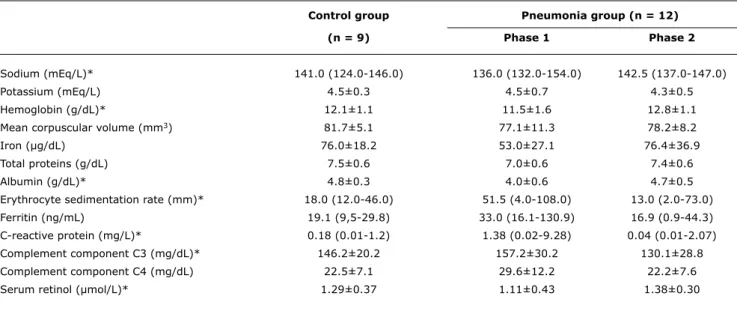
Table 1 shows the laboratory data of the control group, which are within normal values. The comparison between phases 1 and 2 in the pneumonia group showed decreased values for blood hemoglobin and serum sodium and albumin, while there were higher serum values of erythrocyte sedimentation rate, complement component C3 and C-reactive protein in the children during pneumonia. Urinary retinol excretion did not differ between controls (0.16±0.11 µmol/L) and children with pneumonia in phase 1 (0.11±0.06 µmol/L) or phase 2 (0.12±0.08 µmol/L). However, serum retinol levels decreased signiicantly (1.11±0.43 vs. 1.38±0.30 µmol/L, p = 0.04) in phase 1 when compared with phase 2.
Discussion
The present study documented that previously healthy preschool children with pneumonia have decreased levels of hemoglobin, albumin and retinol, while showing several increased serum markers of inlammatory stress compared with a control group. Also, the normalization of serum retinol levels after full recovery from pneumonia suggests that several cases of hypovitaminosis A seen during pneumonia are a systemic manifestation caused by the activation of an acute phase response. Moreover, results show that acute pneumonia in children is associated with decreased serum sodium levels.
Jornal de Pediatria - Vol. 87, No. 5, 2011 459
Control group Pneumonia group (n = 12)
(n = 9) Phase 1 Phase 2
Sodium (mEq/L)* 141.0 (124.0-146.0) 136.0 (132.0-154.0) 142.5 (137.0-147.0)
Potassium (mEq/L) 4.5±0.3 4.5±0.7 4.3±0.5
Hemoglobin (g/dL)* 12.1±1.1 11.5±1.6 12.8±1.1
Mean corpuscular volume (mm3) 81.7±5.1 77.1±11.3 78.2±8.2
Iron (µg/dL) 76.0±18.2 53.0±27.1 76.4±36.9
Total proteins (g/dL) 7.5±0.6 7.0±0.6 7.4±0.6
Albumin (g/dL)* 4.8±0.3 4.0±0.6 4.7±0.5
Erythrocyte sedimentation rate (mm)* 18.0 (12.0-46.0) 51.5 (4.0-108.0) 13.0 (2.0-73.0)
Ferritin (ng/mL) 19.1 (9,5-29.8) 33.0 (16.1-130.9) 16.9 (0.9-44.3)
C-reactive protein (mg/L)* 0.18 (0.01-1.2) 1.38 (0.02-9.28) 0.04 (0.01-2.07)
Complement component C3 (mg/dL)* 146.2±20.2 157.2±30.2 130.1±28.8
Complement component C4 (mg/dL) 22.5±7.1 29.6±12.2 22.2±7.6
Serum retinol (µmol/L)* 1.29±0.37 1.11±0.43 1.38±0.30
table 1 - Laboratory data of the control group and pneumonia group during infection and 45 days after discharge (phases 1 and 2)
* p < 0.05 between phase 1 and phase 2.
characterized by anorexia, fever, peripheral blood neutrophilia, hypoferremia, progressive anemia, hypoalbuminemia, and increases in erythrocyte sedimentation rate, serum levels of C-reactive protein, ferritin, complement component C3, among other acute phase proteins.10 Metabolic derangements
in APR include increase in resting energy expenditure and proteolysis, in addition to hyperglycemia associated with increased glucose production and oxidation, depressed glycogenesis, insulin resistance, and increased glucagon and activities of catecholamines.11 Increases in vasopressin,
renin, and aldosterone activity may also occur and can produce both body luid retention and hyponatremia.12
The transient reduction of serum albumin during the pneumonic process is consistent with the development of acute phase response, and the decrease in serum albumin is often proportional to the severity of the inlammatory response.10 Even though albumin is often used in the
nutritional status assessment, its low levels may be caused by liver disease, nephrotic syndrome, end-stage malignancies, and many acute and chronic infections. During infections, decreased serum albumin levels arise from lower protein intake, reduced liver synthesis, hemodilution, increased protein catabolism or passage of this protein from the intravascular into the interstitial space.12
The decrease of serum retinol seen in this study cannot be attributed to the low intake of vitamin A in the preceding months. Decreased vitamin A absorption is a rare cause of decreased serum retinol levels seen in the children with pneumonia, since they had no diarrhea or history of malabsorption. In the present study, a higher requirement of retinol associated with increased activity of immune cells in pneumonia cannot be excluded. Furthermore, it seems
improbable that augmented urinary retinol excretion played a role in the genesis of low serum retinol levels in our cases, since both pneumonia and control groups excreted similar amounts of retinol in the urine.
Finally, it is not possible to exclude the diminished mobilization of vitamin A from their liver reserves during the acute phase response. Under normal conditions, retinol stored in the liver binds to RBP, a carrier protein which interacts with transthyretin and is involved in the transport of retinol to peripheral tissues.13 As these proteins decrease during the systemic inlammatory response owing to reduced synthesis, there is a progressive reduction of serum retinol in the course of infection.6 The recovery of
infection is likely associated with progressive increase in RBP and retinol serum levels.4
Similarly to our results, da Silva et al.14 have documented plasma retinol levels signiicantly higher after recovery than during pneumonia in 40 hospitalized children (aged 6 months to 5 years). Despite its small sample size, our study had strict inclusion criteria and age group, evaluation of markers of acute phase response and used different techniques to assess nutritional status, including urinary retinol determination. The limitations of this study include the lack of liver biopsy to document the low body reserves of vitamin A, or the performance of a RDR (relative-dose-response) test,15 which allow inferences about retinol liver
reserves.
In conclusion, this study showed that previously healthy preschool children have transient decrease in serum levels of vitamin A during the course of pneumonia, and this seems to be an epiphenomenon of the acute phase response.
460 Jornal de Pediatria - Vol. 87, No. 5, 2011
References
1. Jason J, Archibald LK, Nwanyanwu OC, Sowell AL, Buchanan I, Larned J, et al. Vitamin A levels and immunity in humans. Clin Diagn Lab Immunol. 2002;9:616-21.
2. Stephensen CB. Vitamin A, infection, and immune function. Annu Rev Nutr. 2001;21:167-92.
3. Dudley L, Hussey G, Huskissen J, Kessow G. Vitamin A status, other risk factors and acute respiratory infection morbidity in children. S Afr Med J. 1997;87:65-70.
4. Mitra AK, Alvarez JO, Wahed MA, Fuchs GJ, Stephensen CB. Predictors of serum retinol in children with shigellosis. Am J Clin Nutr. 1998;68:1088-94.
5. Mitra AK, Alvarez JO, Stephensen CB. Increased urinary retinol loss in children with severe infections. Lancet. 1998;351:1033-4. 6. Stephensen CB, Gildengorin G. Serum retinol, the acute phase
response, and the apparent misclassiication of vitamin A status in the third National Health and Nutrition Examination Survey. Am J Clin Nutr. 2000;72:1170-8.
7. West KP Jr. Vitamin A deiciency disorders in children and women. Food Nutr Bull. 2003;24:S78-90.
8. National Center for Health Statistics. Growth curves for children birth-18 years. United States Department of Health Education and Welfare, Vital and Health Statistics; 1977. Series11, Nb.165. 9. Frisancho AR. New norms of upper limb fat and muscle
areas for assessment of nutritional status. Am J Clin Nutr. 1981;34:2540-5.
10. Baumann H, Gauldie J. The acute phase response. Immunol Today. 1994;15:74-80.
11. Mizock BA. Alterations in carbohydrate metabolism during stress: a review of the literature. Am J Med. 1995;98:75-84.
12. Ferreira da Cunha D, Pontes Monteiro J, Modesto dos Santos V, Araújo Oliveira F, Freire de Carvalho da Cunha S. Hyponatremia in acute-phase response syndrome patients in general surgical wards. Am J Nephrol. 2000;20:37-41.
13. Filteau SM, Willumsen JF, Sullivan K, Simmank K, Gamble M. Use of the retinol-binding protein: transthyretin ratio for assessment of vitamin A status during the acute-phase response. Br J Nutr. 2000;83:513-20.
14. da Silva R, Lopes E Jr, Sarni RO, Taddei JA. Níveis plasmáticos de vitamina A em crianças carentes com pneumonia na fase aguda e após recuperação. J Pediatr (Rio J). 2005;81:162-8.
15. Ferraz IS, Daneluzzi JC, Vannucchi H, Jordão Jr AA, Ricco RG, Del Ciampo LA, et al. Zinc serum levels and their association
with vitamin A deiciency in preschool children. J Pediatr (Rio J). 2007;83:512-7.
Correspondence: Selma F. C. Cunha
Departamento de Clínica Médica, FMRP-USP Av. Bandeirantes, 3900 – Monte Alegre CEP 14048-900 – Ribeirão Preto, SP – Brazil Tel.: +55 (16) 3602.3369
Fax: +55 (16) 3633.6695 E-mail: [email protected]