Restorative Dentistry
Veridiana Camilotti(a) Juliana Zilly(a)
Priscilla do Monte Ribeiro Busato(a) Carlos Augusto Nassar(b)
Patrícia Oehlmeyer Nassar(b)
(a) Department of Restorative Dentistry, School
of Dentistry, Universidade Estadual do Oeste do Paraná - UNIOESTE, Cascavel, PR, Brazil.
(b) Department of Periodontics, School of
Dentistry, Universidade Estadual do Oeste do Paraná - UNIOESTE, Cascavel, PR, Brazil.
Corresponding Author: Veridiana Camilotti
E-mail: [email protected]
Received for publication on Feb 10, 2012 Accepted for publication on Apr 03, 2012
Desensitizing treatments for dentin
hypersensitivity: a randomized, split-mouth
clinical trial
Abstract: The aim of this randomized, controlled, split-mouth, clinical study was to differentiate and clinically qualify the effectiveness of different desensitizing agents in the treatment of painful symptoms caused by cervical dentin
hyper-sensitivity (CDH). Two hundred-and-ifty-two teeth of 42 patients were distrib
-uted into seven groups (n = 36): G1 – placebo; G2, G3, G4 and G6 – luoride varnishes; G5 – sodium luoride; G7 – potassium oxalate. Three applications
were made one week apart. A three-score system (Alfa = 0, Bravo = 2, and Char-lie = 3, respectively for no sensitivity, slight sensitivity and high sensitivity) was used to assess CDH after each application and after 30 days. The data were sub-jected to statistical analysis using the Kruskall-Wallis and Dun tests. After the
second week, statistically signiicant differences were observed for all materials compared with the baseline. After 30 days, Group G7 had presented a signiicant
gradual reduction along all the evaluated time intervals. It was concluded that all the desensitizing agents were capable of reducing dentin hypersensitivity, with
the exception of the placebo and the sodium luoride groups.
Descriptors: Dentin Sensitivity; Dentin; Fluorides; Gingival Recession.
Introduction
Cervical dentin hypersensitivity (CDH) is deined as an exaggerated re
-sponse to the stimulation of vital dentin exposed to the oral environment, which causes extreme discomfort to the patient. It is characterized by
short-term, acute pain of variable intensity, which occurs in response to thermal, volatile, tactile, osmotic or chemical stimuli that cannot be attributed to any other type of defect or dental pathology.1 These stimuli are produced by the
ingestion of hot or cold beverages, by contact with acidic foods, or by tooth brushing. Pain may be localized or generalized, affecting one or various tooth surfaces concomitantly, and generally ceases immediately after removing the pain stimulus.2
The etiology and mechanisms underlying the development of dentin
hy-persensitivity have not yet been well explained. Various theories have been propounded in an attempt to explain the mechanism involved in the generation
of pain and transmission of the stimuli through dentin.3 The transmission of
stimuli from exposed dentin to the nerve endings located in the dental pulp
may occur through the odontoblast process or by means of a hydrodynamic mechanism, the latter being considered the most plausible.2
The “Hydrodynamic Theory” proposed by Brännström4 claims that when
loss of enamel and/or cement occurs, the dentinal tubules are exposed to the
oral environment, and the presence of certain stimuli
causes the displacement of luids within the tubules, in -directly stimulating the pulp nerve endings and causing the sensation of pain.
There are various methods available for the treat-ment of dentin hypersensitivity, all with the aim of oblit-erating the dentinal canaliculi.5 Dentinal tubule sealing
can be secured with the use of restorations, dental adhe-sives or the formation of a smeared dentin surface.6
Fluoride varnishes were introduced on the market to
increase the eficiency and permanence of luoride when
in contact with the tooth surface, in order to allow a
slow and continuous release of luoride.7 Varnishes
con-sist of natural resin-based vehicles for luoride, and are
highly adhesive to the tooth structure. They are easy to apply and are low-cost materials.8 The luoride is dis
-solved in an organic solvent, which evaporates when ap-plied, leaving a thin layer of the material covering the
exposed tooth surfaces. The mechanism of action is the deposition of calcium luoride on the tooth surface, with the formation of luorapatite.9 In addition to luoride,
potassium oxalate may be used to treat dentin hyper -sensitivity. It reacts with the calcium of dentin to form
insoluble, acid-resistant calcium oxalate.10
The aim of this study was thus to assess the effec-tiveness of four desensitizing agents in reducing dentin hypersensitivity in a randomized, double-blind, split-mouth clinical trial. The study hypothesis is that there are no differences between the desensitizing treatments.
Methodology
This research was conducted in the city of Cas-cavel, PR, Brazil, with patients from the Dental Clin-ic of the Universidade Estadual do Oeste do Paraná (UNIOESTE), after being approved by the institution’s Ethics Committee on Research Involving Human Be-ings, protocol number 213/2010. The patients signed a Term of Free and Informed Consent and were informed of the characteristics and conditions of the research.
Selection of patients
Forty-two patients between the ages of 18 and 70 and
presenting dentin sensitivity to thermal changes in the
oral environment were selected from the iles of the in
-tegrated clinic at UNIOESTE. Each patient had six teeth
with dentin hypersensitivity, resulting in a total number
of 252 teeth included in the study.
Initial dental sensitivity was assessed using modiied
U.S. Public Health Service criteria,11 a three-score
sys-tem composed by Alpha = 0 (no sensitivity), Bravo = 2 (slight sensitivity) and Charlie = 3 (high sensitivity). The scores were always recorded before and after the application of the test treatments. Each tooth received two stimuli: clinical probing (tactile stimulus) and air blast (thermal evaporative stimulus). The probe stimu-lus was applied under slight manual pressure in the mesiodistal direction on the cervical area of the tooth. The air blast was applied with an air syringe for 1 s at a distance of 1 cm from the tooth surface to avoid
desic-cating the dentin surface. The subjective experience of
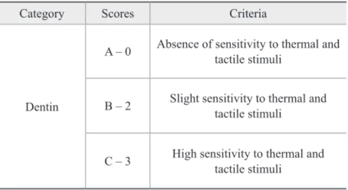
pain reported by the individual was then recorded, as shown in Table 1. The desensitizing agents and placebo
were applied by one experienced operator (a PhD Assis -tant Professor from the Department of Restorative Den-tistry of the institution where the study was conducted). The order in which the teeth were assessed within each
subject was maintained at each visit. The examiner and
the patients did not know which type of treatment cor-responded to each tooth.
The following exclusion criteria were applied: pres -ence of caries, restorations, and ongoing orthodontic or periodontal treatment at the CDH site; patients using medication or presenting systemic diseases; patients who had presented recurrent hypersensitivity in the last 30 days, who were pregnant or breastfeeding, or who
had undergone exogenous dental bleaching within the
previous 3 months. Those included were patients with teeth hypersensitive to air stimulus and good oral hy-giene. The presence of gingival recession and/or non-carious cervical lesions was considered acceptable. The
Table 1 - Criteria for hypersensitivity assessment.
Category Scores Criteria
Dentin
A – 0 Absence of sensitivity to thermal and tactile stimuli
B – 2 Slight sensitivity to thermal and tactile stimuli
sample size was determined by a previous pilot study.
Treatments
After recording the baseline scores, the subjects were randomly and blindly assigned to one of the treatment groups or to the placebo (n = 36 teeth), according to the desensitizing agent used (Table 2). The randomization procedure was conducted before the clinical steps were performed, and was carried out by using sequentially-numbered, opaque, sealed envelopes prepared with un-restricted randomization.12 The name of each treatment
agent and “placebo” was written on a piece of paper that was then sealed inside each envelope before beginning the study. The dental operator who performed all treat-ments then opened an envelope for each case at the be-ginning of each treatment.
If the patient had two lesions side-by-side in the same quadrant (split-mouth), just one of the lesions would re-ceive the treatment at that time. Thus, all patients would have at least one lesion treated per quadrant. Three ap-plications of the material selected for the group were
made for each group and in each patient, with a time interval of one week between applications.
The procedure for each weekly session consisted of
rinsing with water, performing prophylaxis with pro -phylactic paste, isolating with cotton rolls, and conduct-ing dentin dryconduct-ing with an air syrconduct-inge. Application of the materials was made directly to the areas with dentin hy-persensitivity following the manufacturer’s instructions (Table 2).
Dentin sensitivity was assessed at each weekly ses-sion, according to the previously mentioned three-score system, before the material was applied, and using a tri-ple syringe to apply an air/water spray to the teeth under treatment.
Clinical reassessment
Thirty days after the last application of each materi-al, the patients were resubmitted to a sensitivity test per-formed with an air blast and with clinical probing on the
exposed dentin surface of the teeth that had been treated
with the desensitizing agents, and were once again
clas-Groups Material Manufacturer Application Method Composition
G1
(n = 36) Placebo –
Application with a disposable paintbrush, left to
dry for 3 min
Distilled water with thickener
G2
(n = 36) Duraphat
Colgate Oral Pharmaceuticals, Inc., New York, USA
Application with a disposable paintbrush, left to
dry for 3 min
50 mg NaF (sodium luoride) Colophony, ethyl alcohol,
white beeswax
G3
(n = 36) Fluorniz
S.S. White Artigos Dentários Ltda., Rio
de Janeiro, Brazil
5.00 g% sodium luoride, Colophony, toluene sulfonamide, vanillin, saccharine, absolute alcohol
and ethyl alcohol
G4 (n = 36)
Duoluorid XII
FGM Dental Products, Joinville,
Brazil
6% sodium luoride, 6% calcium luoride
and solvents
G5
(n = 36) Flutop
S.S. White Artigos Dentários Ltda., Rio
de Janeiro, Brazil
Application for 4 min
2% sodium luoride, hydroxyethyl cellulose,
sodium hydroxide
G6
(n = 36) Fluorphat
Inodon Laboratório Ind. de Prod. Odontológicos, Curitiba, Brazil
Application with a disposable paintbrush, left to
dry for 3 min
5% sodium luoride, natural varnish and
zirconite powder
G7
(n = 36) Oxa-gel
Art-Dent Ind. e Com. de Prod. Odontológicos Ltda.,
São Paulo, Brazil
Application with a disposable paintbrush, left to
dry for 5 min
3% potassium oxalate monohydrate solution (pH 4),
carboxymethylcellulose gel
siied according to the pain sensation intensity reported
by the patient.
After collecting the inal hypersensitivity data, the
scores were submitted to statistical analysis using the Kruskal-Wallis and Dunn tests. All analyses were per-formed with Sigma Plot 11.0 software for Windows (Eu-ropa Science Ltd., Cambridge, UK).
Results
Table 3 shows the mean scores observed for the ma-terials after each time interval. As of the third week of application, the scores of all the materials used
pre-sented statistically signiicant differences compared to baseline scores, with the exception of the placebo and the sodium luoride groups. Thirty days after the third application, it was observed that the potassium oxalate group had presented a signiicant gradual reduction in
its response to thermal stimuli along the evaluated time intervals. The Placebo and Flutop groups showed no
signiicant reduction in their sensitivity scores after all
the evaluated time intervals.
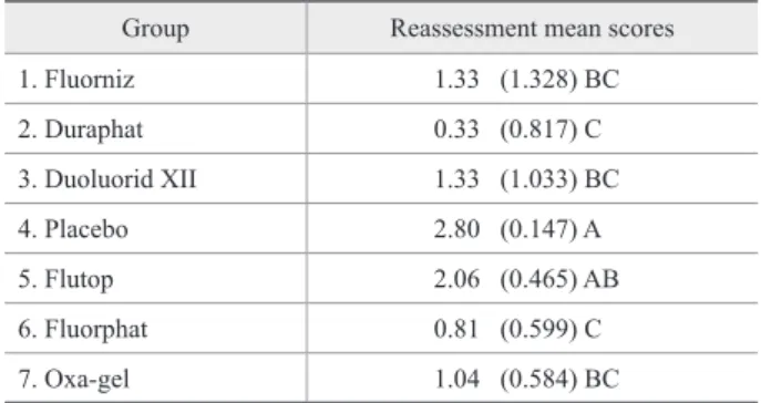
Table 4 shows a comparison of the study groups
at reassessment. Accordingly, it may be observed that Fluorphat (Inodon Laboratório Ind. de Prod. Odon-tológicos, Curitiba, Brazil) and Duraphat (Colgate Oral
Pharmaceuticals, Inc., New York, USA) luoridated var -nishes presented the lowest sensitivity scores. No sensi-tivity reduction was observed in the Placebo Group at reassessment, since its scores were statistically similar to those observed at baseline.
Discussion
Sensitivity assessments were performed on a weekly basis, before and after application of the test materials,
and 30 days after the last application. During this period of time, a reduction was observed in the pain intensity reported by the individuals; however, few subjects re-ported the complete absence of pain. This could be
at-tributed to the reaction that occurs between luoride and the calcium ions in the dentinal luid, which leads to the formation of calcium luoride crystals that are depos -ited in the dentinal tubule openings.13 Since the crystals
formed are of a small size (0.05 µm), a single applica-tion of varnish would not be effective in occluding the dentinal tubules, and multiple applications would thus be required.14 This theory is corroborated by the scores
observed after the irst week of application for Groups
2 (Fluorniz - S.S.White Artigos Dentários Ltda., Rio de
Janeiro, Brazil), 3 (Duraphat), 4 (Duoluorid XII - FGM
Dental Products, Joinville, Brazil) and 6 (Fluorphat). Different results were reported by Lan et al.,15 who
found that a luoride varnish at a concentration of 2%
was capable of diminishing the dentin hypersensitivity
scores after the irst application. Statistically signiicant
Group 1st week 2nd week 3rd week Reassessment (30 days)
1. Fluorniz 3 (0.000) A 1.83(1.033) AB 1.33(1.033) B 1.33 (1.328) B
2. Duraphat 3 (0.000) A 2 (1.095) AB 0.67(1.033) B 0.33 (0.817) B 3. Duoluorid XII 3 (0.000) A 1.83(1.472) AB 1.33(1.033) B 1.33 (1.033) B 4. Placebo 3 (0.000) A 2.92(0.554) A 2.84(0.532) A 2.80 (0.147) A 5. Flutop 3 (0.000) A 2.46(0.614) A 2.33(0.585) A 2.06 (0.465) A 6. Fluorphat 3 (0.000) A 1.88(0.560) AB 0.89(0.587) B 0.81 (0.599) B 7. Oxa-gel 3 (0.000) A 2.06(0.691) AB 1.14(0.569) BC 1.04 (0.584) C
Means followed by different letters indicate significant differences among the experimental conditions in the dif
-ferent time intervals (p < 0.05) within the same group. Table 3 - Mean dentin sensitivity
score values (SD) observed for the study groups.
Table 4 - Comparison among groups at reassessment (initial
score = 3).
Group Reassessment mean scores
1. Fluorniz 1.33 (1.328) BC
2. Duraphat 0.33 (0.817) C
3. Duoluorid XII 1.33 (1.033) BC
4. Placebo 2.80 (0.147) A
5. Flutop 2.06 (0.465) AB
6. Fluorphat 0.81 (0.599) C
7. Oxa-gel 1.04 (0.584) BC
*Means followed by different letters indicate significant differences among
differences were found when the initial assessment (at
irst application) and the third assessment (at third appli -cation) were compared to the reassessment scores after
30 days for all the tested materials, except for the Place -bo and Flutop (S.S.White Artigos Dentários Ltda., Rio de Janeiro, Brazil) groups. Hence, the study hypothesis that there are no differences between the desensitizing treatments was rejected. Different results were found by Hoang-Dao et al.,16 who evaluated three luoridated
varnishes and observed that the action of Duraphat was more effective in diminishing hypersensitivity at
reas-sessment, thirty days after the irst application.
All the Groups in which the luoridated varnishes
were applied presented improvements in dentin
hyper-sensitivity, with a reduction in pain, as expressed by the comparison between the initial and inal means obtained
during and after treatment. When Groups 1, 2, 3 and 6
were compared, there were no statistically signiicant
differences. This made it possible to establish which of
the luoridated varnishes were clinically effective. These
results are in agreement with those of other studies that have also reported a statistically similar action for
differ-ent luoridated varnishes used in the treatmdiffer-ent of hyper -sensitivity after three weeks of application.17,18 Neverthe-less, it is important to point out that Fluorphat (Group 6)
and Duraphat (Group 2) luoridated varnishes presented
statistically different results from those of the other var-nishes tested, with scores close to zero at the time of
re-assessment (Table 4). They were thus considered as the most effective of the luoridated varnishes for the treat -ment of dentin hypersensitivity.
The neutral luoride gel Flutop (Group 5) showed no statistically signiicant reduction in dentin hypersensi -tivity for all the time intervals evaluated. In vitro studies
on this material have shown that the layer of luorides
deposited on the dentin surface is easily displaced and
ultimately removed by oral luids. The action of oral lu -ids is enhanced during salivation and eating,18 thus
sug-gesting why sensitivity may possibly return, and also why these products—so widely used in dentifrices—do not eliminate sensitivity.
The potassium oxalate gel (Group 7), on the other hand, presented a statistically signiicant reduction in sensitivity between the irst and third weeks of evalua -tion. This is in agreement with the study of Pilon et al.19
and Assis et al.,20 who found a signiicant reduction in
dentin hypersensitivity after 21 days of potassium oxa
-late application. This rapid action may be explained by the presence of phytocomplexes formed after the dentin calcium reacts with the potassium oxalate. These com
-plexes make the smear layer dificult to remove during
meals.21
In the present study, the placebo was incapable of
signiicantly decreasing the hypersensitivity scores after
all time intervals, which is in agreement with data found in the literature.12 Further studies with long-term
analy-ses should be conducted to evaluate the real beneits of
desensitizing agents.
Conclusion
Based on the results obtained in this study, it was possible to conclude that all the desensitizing agents were capable of reducing dentin hypersensitivity, with
the exception of the agents in the Placebo and Fluotop groups. Oxa-gel (Art-Dent Ind. e Com. de Prod. Odon -tológicos Ltda., São Paulo, Brazil) showed a decrease
in dentin hypersensitivity from the irst to the second week, but no statistically signiicant difference was ob -served between the scores recorded at the third week and those recorded at reassessment. Considering the reassessment time of 30 days after the last application
of the material, Duraphat and Fluorphat luoridated var -nishes presented the scores closest to zero, suggesting that these two varnishes may have the best effect in the treatment of dentin hypersensitivity.
References
1. Bartold PM. Dentinal hypersensitivity: a review. Aust Dent J. 2006 Sep;51(3):212-8.
2. Orchardson R, Gillam DG. Managing dentin hypersensitivity. J Am Dent Assoc. 2006 Jul;137(7):990-8.
3. Al-Sabbagh M, Brown A, Thomas MV. In-office treatment of den-tinal hypersensitivity. Dent Clin North Am. 2009 Jan;53(1):47-60. 4. Brännström M. [Dentin sensitivity]. Arsb Goteb Tandlak Sallsk.
1964 Apr;21:15-35. Swedish.
5. Jacobsen PL, Bruce G. Clinical dentin hypersensitivity: under-standing the causes and prescribing a treatment. J Contemp Dent Pract. 2001 Feb;2(1):1-12.
6. Lee BS, Kang SH, Wang YL, Lin FH, Lin CP. In vitro study of dentinal tubule occlusion with sol-gel DP-bioglass for treatment of dentin hypersensitivity. Dent Mater J. 2007 Jan;26(1):52-61 7. Cunha-Cruz J, Wataha JC, Zhou L, Manning W, Trantow M,
choices made by dentists of the northwest precedent network. J Am Dent Assoc. 2010 Sep;141(9):1097-105.
8. Cummins D. Recent advances in dentin hypersensitivity: clinically proven treatments for instant and lasting sensitivity relief. Am J Dent. 2010 May;23(A):3A-13A.
9. Lawson K, Gross KB, Overman PR, Anderson D. Effectiveness of chlorhexidine and sodium fluoride in reducing dentin hypersensi-tivity. Dent Hyg. 1991 Sep;65(7):340-4.
10. Cunha-Cruz J, Stout JR, Heaton LJ, Wataha JC. Dentin Hyper-Dentin Hyper-sensitivity and Oxalates: a Systematic Review. J Dent Res. 2011 Mar;90(3):304-10.
11. Bayne SC, Schmalz G. Reprinting the classic article on USPHS evaluation methods for measuring the clinical research perfor-mance of restorative materials. Clin Oral Invest. 2005:9:209-14 [cited 2011 Jul 20]. Available from: http://deepblue.lib.umich.edu/ bitstream/2027.42/47873/1/784_2005_Article_17.pdf. doi 10.1007/ s00784-005-0017-0.
12. Ozen T, Orhan K, Avsever H, Tunca YM, Ulker AE, Akyol M. Dentin hypersensitivity: a randomized clinical comparison of three different agents in a short-term treatment period. Oper Dent. 2009 Jul-Aug;34(4):392-8.
13. Absi EG, Addy M, Adams D. Dentin hypersensitivity: A study of the patency of dentinal tubules in sensitive and nonsensitive cervical dentin. J Clin Periodontol. 1987 May;14(5):280-5. 14. Liu HC, Lin CP, Lan WH. Sealing depth of Nd:YAG laser on human
dentinal tubules. J Endod. 1997 Nov;23(11):691-3.
15. Lan WH, Liu HC, Lin CP. The combined occluding effect of sodium fluoride varnish and Nd:YAG laser irradiation on human dentinal tubules. J Endod. 1999 Jun;25(6):424-6.
16. Hoang-Dao BT, Hoang-Tu T, Tran-Thi NN, Koubi J, Camps J, About I. Clinical efficiency of a natural resin fluoride varnish (Shellac F) in reducing dentin hypersensitivity. J Oral Rehabil. 2009 Feb;36(2):124-31.
17. Ritter AV, Dias WL, Miguez P, Caplan DJ, Swift EJ Jr. Treating cervical dentin hypersensitivity with fluoride varnish: a random-ized clinical study. J Am Dent Assoc. 2006 Jul;137(7):1013-20. 18. Aranha AC, Pimenta LA, Marchi GM. Clinical evaluation of
de-sensitizing treatments for cervical dentin hypersensitivity. Braz Oral Res. 2009 Jul-Sep;23(3):333-9.
19. Pillon FL, Romani IG, Schmidt ER. Effect of a 3% potassium oxa-late topical application on dentinal hypersensitivity after subgingi-val scaling and root planing. J Periodontol. 2004 Nov;75(11):1461-4. 20. Assis JS, Rodrigues LK, Fonteles CS, Colares RC, Souza AM,
Santiago SL. Dentin hypersensitivity after treatment with desen-Dentin hypersensitivity after treatment with desen-sitizing agents: a randomized, double-blind, split-mouth clinical trial. Braz Dent J. 2011 Feb;22(2):157-61.