CEREBRAL EVOKED POTENTIALS IN HUMAN
CHRONIC CHAGAS' DISEASE
O. M. GENOVESE — OLGA P. SANZ — J. CORREALE
MARCELA GARCIA ERRO — R. E. P. SICA
S U M M A R Y — S e v e n t y f i v e p a t i e n t s w i t h t h e d i a g n o s i s o f c h r o n i c C h a g a s ' d i s e a s e w e r e s t u d i e d b y e m p l o y i n g E P s t e c h n i q u e s . T w o o f t h e m h a d d e l a y e d a r r i v a l o f t h e s i g n a l t o t h e E r b ' s p o i n t a n d o n e t o t h e s p i n a l c o r d w h e n l o o k i n g a t S E P s . T W o p a t i e n t s h a d i n c r e m e n t o f t h e t i m e i n t e r v a l b e t w e e n w a v e s 1st a n d I I I r d , w h e n s t u d y i n g P E A T s . T h e s e f i n d i n g s w e r e i n t e r p r e t e d a s d u e t o p e r i p h e r a l n e r v e f i b e r s d a m a g e , a f e a t u r e d e s c r i b e d in p r e v i o u s p a p e r s . T h e , m o s t s t r i k i n g f i n d i n g w a s t h e p r o l o n g e d t i m e i n t e r v a l b e t w e e n w a v e s N13 a n d N 2 0 ( S E P s ) f o u n d in t w o p a t i e n t s a n d b e t w e e n w a v e s I l l r d a n d V t h ( P E A T ) s e e n in 7 a f f e c t e d s u b j e c t s . T h e s e o b s e r v a t i o n s s u g g e s t e d t h e d e v e l o p m e n t o f s o m e s o r t o f C N S i n v o l v e m e n t , p e r h a p s r e l a t e d t o m y e l i n d a m a g e , i n p a t i e n t s w h o r e a c h e d t h e c h r o n i c s t a t e o f t h e i n f e c t i o n .
Estudio de potenciales evocados cerebrales en la enfermedad de Chagas crónica humana.
R E S Ú M E N — S e t e n t a y c i n c o p a c i e n t e s c o n el d i a g n ó s t i c o d e e n f e r m e d a d d e C h a g a s c r ó n i c a f u e r o n e s t u d i a d o s c o n el e m p l e o d e l o s p o t e n c i a l e s e v o c a d o s c e r e b r a l e s . T r e s d e e l l o s t u v i e r o n r e t a r d o d e l a r r i b o d e l a s e n a l a) p u n t o d e E r b ó a l i n g r e s o m e d u l a r e n l o s p o t e n c i a l e s e v o c a d o s s o m a t o s e n s i t i v o s ( P E S S ) . D o s p a c i e n t e s p r e s e n t a r o n i n c r e m e n t o d e l i n t e r v a l o e n t r e l a s o n d a s I y I I I e n l o s p o t e n c i a l e s e v o c a d o s a u d i t i v o s d e t r o n c o ( P E A T ) . E s t o s h a l l a z g o s f u e r o n i n t e r p r e t a d o s c o m o d e b i d o s al d a n o e n l a f i b r a n e r v i o s a p e r i f é r i c a , u n h e c h o d e s c r i p t o e n t r a b a j o s p r e v i o s . E l h a l l a z g o m a s s o r p r e n d e n t e f u e la p r o l o n g a c i ó n d e l i n t e r v a l o d e t i e m p o e n t r e l a s o n d a s N 1 3 - N 2 0 en l o s P E S S , h a l l a n d o s e e n d o s p a c i e n t e s y e n t r e l a s o n d a s I I I y V e n l o s P E A T e n 7 s u j e t o s . E s t a s o b s e r v a c i o n e s s u g i e r e n d e a l g u n m o d o , el c o m p r o m i s o d e s i s t e m a n e r v i o s o c e n t r a l , q u i z á s r e l a c i o n a d o al d a ñ o d e l a m i e l i n a , e n p a c i e n t e s e n e s t a d i o c r ó n i c o d e l a i n f e c c i ó n .
Nervous system damage is known to occur in chronic Chagas' disease either
in humans 1.2 or in experimental animals 3,4. Much of the work has been done searching
for peripheral nervous system involvement in this illness, and currently evidences have
been accumulated supporting the insult of the spinal alpha motoneurone soma 5, the
posterior root ganglia neurones 6 and the peripheral nerve fibers 6. Howewer, as far
as we know none study has been carried out in humans looking for an eventual damage
of their central nervous system ( C N S ) , though detailed analysis of C N S changes has
been carried out in animals chronically infected, in which loss of Purkinje cells 3 and
of hypothalamic neurones 2 have been described.
The aim of the present investigation has been to make a gross approach to the
CNS of patients with chronic Chagas' disease by employing a non-invasive
electro-physiological method as a first attempt for detecting eventual existing abnormalities.
S e c c i ó n E l e c t r o n e u r o f i s i o l o g í a C l í n i c a , D i v i s i ó n N e u r o l o g í a , H o s p i t a l R a m o s M e j í a , B u e n o s A i r e s .
M A T E R I A L , A N D M E T H O D S
Patients — The study involved 75 subjects with the diagnosis of chronic Chagas' disease made at the Instituto Nacional de Diagnostico e Investigación de la Enfermedad de Chagas in Buenos Aires. F o r these purposes three serological tests were employed, namely: 1. immunofluorescence; 2. complement fixation; 3. haemoagglutination. Only were accepted for this investigation those patients w h o showed positiveness of, at the least, two tests. Thirty were females and 45 were males, their ages ranged between 18 and 57 years. Coincidental causes of neurological disorders were eliminated from the study by rejecting patients over 60 years old ( ? ) and others who had had toxic or metabolic disorders known to induce nervous system damage. Those patients with any genetic disease or other parasitic illness were also excluded. Controls — A total of 54 healthy and non selected subjects, not infected with Trypanosoma crwzi, whose ages ranged between 18 and 59 years, served as controls.
Electrophysiological techniques — Evoked potentials were recorded by employing a A T I
(type 960/4) averager machine.
1. Somatosensory evoked potentials ( S E P s ) . Stimulation: Electrical stimuli were applied percutaneously to the median nerve at the wrist. The pulses were 0.1 ms duration and their intensity w a s progressively increased until a weak twitch of the thenar muscles could be observed. T h e number of stimuli delivered was the necessary to evoke a well defined S E P and varied between 120 and 500. The frequency of the discharge was 2 H z . Recording: Fine platinum electrodes were employed as recording and reference electrodes. Recording electrodes were positioned at three different locations according to the type of recorded potential: a) contralateral (to the stimulus) somatosensory area (C3 or C4) (71 pacients), b) nuchal midline C'6 (40 patients), and c) Erb's point ipsilateral to the stimulus (40 patients). Scalp and spine electrodes were refered to another electrode situated at F z position, while the E r b electrode w a s coupled to another electrode positioned at the contralateral C3 or C4 locations.
2. Brain stem auditory evoked potentials. Altogether 30 patients were studied with this technique. Stimulation: Conventional audiometric earphones were applied to both ears. Monoaural stimulation, employing click stimuli with white masking noise delivered to the opposite ear was employed. The repetition rate was 11 H z and the click intensity was adjusted to 80 dB. T w o thousand pulses were delivered for each recording. Recording: The stigmatic electrode was a fine platinum needle inserted in the scalp at Cz position refered to an electrode placed at A l or A 2 locations ipsilateral to the stimulus. Every patient was submitted to audiometric evoked responses previously to the brain stem auditory evoked potentials investigations; all the figures obtained were normal.
3. Visual evoked potentials. T w e n t y five patients were submittted to this particular inves-tigation. Stimulation: The subject was sitting in a comfortable chair and positioned 1 m from a T V screen with one eye patched and the gaze fixed on a dot in the center of the screen. The screen subtended an angle of 15. Check board pattern was reversed one every second and 50 stimuli were delivered. Recording: The recording electrode was also a fine platinum electrode which was positioned at Oz and refered to another needle situated at Cz.
Treatment of the results — Difference between two mean values was estimated by the Student 't' test. Throughout the text means are given with 1 S D .
R E S U L T S
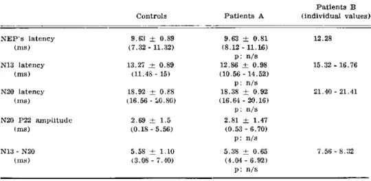
1. Somatosensory evoked potential — W h e n individual values were analyzed, it could be seen that some few patients were situated outside of the control range. So, one of
them had a prolonged N9 wave (12.28 m s ) , two others disclosed prolonged N13 (15.32 ms and 16.76 m s ) and N20 (21.40 ms and 21.41 m s ) waves, while other two showed increased intervals between waves N13 and N20 (7.56 ms and 8.32 m s ) . The difference between these individual values and the control means were of more than two SD, except for a single figure (7.56 m s ) which differed from the control mean by 1.78 S D (for comparison, see
Table 1 ) . Both patients snowing prolonged central conduction time between N13 and N20 waves disclosed normal brain stem potentials when explored with auditory stimulation. All the other patients had values whithin the control limits and, when they were compared with controls, no differences were found between uaired means for each point of recording
276 Arq Neuro-Psiquiat (Sâo Paulo) iff(3) 1989
Patients B Controls Patients A (individual values)
N E P ' s latency (ms)
N13 latency (ms)
N20 latency (ms)
N20 P22 amplitude (ms)
9.63 ± 0.89 (7.32 - 11.32)
13.27 ± 0.89 (11.48 - 15)
18.92 ± 0.88 (16.56 - 20.80)
2.69 ± 1.5 (0.18 - 5.56)
9.63 ± 0.81 (8.12 - 11.16)
p: n/s 12.86 ± 0.98 ( 1 0 . 5 6 - 14.52)
p: n / s 18.38 ± 0.92 (16.64 - 20.16)
p : n/s 2.81 ± 1.47 (0.53 - 6.70)
p : n/s
12.28
15.32 - 16.76
21.40 - 21.41
N13 - N20 (ms)
5.58 ± 1.10 (3.08 - 7.40)
5.38 ± 0.65 (4.04 - 6.92)
p: n/s
7.56 - 8.32
Table 1 — Somatosensory evoked potentials in patients with chronic Chagas' disease. A: patients with values within the control range. B: patients with values out of the control range. NEP's: nerve evoked potential, recorded at the Erb's point. Between brackets the lower and upper limits.
Latency [nisj
W a v e Controls Patients A
Patients B (individual values)
l 1.44 ± 0.14
(1.2 - 1.7)
1.47 ± 0.12 (1.18 - 1.7)
p: n/s
11 2.57 ± 0.18
(2.2 - 2 . 8 )
2.57 ± 0.21 (1.8 - 2 . 8 )
p : n/s
3.01 - 3.04
III 3.66 ± 0.17
(3.3 - 4.1)
3.66 ± 0.23 ( 2 . 6 - 4.1)
p: n/s
I V 4.81 ± 0.19
( 4 . 5 _ 5.1)
4.81 ± 0.26 (3.9 - 5.1)
p: n/s
5.36 - 5.37
V 5.54 ± 0.19
( 5 . 3 - 5.9)
5.59 ± 0.23 ( 5 . 0 - 5 . 9 )
p: n/s
6.36
IU-V
Interval [ m s ] I - I I I
5.48 ± 0.24 ( 5 . 1 - 5.9)
2.22 ± 0.15 (1.9 - 2.5)
5.34 ± 0.24 (4.86 - 5.7)
p: n/s
2.17 ± 0.21 ( 1 . 2 - 2 . 5 )
p : n/s
2.60 - 2.70
I I I - V 1.85 ± 0.16
( 1 . 6 - 2 . 1 )
1.77 ± 0.25 ( 1 . 2 - 2 . 1 )
p : n/s
2.20 - 2.22 - 2.30
2.30 - 2.60 - 2.90 3.00
Evoked potential in Chagas' disease 277
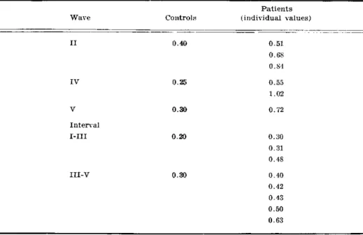
2. Brain steam auditory evoked potential — From the patients studied with this technique, 12 of them had one or more abnormal responses. T w o patients showed prolonged latencies of the I l n d (3.01 and 3.04 m s ) and I V t h (5.37 and 5.86 m s ) waves; one of them had also a delayed Vth wave (6.36 m s ) on one side; both patients had a slightly bilaterally prolonged I s t - I I I r d waves interval (2.6 and 2.7 ms, respectively), while the time elapsing between waves I l l r d and Vth was normal in both of them. Seven other patients showed increment of the intervals between waves I l l r d and V t h on one side (2.2, 2.22, 2.3, 2.3, 2.6, 2.9 and 3.0 ms, respectively); here too, the difference between these individual values and the control means were of more than two S D (for comparison, see Table 2 ) . W h e n com-paring both sides, it was observed that good correspondence in latency values could be found in controls.; so, the maximal discordance amounted to 0.4 ms for the second wave (Table 3 ) ; when the same procedure was followed with patients it was noted that some of them were beyond the normal maximal limits (Table 3 ) . W h e n working up the same sort of test for the intervals between waves 1st and I l l r d and between waves I l l r d and Vth, it was noted that altogether 8 patients were outside the normal values (Table 3 ) . The remainder 18 patients had values well within the control range and their mean aid not significantly difler from controls (Table 2 ) .
3. Visual evoked potential — None of the studied patients had abnormal values. Their means did not differ from the normal population (Table 4 ) .
Patients
W a v e Controls (individual values)
I I 0.40 0.51
0.68
0.84
I V 0.25 0.55
1.02
V 0.30 0.72
Interval
I - I I I 0 . 2 0 0.30
0.31
0.48
I I I - V 0.30 0.40
0.42
0.43
0.50
0.63
Table 3 — Differences between waves latencies and inter-wave intervals expressed in ms. Maximal values are given for controls in the central column. On the right hand column only the patients who had values above the normal upper limit. None patient showed abnormal values for waves I and III.
Controls Patients
P 100 latency 98.4 ± 9.2 91.22 ± 7.23 n/s
( m s ) (81 - 113) (79 - 112)
P 100 amplitude 9.7 ± 5.69 5 . 7 ± 3.75 n/s
(uV) ( 1 . 5 - 1 8 ) ( 2 . 5 - 1 8 )
C O M M E N T S
Scatter and mild abnormalities were observed in the EPs obtained in patients with chronic Chagas' disease. Some of them, such as the delayed arrival of the signal to the Erb's point ( N 9 , S E P ) or to the spinal cord ( N 1 3 , S E P ) may be related to the impairment of the sensory fibers which was formerly recognized by Sica et al.6 in patients affected by this condition. However, only three patients showed these abnor-malities; this seems to be at variance with the findings of Sica et al. 6
who working on the peripheral nerve found that about 4 5 % of the studied population had some sort of impairment This discrepancy may be attributed to the different techniques < m-ployed, being more sensitive for detecting sensory peripheral nerve damage the one used by Sica et al., while the methodology employed in the present paper allows to disclose just gross alterations in those fibers.
The prolonged interval between waves 1st and Illrd may partially be due to involvement of the 8th nerve, despite that same authors have recently postulated that involvement of the pons and/or medulla may produce similar findings 8.
However, the most importante observation in this study was the prolonged interval seen between waves N13 and N20 in two patients when looking at SEPs and between the Illrd and Vth waves in 7 affected subjects when studying P E A T s . Both of these findings suggest that C N S may be altered in some patients who reached the chronic state of the infection. A s far as we know, this is the first reference to this possibility in humans and might be related to some sort of impairement of the central myelin. Nevertheless, this tneoretical myelin damage has to be quite mild, because none of the affected suojects showed clinical abnormal features.
Owe to the type of approach done and the technique used in this first study, no clues could be obtained tor inferring the pathogenesis which may underlie those findings.
Acknowledgment — This investigation received financial support from the U N D P / W o r l c i B a n k / W H O Special Programme for Research and Training in Tropical Diseases.
R E F E R E N C E S
1. B r a n d ã o H J , Z u l i a n R — N e r v e cell d e p o p u l a t i o n in c h r o n i c C h a g a s ' d i s e a s e : a q u a n t i t a t i v e s t u d y i n t h e c e r e b e l l u m . R e v I n s t M e d T r o p S ã o P a u l o 8 :281, 1966.
2. B r i t t o C o s t a R , G a l l i n a R A — H i p o t á l a m o a n t e r i o r n a m o l é s t i a d e C h a g a s h u m a n a . R e v I n s t M e d T r o p S ã o P a u l o 13 : 92, 1971.
3. J a r d i m E — A l t e r a ç õ e s q u a n t i t a t i v a s d a s c é l u l a s d e P u r k i n j e n a m o l é s t i a d e C h a g a s e x p e r i m e n t a l n o c a m u n d o n g o . A r q N e u r o - P s i q u i a t ( S ã o P a u l o ) 25 : 199, 1967.
4. J a r d i m E - - M o l é s t i a d e C h a g a s e x p e r i m e n t a l n o r a t o : p a r a s i t i s m o d o n ú c l e o d e I I I p a r c r a n i a n o . R e v I n s t M e d T r o p S ã o P a u l o 13 : 405, 1971.
5. S a n z O P , R a t u s n u A F , A r i s t i m u s o G G , O ' N e i l l E M , S i c a R E P — A n e l e c t r o p h y s i o l o g i c a l i n v e s t i g a t i o n o f s k e l e t a l m u s c l e in h u m a n c h r o n i c C h a g a s ' d i s e a s e . A r q N e u r o - P s i q u i a t ( S ã o P a u l o ) 36 : 319, 1978.
6. S i c a R E P , F i l i p i n i D , P a n i z z a M, F u m o T , B a s s o S, L a z z a r i J, Molina. H A — I n v o l v e m e n t of t h e p e r i p h e r a l s e n s o r y n e r v o u s s y s t e m in h u m a n c h r o n i c C h a g a s ' d i s e a s e . M e d i c i n a ( B u e n o s A i r e s ) 46 : 662, 1986.
7. S i c a R E P , S a n z O P , C o l o m b i A — T h e e f f e c t s o f a g e i n g u p o n t h e h u m a n s o l e u s m u s c l e : a n e l e c t r o p h y s i o l o g i c a l s t u d y . M e d i c i n a ( B u e n o s A i r e s ) 36 : 443, 1976.