Vol-7, Special Issue-Number2-April, 2016, pp1137-1147 http://www.bipublication.com
e cl ti Ar h rc ea es R
TNF-
α pro
-inflammatory cytokinemodulates CD44 expression in human
lung epithelial cell line (A549) treated with Temporin-Ra
Maryam Hooshmand1, Ahmad Asoodeh1, 2, * And Fatemeh Behnam Rassouli2 1
Department of Chemistry, Faculty of Science, Ferdowsi University of Mashhad, Mashhad, Iran; 2
Cellular and Molecular Research Group, Institute of Biotechnology, Ferdowsi University of Mashhad, Mashhad, Iran;
* Corresponding author: A. Asoodeh, Email: [email protected], Tel/Fax: +98 51 38795457
ABSTRACT
Antimicrobial peptides (AMPs) are molecules present in innate immune systems of vertebrates and invertebrates. These small peptides inhibit the growth of pathogens invading host's body. In this study, the toxicity effect of Temporin-Ra (T-Ra) antimicrobial peptide on A549 cell line was investigated by MTT assay. Furthermore, the toxicity of T-Ra peptide on human's red and white blood cells was investigated. Gene expression levels of TNF-α (tumor necrosis factor-alpha)aspro-inflammatory cytokine, and CD44 cancer marker were investigated by real time- PCR, 48 h after treatment of A549 cells by different concentrations of peptide. Moreover, the production of reactive oxygen species was studied by flow cytometer. According to our results, T-Ra viability of A549 cells decreased up to 15%, while had no hemolytic and cytotoxic effects on human blood cells. In addition, T-Ra increased the expression of pro- inflammatory cytokine (TNF-α) at the highest concentration (30 µg/ml), while decreased gene expression of CD44 cancer marker. Production of reactive oxygen species (ROS) in A549 cells treated by T-Ra significantly increased. In conclusion, our results revealed that T-Ra could induce the expression of TNF-α, which consequently decrease CD44 expression, increase the production of reactive oxygen species, and as a result induced death in A549 cell lines.
Keywords: Flow cytometer, Reactive oxygen species, Real Time-PCR, Temporin-Ra.
Abbreviations: AMPs, antimicrobial peptides; Temporin-Ra, T-Ra; TNF-α, tumor necrosis factor alpha; ROS, reactive oxygen species; FBS, fetal bovine serum; DMSO, dimethyl sulphoxide; PBS, phosphate buffer saline; HBSS, hank's buffered salt solution.
1. INTRODUCTION
In spite of recent advances in treatment modalities, cancer remains a major source of deleterious and mortalitywhole over the world [1].Mortality from cancer is about to surpass from cardiovascular diseases in near future [2] and [3]. Cancer is characterized by uncontrolled division of cells and the ability of these cells to invade other tissues leading to the formation of tumor mass, vascularization, and metastasis. In spite of great advances in cancer therapy, there is considerable current interest in developing
future, capturing the large share of the cancer therapeutic market[5]. The biological treatment option against cancer includes the use of proteins, monoclonal antibodies, and peptides [6].Many natural or synthetic cationic peptides have been reported to show anticancer activity with characteristics including the ability to kill target cells rapidly, the broad spectrum of activity, and the specificity for cancer cells. Compared with the traditional cancer treatments such as chemotherapy or radioactive treatment, peptides with high specificity against cancer cells may present the way of killing cancer cells while protecting normal cells and helping patients to recover rapidly [7]. A growing number of studies have shown that antimicrobial peptides (AMPs) have evolved as integral components of strategic and carefully regulated mechanisms of immunity against infection and cancer [8]. Antimicrobial peptides (AMPs) are oligopeptides with a varying number of amino acids [6]. Antimicrobial peptides (AMPs) are a part of the innate immune defense mechanism of many organisms.
In addition to direct involvement in innate immunity, AMPs have also been found to influence host’s inflammatory responses during an infection.Lipopolysaccharide (LPS) molecules are released from bacteria as a result of antibiotic treatment or host immunity, can induce AMP production in mammals. The property of AMPs in destruction of membrane structure or indirectly trigger the cascade consequences in pathogens is believed to play significant role in anticancer activity as well [4]. Employment of AMPs as anticancer agents either alone or in combination with other conventional drugs is being considered as a therapeutic strategy ([9], [10]and [11]). Temporin-Ra (T-Ra) isa 14-amino acid antimicrobial peptide belonging toTemporin
family with the sequence of
FLKPLFNAALKLLP (1585.1 Da, pI 10.0) which previously isolated from the skin secretions of the Ranaridibunda[12]. Previous study showed that Temporin-Ra displayed significant antimicrobial activity against both gram-positive and gram-negative bacteria[12] and was able to increase inflammatory reagents
such as IL-8 and IL-1β [13].This report, focused on cytotoxicity effect of Temporin-Ra peptide on human adenocarcinoma lung cancer cells (A549). Furthermore,the ability of this peptide in expression of TNF-α pro-inflammatory cytokine and CD44 cancer marker genes was investigated. Also, the cytotoxic and hemolytic effect of Temporin-Ra peptide on human blood cells was studied. Moreover, the production of reactive oxygen species induced by Temporin-Ra in A549 cells was examined.
2. MATERIALS AND METHODS 2.1. Cell culture
A549 human adenocarcinoma lung cancer cell line was prepared from Pastor Institute (Tehran, Iran) by accession number of C-137. Cells were cultured in RPMI-1640 (Biosera, East Sussex, UK) medium containing 10% heat-inactivated FBS (Gibco, Grand Island, NY, USA) and 100
µg/mL streptomycin and 100 U/mL penicillin
(Biosera, East Sussex, UK) [14].
2.2. MTT assay
The cytotoxic effect of T-Ra peptide on A549 cell line was studied byMTT assay. 1 × 10⁴ cells were seeded in 96-well plate andafter 24 h-incubation, the cells was treated at 15 and 30
µg/mL of Temporin-Ra. After passing 24 and 48 h- cell treatment, volume of 20 µl of MTT solution (Merck, Quebec, Canada)was added to each well. The cells were incubated in darkness for 4 h, after the indicated time, culture medium was removed and 150 µl of dimethyl sulphoxide (DMSO) was added to each well to dissolve the formazan crystals. Absorbance at 570 nm was measured by using a Bio-Tekmicro plate reader (Bio-Tek Instruments, Winooski, VT, USA). Non-treated cells were considered as control in cell viability assay[15]. All MTT tests were carried out five times. Statistical analysis was performed by one-way ANOVA test with significant differences between mean values at p<0.05. Percentage of cytotoxicity of Temporin-Ra was calculated by fallowing equation (2.1): Equation 2.1
2.3. Hemolysis assay
A possible limitation to the development of antimicrobial peptides as antibiotics is their potential to cause injury to mammalian cell membranes. To assess hemolytic activity of Temporin-Ra,human erythrocytes weretreated by indicated concentrations of Temporin-Ra for 8 h on blood agar platesand measured through reading the optical densityof releasing hemoglobin from thetreated erythrocytes. Triton X-100 (0.1% V/W) was considered as positive control of 100% hemolysis and PBS as a negative control of 0% hemolysis.The hemolysis effect of Temporin-Raon blood agar plates containing 7% of prepared human erythrocytes was likewise measured. Some wellswere created in the medium by a puncher and5 µL of various concentrations of Temporin-Ra was added to each well.The radios of hemolytic halos formed around of each well were measured[16]. The hemolysis effect of Temporin-Ra via absorbance
was measured by addition of 10 µL of different concentrations of Temporin-Ra to 190
µLprepared human erythrocytes. The absorbance of erythrocytes was measured at 576 nm[17].
2.4. Cytotoxic effect on human leukocytes To investigate the cytotoxic effect of Temporin-Ra concentrations on human leukocytes, at first the leukocytes were isolated from human plasma. After centrifugation of heparinized blood at 1,300 rpm, the supernatant was removed and PBS was added to the sediment. 3 mLficoll(Biosera, East Sussex, UK) was added carefully to the solution that formed a layer whichcontained leukocytes. The layer was washed three times with HBSS solution (Biosera, East Sussex, UK). At last, isolated leukocytes resuspendedin RPMI 1640 [18]. To examine the Temprorin-Ra toxicity on human leukocytes, 1× 10⁴ leukocytes has counted and seeded in 96-well plate and Temporin-Ra solution at final concentration was added to each well. The viability of cells was assessed viatrypan blue staining by followingequation (2.2):
Equation 2.2 Cell viability (%)= (average of alive cells/average of total cells)×
100
2.5. Examining ROS levels
To investigate the production of reactive oxygen species (ROS) in A549 cells,exposed to different concentrations ofTemporin-Ra peptide,2′, 7′- dichlorohydrofluorescein-diacetate (DCFH-DA) a fluorescent, cell-permeable probe was utilized. 2 × 10⁵ cells were seeded in a 96-well plate and after 24 h, forenamed concentrations of Temporin-Ra were added to wells. 50 µL of
20µM DCFH-DA probe (Titrachem, Iran) was added to each well except controlsample.Cells were transferred to flow cytometer tubes and the fluorescence intensity was measured at 488 nm excitation and 520 nm emission wavelengths[19,[20].
2.6. RNA isolation and Real time PCR
A549 cells cultured with different concentrations of Temporin-Ra were harvested after 48 h. Total RNA was purified using total RNA isolation kit (Dena ZistAsia, Mashhad, Iran).Each sample was treated with DNase I (1U/µl) (Gibco BRL, Rockville, MD, USA) to eliminate any possible DNA contamination and subsequently tested for presence of DNA using polymerase chain reaction (PCR) amplification with the reverse transcription (RT) step. Total RNA concentration was determined from spectrophotometric optical density measurement (ratio of 260 to 280 nm). The ratio between the spectrophotometric readings at 260 nm and 280 nm (OD260/OD280) was used to provide an estimate of the purity of the nucleic acid[21]and the ratio in all samples ranged between 1.7 and 2.0.
published resources and ordered from Dena ZistAsia (Table1).
Table 1. Primers used for gene expression
Product size (bp)
Primer sequence(5’-3’)
Annealing conditions (°C
- Second)
Primer direction Gene
NCBI address
127
134
176 GAAGGCTGGGGCTCATTTGA
GCTGATGATCTTGAGGCTGTTGT
AGCCCATGTTGTAGCAAACC TGAGGTACAGGCCCTCTGAT
CGGACACCATGGACAAGTTT GAAAGCCTTGCAGAGGTCAG
60 – 15
55 – 15
59 – 30 Forward
Reverse
Forward Reverse
Reverse Forward
h
GAPDH
h
GAPDH
h
TNF-
h
TNF-
h CD44
h CD44
NM_001289746.1
NM_000594.3
XM_006718390.2
The resulting amplicons were examined by melting peaks and 1% agarose gel electrophoresis. The relative gene expression level of each target gene was calculated using the threshold cycle (2−ΔΔCT) method. GAPDH was used as a housekeeping gene for normalization, and untreated cells were used as a reference[22].
2.7. Statistical analysis
The statistical significance was assessed by SPSS software, one way ANOVA and LSD test. All data were presented as mean ± SD and p ≤ 0.05 was considered significant for all comparisons.
3. RESULTS
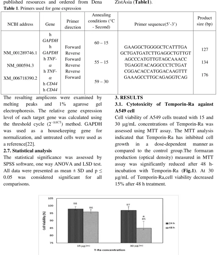
3.1. Cytotoxicity of Temporin-Ra against A549 cell
Cell viability of A549 cells treated with 15 and
30 µg/mL concentrations of Temporin-Ra was assessed using MTT assay. The MTT analysis indicated that Temporin-Ra has inhibited cell growth in a dose-dependent manner as compared to the control group.The formazan production (optical density) measured in MTT assay was significantly reduced after 48 h-incubation with Temporin-Ra (Fig.1). At 30
µg/mL of Temporin-Ra,cell viability decreased 15% after 48 h treatment.
Fig. 1. Cytotoxic effect of Temporin-Ra (T-Ra) on A549 cells using MTT assay.
The cells were initially maintained in a humidified incubator at 37 °C for 24 h and 48 h, and T-Ra was added at different concentrations. The percentage of viable cells was calculated as described in part 2.2 the results are shown as means ± standard deviations of two independent experiments performed in triplicate. *P < 0.05, one-way ANOVA test.
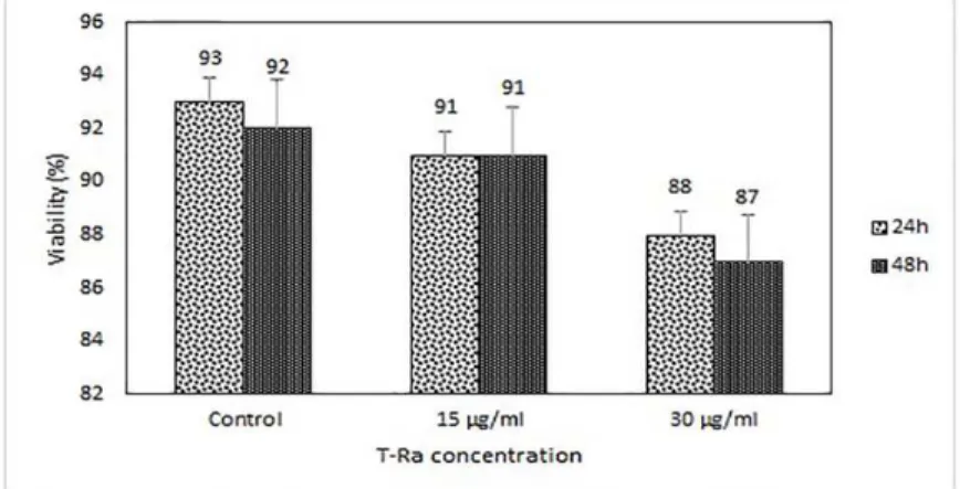
3.2. Hemolytic assay of Temporin-Ra
of the hemolytic halos of each well were measured in triplicate in comparison with the radios of Triton X-100. Temporin-Ra had modest hemolytic activity against human
erythrocytes. The hemolytic effect of Temporin-Ra via optical density of treated erythrocytes showed a non-significant hemolytic activity compared with positive control.
Fig. 2a.Fig. 2.Hemolytic activity of Temporin-Ra. Human erythrocytes were incubated with various concentration of T-Ra peptide. Values are means of three hemolytic halo radiuses. The results are shown as means ± standard deviations of different studies considered in double.
Fig. 2b.Hemolytic activity of T-Ra based on releasing hemoglobin from human red blood cells via optical density. Values are optical density of human erythrocytes treated with different concentrations of T-Ra. The results are shown as means ± standard deviations of different studies considered in double. 3.3 Cytotoxic effect of Temporin-Ra on human leukocytes
Cytotoxic effect of peptide on human leukocytes was carried outby trypan blue staining after 24 and 48 h cell treatment. Reduction of cell viability at the highest time and concentration was only 10% in compare to control.Temporin-Ra showed no significant cytotoxicityon human leukocytes (Fig.3).
Fig. 3.Cytotoxic effect of T-Ra peptide on human leukocytes. Leukocytes were incubated for 24 h and
means ± standard deviations of two independent experiments considered in triplicate by using one-way ANOVA test.
3.4. Reactive oxygen species (ROS) production in A549 cells affectedby T-Rapeptide
We assessed ROS production in response to Temporin-Ra peptide in A549 cells using DCFH-DA probeandflow cytometer after 24 and 48 h. Temporin-Ra significantly increased the ROS production in a time dependent way as determined by fluorescence emission (Fig.4).Temporin-Ra significantly increased ROS production after 48 h at 15 and30 µg/mLin comparison with control.
Fig. 4.Temporin-Ra-inducing ROS generation was detected using DCFH-DA fluorophore.
The cells were incubated for 24 and 48 h with Temporin-Ra. Then, 50 µL of DCFH-DA dissolved in PBS was added. The results were shown as fluorescence emission indicating ROS production. The right shift of the curve implies fluorescence emission increase. GM, CV and M1 show area under the curve, cyclic voltammetry and normalization scale of samples, respectively.
3.5. Gene expression changes induced by Temporin-Ra
The expression of TNF-α pro-inflammatory cytokine and CD44 cancer marker upon treatment of A549 cells for 48 h was quantified by real time PCR. Results showed that upon treatment of A549 cells by Temporin-Ra at 30
µg/mL concentrations, TNF-α was significantly up-regulated (P < 0.05) about 5-foldcompared to control. Interestingly, on the other hand, the expression of CD44 had significant 2-fold down-regulation (P < 0.05) compared to control (Fig.5).
cDNA synthesized from isolated RNA. GAPDH was used as a housekeeping gene for normalization. Untreated cells were used as a reference. The relative gene expression level of each tested gene was calculated using the 2−ΔΔCT Pffafl’s method and is presented in log2 form. Data represent mean relative gene expression levels ± standard deviations performed from three independent experiments. *P < 0.05 and **P < 0.01, one-way ANOVA test.
3.6. Real time PCR products gel electrophoresis
To examine the signaling events induced or modulated by Temporin-Ra peptide, the products of real timePCR for CD44 and TNF-αgenesin A549 cells in the presence or absence of Temporin-Rapeptide were analyzed on 1% agarose gel (Fig.6aand 6b). Results showed that Temporin-Raat30g/mL enhanced TNF-α expressionafter 48 h (Fig.6a). Whereas, increase of TNF-α upon treatment of A549 cells with Temporin-Ra for 48 h, decreased CD44 expression (Fig.6b).
Fig. 6a.The effects of T-Ra peptide on gene expressions in A549 cells.Presents gene expression of TNF-α.
Fig. 6b.The effects of T-Ra peptide on gene expressions in A549 cells.Presents CD44 gene expression.
4. DISCUSSION
Although antimicrobial peptides have been essentially studied and developed as potential alternatives for fighting infectious diseases, their use as anticancer peptides in cancer therapy has been regarded as a therapeutic strategy to explore. As human cancer remains a cause of high mortality worldwide, an urgent need of new, selective, and more efficient drugs is evident. Even thoughantimicrobial peptides are expected to be selective toward tumor cells without impairing the normal body
physiological functions, the development of selective antimicrobial peptides has been a challenge. Antimicrobial peptides are unique molecules when compared to the actual chemotherapeutic arsenal available for cancer treatment and display a variety of modes of action which in some types of cancer seem to co-exist[23].
after 48 h, significantly decreased cell viability about 15% which is consistent with previous reports. Wang et al.(2013) studied MDA-MB-231 and MCF-7 breast cancer cells after one
hour exposure to 40 µM temporin-1CEa. The percentages of cells viability assessed by MTT assay were 22% ±4% for MCF-7 cell line, and 61% ±2% for MDA-MB-231 cell line[24]. Homayouni-Tabrizi et al. (2015)showed that Brevinin-2R showed no significant toxic effects on HepG2 cells and inhibited the cell growth about 5% to 25% at concentrations of 10 µg/ml
to 40µg/ml[25].
In present study, we determined the hemolysis effect of Temporin-Ra at 15 and 30 µg/ml concentrations on human erythrocytes. Results showed that T-Ra has no significant hemolytic activity against human erythrocytes. A vast number of studies were conducted to assay the hemolytic effect of antimicrobial peptides on human erythrocytes. For instance, Stark et al.(2002)investigated hemolytic activities of 4F, F17-6R, F25-6K and F17-6R peptides against rabbit and human erythrocytes at 50 µMand 200 µM. Little or no hemolysis was detected[26]. Silva et al.(2012)investigated the linear peptide Cn-AMP1isolated from coconut water (Cocosnucifera) against human erythrocytes and the peptide showed no hemolytic activity[27]. We also examined the cytotoxicity of Temporin-Ra on human leukocytes. Temporin-Temporin-Ra at15 and
30 µg/mlhad no significant cytotoxicity on human leukocytes.Previous studies are agreementto our PR-39 and NK-lysin) for their cytotoxic effects on blood mononuclear cells. None of these peptides exhibited significant cytotoxic effect on resting lymphocytes isolated either from peripheral blood or from spleen with the exception of high concentrations of NK-lysin[28]. Pacor et al.(2002)have also studied
the cytotoxic activity of three artificial α-helical antimicrobial peptidesP19(8), D-P19(9/B) and L-P19(9/B)at 16 µM, 40 µM and 160 µM concentrations on mouse primary cultures of resting or activated lymphocytes. Damage to lymphocytes does not appear to be statistically
significant up to 16 µM peptide for P19(8)[29]. Inthis study, production levels of reactive oxygen species in A549 cells treated with
Temporin-Ra were determined using -2′,7′ -dichlorodihydrofluorescein diacetateand flow cytometer. Temporin-Ra significantly increased ROS production in these cells. Several studies demonstrated that a number of AMPs have the ability to induce ROS production in cancer cells;for instance, Alalwani et al. (2010)demonstrated that LL-37 triggers ROS production in human neutrophils. They assessed ROS production in response to LL-37 at 5, 10,
15, 20 µg/ml using flow cytometer. LL-37 significantly increased ROS triggered in a dose-dependent way[30]. Paredes-Gameroa et al. (2012)studied the level of ROS production in the human erythroleukemia K562 cell line by gomesin, protegrin, tachyplesin and polyphemusin II along with their linear analogs. Only protegrin showed a significant increase in ROS production[31].
It has been reported that epithelial cells can produce antimicrobial peptides once exposing to invading microbes[32]. In addition to bactericidal activity, AMPs increase the production of cytokines and chemokines; thereby, showing an important role of AMPs in the immune response[33].
The effect of Temporin-Ra on gene expression of pro-inflammatory cytokine TNF-α in A549 cell line was studied at various concentrations. A549 cells were treated with 15and 30 µg/mL of Ra for 48 h. The results demonstrated that T-Racan influence the gene expression of inflammatory factor. The results demonstrated an increase of TNF-α mRNA expression following 48 h induction with 30µg/mLof the peptide with approximately 5-fold increase in gene expression. A vast number of studies were conducted to assay the effect of antimicrobial peptides on TNF-α gene expression. For example,Mookherjee et al. (2006)examined temporal transcriptional profile and identified differentially expressed genes in LPS-stimulated monocytes in the presence or absence of LL-37. LL-37 inhibited TNF-α induced expression by bacteria lipopolysaccharide[34]. In another study, Brandenburg et al. (2010)examined rCRAMP antimicrobial peptide at 2, 5 and 10
gene expressions of IL-10, TNF-α, IL-6 and IL-1β[35].
We also examined gene expression of CD44 cancer marker in Tempoin-Ra-A549 treated cells for 48 h. CD44 expression was significantly down-regulated compared to control at 30 µg/mL of the peptide. Our results demonstrate that down-regulation of the CD44 cancer marker was associated with TNF-α pro-inflammatory cytokine up-regulation. Preliminary experiments confirm our results.
For instance, Muthukumaran et
al.(2006)indicated that TNF-αcould regulate CD44 expression differentially on TNF-α treatment of SKA and SKOV3 cells based on its ability to activate JNK pathway[36].Stadlmann et al. (2003)also analyzed the effects of the pro-inflammatory cytokines, interleukin-1beta (IL-1beta) and tumor necrosis factor-alpha (TNF-α) on morphology and expression of adhesion molecules such as CD44 cancer marker in human peritoneal mesothelial cells (HPMC). IL-1beta furthermore up-regulated the expression of the adhesion molecule CD44 while expression of adhesion molecule was down-regulated by TNF-alpha[37].
5. CONCLUSION
In summary,our in vitro study onTemporin-Ra peptide demonstrated that this peptide had no hemolytic activity on human erythrocytes as well as human leukocytes indicating on its potential application as a natural agent while it had a 15% toxicity on A549 cell viability line after 48 h. Furthermore,Temporin-Ra could up-regulategene expression of TNF-α pro -inflammatory cytokine in A549 cells and this up-regulation leads to CD44 cancer marker down-regulationafter 48 h-treatment. Finally, Temporin-Ra induced production of reactive oxygen species in A549 cells after 48 h which is consequent of TNF-α's increase. Owing to no significant cytotoxic effect of T-Ra peptide on normal tested cells and its impact on gene regulation, demonstratethe potential application of T-Ra peptide as a natural agent while its cancer therapy property needs further investigations.
ACKNOWLEDGMENT
This work was supported by Ferdowsi University of Mashhad Research and Technology council (grant number: 3/38441, 14/06/1394). The authors would like to thank them.
Conflicts of Interest
Authors declare that there is no conflict of interest.
REFERENCES
1. Hoskin, D.W. and Ramamoorthy, A. (2008) Studies on anticancer activities of antimicrobial peptides. Biochimicaet
BiophysicaActa
(BBA)-Biomembranes. 1778(2), 357-375.
2. Jemal, A., Bray, F., Center, M.M., Ferlay, J., Ward, E. and Forman, D. (2011) Global cancer statistics. CA: Cancer J. Clin. 61(2), 69-90.
3. Garcia, M., Jemal, A., Ward, E.M., Center, M.M., Hao, Y., Siegel, R.L., Thun, M.J. (2007) Global Cancer Facts and Figures American Cancer Society. Atlanta
4. Liu, X., Li, Y., Li, Z., Lan, X., Leung, P.H.M., Li, J., Yang, M., Ko, F. and Qin, L. (2015) Mechanism of Anticancer Effects of Antimicrobial Peptides. Journal of Fiber Bioengineering and Informatics. 8(1), 25-36.
5. Enbäck, J. and Laakkonen, P. (2007)Tumour-homing peptides: tools for targeting, imaging and destruction. Biochem. Soc. Trans. 35(4), 780-783.
6. Thundimadathil, J. (2012) Cancer treatment using peptides: current therapies and future prospects. J Amino Acids.1-13
7. Huang, Y.B., Wang, X.F., Wang, H.Y., Liu, Y. and Chen, Y. (2011) Studies on mechanism of action of anticancer peptides by modulation of hydrophobicity within a defined structural framework. Mol. Cancer Ther. 10(3), 416-426.
9. H anahan, D. and Weinberg, R.A. (2000) The
hallmarks of cancer. Cell.100(1), 57-70. 10.Torcato, I.M., Huang, Y.H., Franquelim,
H.G., Gaspar, D., Craik, D.J., Castanho, M.A. and Henriques, S.T. (2013) Design and characterization of novel antimicrobial peptides, R-BP100 and RW-BP100, with activity against negative and Gram-positive
bacteria. Biochim.Biophys.Acta., 1828(3), 944-955.
11. Zhu, X., Zhang, L., Wang, J., Ma, Z., Xu, W., Li, J. and Shan, A. (2015) Characterization of antimicrobial activity and mechanisms of low amphipathic peptides
with different α-helical propensity. ActaBiomater. 18, 155-167.
12. Asoodeh, A., Zardini, H.Z. and Chamani, J. (2012) Identification and characterization of two novel antimicrobial peptides, temporin‐Ra and temporin‐Rb, from skin secretions of the marsh frog (Ranaridibunda). J. Pept. Sci., 18(1), 10-16. 13.Asadi, F., Asoodeh, A., Kashef, R.,
Housaindokht, M.R., Haghparast, A. and Chamani, J. (2013) The effect of antimicrobial peptide Temporin-Ra on cell viability and gene expression of pro-inflammatory factors in A549 cell line.Int J Pept Res Ther.19(4), 373-380.
14. Castro, M.A.A., Schwartsmann, G. and Moreira, J.C.F. (2001) Intercellular contact-dependent survival of human A549, NCI-H596 and NCI-H520 non-small cell lung carcinoma cell lines. Braz J. Med. Biol. Res. 34(8), 1007-1013.
15. Anghel, R., Jitaru, D., Badescu, L., Ciocoiu, M. and Badescu, M., 2014. The cytotoxic effect of Cecropin A and Cecropin B on the MDA-MB-231 and M14K tumour cell lines.JBiSE.504-515.
16.Munk, J.K., Ritz, C., Fliedner, F.P., Frimodt-Møller, N. and Hansen, P.R. (2014) Novel method to identify the optimal antimicrobial peptide in a combination matrix, using anoplin as an example. Antimicrob.Agents Chemother. 58(2), 1063-1070.
17.Murphy, G.L., Whitworth, L.C., Clinkenbeard, K.D. and Clinkenbeard, P.A. (1995) Hemolytic activity of the Pasteurellahaemolyticaleukotoxin. Infect. Immun. 63(8), 3209-3212.
18. Fuss, I.J., Kanof, M.E., Smith, P.D. and Zola, H. (2009) Isolation of whole mononuclear cells from peripheral blood and cord blood. John Wiley & Sons, inc. (pp 7-1).
19. Li, Y., Huang, H., Xue, L., Zhuang, D., Cheng, X., Cui, L. and Zhang, H. (2015) Effects of MC-LR on ROS level in human bronchial epithelia cells and Chinese hamster ovary cells. Life Sci. 12(5), 170-173.
20. Maurya, D.K., Nandakumar, N. and Devasagayam, T.P.A. (2010) Anticancer property of gallic acid in A549, a human lung adenocarcinoma cell line, and possible mechanisms. Clin.Biochem. N. 8548(1), 85-90.
21. Berti, R., Williams, A.J., Moffett, J.R., Hale, S.L., Velarde, L.C., Elliott, P.J., Yao, C., Dave, J.R. and Tortella, F.C. (2002) Quantitative Real-Time RT—PCR Analysis of Inflammatory Gene Expression Associated with Ischemia—Reperfusion Brain Injury. J Cereb Blood Flow Metab.22(9), 1068-1079. 22. Peinnequin, A., Mouret, C., Birot, O.,
Alonso, A., Mathieu, J., Clarençon, D., Agay, D., Chancerelle, Y. and Multon, E. (2004) Rat pro-inflammatory cytokine and cytokine related mRNA quantification by real-time polymerase chain reaction using SYBR green. BMC immunology,5(1), 1. 23. Gaspar, D., Veiga, A.S. and
Castanho, M.A. (2014) From antimicrobial to anticancer peptides. A review. New edge of antibiotic development: antimicrobial peptides and corresponding resistance. 24. 24. Wang, C., Tian, L.L., Li, S., Li, H.B.,
Zhou, Y., Wang, H., Yang, Q.Z., Ma, L.J. and Shang, D.J. (2013) Rapid cytotoxicity of antimicrobial peptide tempoprin-1CEa in breast cancer cells through membrane destruction and intracellular calcium mechanism. PLoS One, 8(4), e60462.
(2015) Antimicrobial peptide Brevinin-2R induces the secretion of a pro-inflammatory cytokine in HepG2 cells. Basic Res. Med. Sci, 2(2), 23-29.
26. Stark, M., Liu, L.P. and Deber, C.M. (2002) Cationic hydrophobic peptides with antimicrobial activity. Antimicrob.Agents Chemother.46(11), 3585-3590.
27. Silva, O.N., Porto, W.F., Migliolo, L., Mandal, S.M., Gomes, D.G., Holanda, H.H., Silva, R.S., Dias, S.C., Costa, M.P., Costa, C.R. and Silva, M.R. (2012) Cn‐AMP1: A new promiscuous peptide with potential for microbial infections treatment. J Pept. Sci. 98(4), 322-331.
28. Catrina, S.B., Refai, E. and Andersson, M. (2009)The cytotoxic effects of the anti‐bacterial peptides on leukocytes. J. Pept. Sci. 15(12), 842-848.
29. Pacor, S., Giangaspero, A., Bacac, M., Sava, G. and Tossi, A. (2002) Analysis of the cytotoxicity of synthetic antimicrobial peptides on mouse leucocytes: implications for systemic use. J. Antimicrob. Agents Chemother.50(3), b339-348.
30. Alalwani, S.M., Sierigk, J., Herr, C., Pinkenburg, O., Gallo, R., Vogelmeier, C. and Bals, R. (2010) The antimicrobial peptide LL‐37 modulates the inflammatory and host defense response of human neutrophils. Eur. J. Immunol, 40(4), 1118-1126.
31. Paredes-Gamero, E.J., Martins, M.N., Cappabianco, F.A., Ide, J.S. and Miranda, A. (2012) Characterization of dual effects induced by antimicrobial peptides: regulated cell death or membrane disruption. Biochim.Biophys.Acta, 1820(7), 1062-1072. 32. Zasloff, M., 2007. Antimicrobial
peptides, innate immunity, and the normally sterile urinary tract. J. Am. Soc. Nephrol. 18(11), 2810-2816.
33. Elssner, A., Duncan, M., Gavrilin, M. and Wewers, M.D. (2004) A novel P2X7 receptor activator, the human cathelicidin-derived peptide LL37, induces IL-1β processing and release. J. Imm. 172(8), 4987-4994.
34. Mookherjee, N., Brown, K.L., Bowdish, D.M., Doria, S., Falsafi, R., Hokamp, K., Roche, F.M., Mu, R., Doho, G.H., Pistolic, J. and Powers, J.P. (2006) Modulation of the TLR-mediated inflammatory response by the endogenous human host defense peptide LL-37. J. Imm. 176(4), pp.2455-2464.
35. Brandenburg, L.O., Jansen, S., Wruck, C.J., Lucius, R. and Pufe, T. (2010) Antimicrobial peptide rCRAMP induced glial cell activation through P2Y receptor signalling pathways. Mol. Immunol. 47(10), 1905-1913.
36. Muthukumaran, N., Miletti-González, K.E., Ravindranath, A.K. and Rodríguez-Rodríguez, L. (2006) Tumor necrosis
factor-α differentially modulates CD44 expression in ovarian cancer cells. Mol. Cancer. Ther. 4(8), 511-520.