www.bjournal.com.br
Volume 44 (2) 84-181 February 2011
doi:
Braz J Med Biol Res, February 2011, Volume 44(2) 140-148
10.1590/S0100-879X2010007500145
Enhanced inhibition of murine prostatic carcinoma growth by
immunization with or administration of viable human umbilical
vein endothelial cells and CRM197
Huiyong Zhang, Yong Lu, Didier Mekoo, Yu Zhang, Jing Fang, Rongyue Cao and Jingjing
Liu
Institutional Sponsors
The Brazilian Journal of Medical and Biological Research is partially financed by
Enhanced
inhibition of murine prostatic
carcinoma growth by immunization with
or administration of viable human umbilical
vein endothelial cells and CRM197
Huiyong Zhang
1*, Yong Lu
2*, Didier Mekoo
2, Yu Zhang
2, Jing Fang
3,
Rongyue Cao
2and Jingjing Liu
21Department of Life Science and Biotechnology, Xinxiang Medical University, Xinxiang, China 2The Minigene Pharmacy Laboratory, Biopharmaceutical College, China Pharmaceutical University,
Nanjing, China
3Institute of Chemical Industry of Forest Products, CAF, Nanjing, China
Abstract
Vaccination with xenogeneic and syngeneic endothelial cells is effective for inhibiting tumor growth. Nontoxic diphtheria toxin
(CRM197), as an immunogen or as a specific inhibitor of heparin-binding EGF-like growth factor, has shown promising antitumor
activity. Therefore, immunization with or administration of viable human umbilical vein endothelial cells (HUVECs) combined with
CRM197 could have an enhancedantitumor effect. Six-week-old C57BL/6J male mice were vaccinated with viable HUVECs,
1 x 10⁶ viable HUVECs combined with 100 μg CRM197, or 100 μg CRM197 alone by ip injections once a week for 4
consecu-tive weeks. RM-1 cells (5 x 10⁵) were inoculated by sc injection as a preventive procedure. During the therapeutic procedure,
6-week-old male C57BL/6J mice were challenged with 1 x 10⁵ RM-1 cells, then injected sc with 1 x 10⁶ viable HUVECs, 1 x
106 viable HUVECs + 100 μg CRM197, and 100 μg CRM197 alone twice a week for 4 consecutive weeks. Tumor volume and
life span were monitored. We also investigated the effects of immunization with HUVECs on the aortic arch wall and on wound healing. Vaccination with or administration of viable HUVECs+CRM197 enhanced theinhibition of RM-1 prostatic carcinoma by 24 and 29%, respectively, and prolonged the life span for 3 and 4 days, respectively, compared with those of only vac-cination or administration with viable HUVECsof tumor-bearing C57BL/6J mice. Furthermore, HUVEC immunization caused some damage to the aortic arch wall but did not have remarkable effects on the rate of wound healing; the wounds healed in approximately 13 days. Treatment with CRM197 in combination with viable HUVECs resulted in a marked enhancement of the antitumor effect in the preventive or therapeutic treatment for prostatic carcinoma in vivo, suggesting a novel combination for anti-cancer therapy.
Key words: RM-1 prostatic carcinoma; CRM197; Immunization; Human umbilical vein endothelial cells
Introduction
Correspondence: Jingjing Liu or Rongyue Cao, Minigene Pharmacy Lab., China Pharmaceutical University, Tongjia Xiang 24, Nanjing 210009, Jiangsu, China. Fax: +55-11-86-25-320-4240. E-mail: [email protected]
*These authors contributed equally to this study.
Received December 16, 2009. Accepted December 6, 2010. Available online December 17, 2010. Published February 7, 2011.
Angiogenesis is a physiological process involving the growth of new blood vessels from pre-existing ones. In adults, angiogenesis is almost quiescent, except under conditions such as wound healing and the menstrual cycle. However, it is also necessary and required for tumor development and metastasis (1-3). Therefore,
anti-angiogenic therapies applied in an attempt to fight cancer
and malignancies have been investigated intensively since the 1970’s (4-7). The process of angiogenesis is a
umbili-Carcinoma inhibition by immunization with HUVEC and CRM197 141
cal vein endothelial cells (HUVECs) as a vaccine to treat cancer patients with brain tumors achieved promising results (13), underscoring the clinical importance of such study. However, there are still no reports on immuniza-tion with HUVECs to treat prostatic carcinoma. Chen et al. (12) reported that a viable HUVEC immunization can induce a stronger cytotoxic T lymphocyte response than
paraformaldehyde-fixed ones. On this basis, we tested
whether immunization with viable HUVECs could inhibit the growth of prostatic carcinoma.
CRM197 is the product of a single missense mutation
(Gly52 to Glu) within fragment A of the diphtheria toxin
(14,15). Recently, CRM197 (58,422 Mr) has been shown to induce very weak toxicity to some cell strains, but it still shares immunological properties with the native diphtheria toxin (16,17). CRM197 commonly acts as an immunological adjuvant or as a carrier protein for vaccination (18,19), or as an inhibitor of heparin-binding epidermal growth factor
(HB-EGF). More recently, as an immunogen, CRM197 has
also shown promising antitumor activity (20,21). Therefore, it is possible that immunization combining viable HUVECs with CRM197 may enhancethe antitumor effects during tumor immunotherapy.
In addition, CRM197 can also bind a specific membrane receptor, the HB-EGF-like growth factor (22,23), which is a
member of the superfamily of growth factors that competes
for the epidermal growth factor receptor. HB-EGF has been
tested especially in cancer and has been shown to play a key role in the acquisition of malignant phenotypes.
More-over, HB-EGF expression is essential for tumor formation
in cancer-derived cell lines (24-26).
CRM197 has been used as an inhibitor of HB-EGF to
treat cancer in experimental animal studies (20,27,28). Mice bearing xenografted tumors, which were treated with CRM197 showed a remarkable suppression of tumor growth (27), and a clinical trial of CRM197 in patients with advanced cancer also showed a promising antitumor effect (20).Increasing evidence suggests a critical role for HB-EGF against prostatic carcinoma growth and tumor progression
(29,30). Thus, blocking the function of HB-EGF expressed
on the tumor cell by administering CRM197 and simulta-neously blocking the blood supply to prostatic carcinoma using immunization in combination with EC is expected to have a promising therapeutic effect.
The use of endothelial cells as a vaccine to treat can-cer has been investigated for almost 10 years. However, there arehardly any reports about their effects on wound healing and on the vascular system. In the present study, we investigated the effect of vaccination or administration of CRM197 by mixing it with viable HUVECs in terms of enhancing the inhibitory ability against RM-1 prostatic carcinoma and prolonging the life span of C57BL/6J tumor-bearing mice. We then compared its effects with those observed in mice vaccinated with or receiving viable HUVEC or CRM197 alone.
Material and Methods
Cell lines and mice
RM-1 cells were purchased from the Shanghai Institute of Cell Biology (Shanghai, China) and cultured in RPMI 1640 supplemented with 10% newborn calf serum and 1%
antibiotics (i.e., 100 U/mL penicillin G, 100 µg/mL strep -tomycin sulfate). HUVECs, endothelial cell medium, and poly-L-lysine were from ScienCell (USA) and were cultured
in a poly-L-lysine-coated flask at 37°C in an atmosphere of 95% air and 5% CO2. For all experiments, 6-week-old C57BL/6J male mice were purchased from the Model Animal Research Center of Nanjing University and housed
in our laboratory under specific pathogen-free conditions.
All procedures in the animal experiments were approved by the Animal Study Committee of the Institute of China Pharmaceutical University.
Vaccine preparation and effect of vaccination on the growth of murine prostatic carcinoma
Thirty-two C57BL/6J mice were randomly divided into four groups of 8 animals each, which were treated with phos-phate-buffered saline (PBS), HUVECs, HUVECs+CRM197, and CRM197,respectively. Subconfluent HUVEC cells were harvested by digesting with 0.25% trypsin and washing three times with PBS. HUVECs (1 x 10⁶) suspended in 200 μL PBS were administered intraperitoneally (ip) once a week for 4 consecutive weeks. Also, 1 x 10⁶ HUVECs mixed with 100
μg CRM197 (from Shanghai Institute of Biological Products) in 200 μL PBS, 100 μg CRM197 in 200 μL PBS, and 200 μL PBS were administered in the same way. Sera were
sampled at various times for later analyses of antibodies.
One week after the last immunization, C57BL/6J mice were
inoculated subcutaneously (sc) with 5 x 10⁵ RM-1 cells on the back. When tumors became palpable, tumor volumes were measured every day until the mice died.
Tumor dimensions were measured with calipers, and tumor volumes were calculated using the following formula: tumor volume (mm3) = length x width2 x 0.52. The life span of the animals was also recorded.
Administration of CRM197 and viable HUVECs
Prior to administration,32C57BL/6J mice were chal-lenged with 1 x 10⁵ RM-1 cells and randomly divided into the PBS group (control), viable HUVEC group, viable HUVECs+CRM197 group, and CRM197 group (N = 8 in each group). Methods for the preparation of HUVECs, vi-able HUVECs+CRM197, and CRM197 were the same as thosefor the preventive procedure. The mice were injected
Detection of CRM197 antibody by ELISA
An ELISA was performed to detect the anti-CRM197 antibody levels in the immune sera, as described by Yankai et al. (31). Briefly, 96-well flat-bottomed ELISA plates (Co
-star, USA) were coated with 100 μL/well CRM197 proteins (10 μg/well) and kept overnight at 4°C. Plates were blocked
with PBS containing 5% (w/v) bovine serum albumin (BSA,
Sigma) and then incubated with 100 μL/well 1:100 dilutions
of sera collected from immunized animals in the different vaccinated groups and from the control. A secondary
HRP-conjugated goat anti-mouse IgG (Boster Biological
Technology, China) was used for substrate reaction. Ab-sorbance was measured at a wavelength of 450 nm. Each measurement was carried out in duplicate.
Effect of immune sera on HUVEC proliferation in
vitro
Subconfluent HUVECs were seeded in a 96-well plate
(104 cells, 100 μL/well) and cultured overnight. Thereafter,
10 μL of the immune sera from the three vaccinated groups
and the PBS group were added, followed by incubation at
37°C for 24 h in the presence of 10 μL guinea pig serum
as the source of complement. At the end of incubation,
20 μL 3-(4,5-dimethylthiazol-2-yl)-2,5-diphenyltetrazolium
bromide (MTT, 5 mg/mL) was added. Cells were cultured for4 h at 37°C. Finally, the medium was discarded and 100
μL DMSO was added for cell lysis to measure absorbance
at 570 nm. Each experiment was performed in triplicate and the mean value was used as the representative value for each experiment.
The inhibition rate (IR) was calculated according to the following formula: IR (%) = [1 - (absorption value of experi-mental group / absorption value of control group)] x 100%. RM-1 cells were used as the parallel control.
Immunolabeling and confocal laser microscopy
HUVECs (2 x 10⁵) were seeded on a poly-L-lysine-coated coverslip and then cultured overnight in 6-well cell culture plates. Cells grown on the coverslip were washed three times with PBS and blocked with 5% BSA/PBS for 1 h
at room temperature, followed by incubation for 1 h at 37°C
with antisera from the PBS, HUVEC, HUVEC+CRM197, and CRM197 groups, respectively. After washing with 0.1%
Tween-20/PBS, 10 μg/mL fluorescein-conjugated goat anti-mouse IgG (Boster Biological Technology, China) diluted in
2% BSA and 0.1% Tween-20/PBS was applied, followed by
incubation for 1 h at 37°C. After three additional washes with
0.1% Tween-20/PBS, cells were observed with a confocal
laser scanning microscope. One coverslip with HUVEC (but
serum-free) served as a negative control.
Wound healing model
Eight mice were vaccinated with viable HUVECs as described above, with eight other mice left untreated and
used as control. One week after the last vaccination, mice
were sprayed with 75% ethanol to prevent infection and then anesthetized, and about 0.5-cm2 wounds were made on the hind limbs with eye scissors in a sterile environment. Wounds were then wrapped with a sterile bandage, and rates of wound healing were monitored daily.
Aortic arch
Four groups (N = 3) of mice were vaccinated with PBS, HUVECs, HUVECs+CRM197, and CRM197. The immuni-zation methods were the same as previously described. After
four vaccinations, mice were sacrificed and the aortas were
collected. The fat surrounding the aortas was removed, the
aortas were opened with eye scissor and fixed in situ with 2% formaldehyde plus 2% glutaformaldehyde. Samples were subjected to the critical drying point process and gold coated prior to observation by scanning electron microscopy (SEM, Carl Zeiss SMT). Three aortas from each group were evaluated and photographed.
Statistical analysis
The survival data were analyzed using the life tables of survival provided by the SPSS 11.0 software. Further
multivariance data were analyzed by one-way ANOVA with
the same software and the Student t-test was used for all other comparisons. A P value of <0.05 was considered to
be statistically significant.
Results
Vaccination with viable HUVECs+CRM197 prevented the development of prostatic carcinoma and
prolonged the life span of the tumor-bearing mice
In the preventive procedure, after four vaccinations with viable HUVECs, viable HUVECs+CRM197, and CRM197, 5 x 10⁵ RM-1 cells were inoculated sc on the animal’s back. Tumor measurements were started when the tumor became palpable and continued until the mice died. The results (Figure 1A) show that vaccination with viable HUVECs, HUVECs+CRM197, or CRM197 retarded the development of murine prostatic carcinoma compared to the PBS-treated group. Tumor volumes in mice immunized with viable HU-VECs, CRM197, and HUVECs+CRM197 on day 24 after tumor inoculation were only 48, 78, and 24% of those of the PBS-treated group, respectively. HUVEC+CRM197 immunization showed the strongest tumor inhibition among the three vaccinations (Figure 1A). All datawere significantly different between groups (P < 0.05).
Carcinoma inhibition by immunization with HUVEC and CRM197 143
Induction of therapeutic antitumor activity
Prior to vaccination, C57BL/6J mice were challenged with 1 x 10⁵ RM-1 cells, and then injected sc with viable HUVECs, CRM197, and HUVECs+CRM197 twice a week for 4 consecutive weeks. Figure 2A shows that administration of viable HUVECs, viable HUVECs+CRM197, or CRM197 could also inhibit the growth of prostatic carcinoma. Tumor volumes of mice treated with viable HUVECs, CRM197,
and HUVECs+CRM197 on day 26 after tumor inoculation were 54, 52, and 25% of those of the PBS-treated group,
respectively. There were significant differences between
the HUVEC+CRM197 group and the two other treatment
groups. However, no significant difference was found be -tween the CRM197- and viable HUVEC-treated groups.
These two groups differed significantly from the PBS group
(P < 0.05; Figure 2A).
Figure 1. Tumor growth and survival rates in the preventive procedure. A, Vaccination with viable HUVECs mixed with CRM197 fur-ther retarded tumor growth in the preventive process. C57BL/6J mice were immunized with viable HUVECs, HUVECs combined with CRM197, CRM197, or PBS weekly for four consecutive weeks. Then, they were challenged with 5 x 10⁵ RM-1 cells (N = 8). Tumor
volumes were monitored until death occurred. The difference in tumor volume was significantly different between the four groups
on days 22, 23, and 24 (P < 0.05, Student t-test). Data are reported as means ± SD. B, Viable HUVECs combined with
CRM197-immunized mice survived significantly longer than mice in the other three groups. HUVECs = human umbilical vein endothelial cells;
CRM197 = nontoxic diphtheria toxin.
Figure 2. Tumor growth and survival rates in the therapeutic procedure. Prior to administration, C57BL/6J mice were challenged with 1 x 10⁵ RM-1 cells and then randomly divided into the PBS (control), viable HUVEC, HUVEC+CRM197, and CRM197 groups (N = 8
in each group). The mice were then injected sc with PBS, viable HUVECs, viable HUVECs combined with CRM197, and CRM197, respectively, twice a week for 4 successive weeks. A, Administration of viable HUVECs combined with CRM197 further retarded tumor
growth in the therapeutic process. There were significant differences between the HUVEC+CRM197 group and the two other treatment
It can also be seen in Figure 2B that administration of viable HUVECs and CRM197 extended the life span of tumor-bearing mice. The average number of survival days for PBS, CRM197, viable-HUVEC, and HUVEC+CRM197 groups were 27.8, 33, 32.8, and 37, respectively.
Measurement of CRM197 antibody level by ELISA
To investigate the CRM197 antibody level in the
pre-ventive procedure, we compared the CRM197-specific immunoglobulin G (IgG) levels in sera collected from im -munized mice using ELISA (Figure 3). Serum samples were
collected during the first, second, third, and fourth weeks after the initial immunization. The anti-CRM197 IgG levels
of the CRM197 group and the viable HUVEC+CRM197 group were greatly increased after the initial immunization compared with the PBS group and viable HUVEC group. However, after additional immunizations, the anti-CRM197
IgG level was the same, although there were three additional
immunizations.
Immune sera from the HUVEC+CRM197 group can improve inhibition of HUVEC proliferation compared to the sera from the other two vaccinated groups
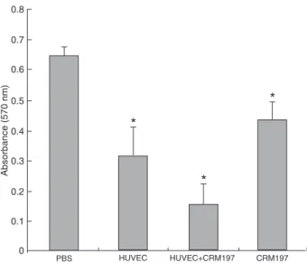
To evaluate the inhibitory effect of sera from immunized mice on the proliferation of HUVECs in vitro, cultured HUVECs were incubated with pre-immunized serum. Final vaccinated antisera (N = 8) were from mice immunized with HUVECs, HUVECs+CRM197, and CRM197. MTT results showed that sera from mice immunized with HUVECs, HUVECs+CRM197, and CRM197 all inhibited HUVEC proliferation (Figure 4). The rates of inhibition of sera from the HUVEC, HUVEC+CRM197, and CRM197 groups rela-tive to the sera from PBS group were 51.1, 68.2, 32.5% (P < 0.01), respectively. However, the anti-HUVEC sera also showed less than 10% inhibition against the proliferation of RM-1 compared to sera from the PBS group.
Antisera reacted with membrane proteins of HUVECs
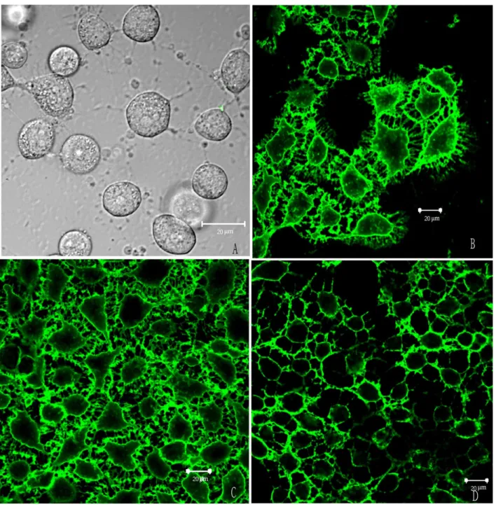
To determine whether the antisera from the three dif-ferent vaccinated groups reacted with HUVECs, immuno-cytochemistry experiments were performed using confocal laser scanning microscopy. The results showed that all antisera, except those from the PBS group, reacted with the HUVEC membrane (Figure 5).
Effect of HUVEC immunization on the aorta’s endothelium and wound healing
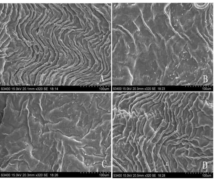
To study the potential effect of HUVEC immunization on the vascular system, we observed the inner wall of the aorta, isolated from the control and the vaccinated groups, by SEM. The aorta’s endothelia from the PBS group and the CRM197-immunized group showed regular aortic plica, whereas HUVEC immunization caused its disappearance in the other two groups (Figure 6). Surprisingly, when the
influence of HUVEC on wound healing was monitored,
Figure 3. Detection of anti-CRM197 antibodies in immunized mice. Anti-CRM197 antibodies were detected by ELISA as early as 1 week after immunization in the sera of animals immunized with CRM197 and other immunized groups. Antibody levels peaked 2 weeks after immunization. The anti-CRM197 level from the groups vaccinated with CRM197 (CRM197 alone and
CRM197+viable HUVECs) showed a significant increase com -pared with those from the PBS group and the HUVEC group (*P < 0.05, Student t-test), but there were no differences between these two groups, which were vaccinated with CRM197 or HUVECs+CRM197 (N = 8 animals per group). Data are reported as means ± SD. HUVECs = human umbilical vein endothelial cells; CRM197 = nontoxic diphtheria toxin.
Figure 4. Effect of immune sera on HUVEC proliferation. Immune sera isolated from viable HUVEC+CRM197-immunized mice had the strongest ability to inhibit the proliferation of HUVECs in vitro.
Immune sera (10 μL) from immunized groups and guinea pig sera (10 μL) were added to the HUVEC culture medium in a final volume of 120 μL/well. Except for the PBS groups, all the immu
-nized sera showed significant inhibition against the proliferation
Carcinoma inhibition by immunization with HUVEC and CRM197 145
cancer treatment only depends on blocking one specific growth factor have not been very successful (33). Other reports have shown that some proteins, including αvβ3
integrin and receptors of certain angiogenic growth fac-tors, are expressed on proliferating ECs but are not easily detected in the normal quiescent vascular endothelium (2,3,5,34). Proteins expressed on the endothelium of new vessels in mice, humans, and other species are homologous HUVEC-vaccinated mice recovered at the same rates as
those from the PBS group. The whole healing process lasted approximately 13 days (data not shown).
Discussion
Since angiogenesis is a complex process involving multiple growth factors (32), studies aiming to show that
(11). Chen et al. (12) reported that anti-HUVEC serum can bind tightly to the blood vessels within the tumors. This effect, above all, provides a rational explanation for using HUVECs as a vaccine to inhibit tumor growth. Compared
with specific tumor cell vaccines, endothelial cells have a
broad spectrum of anti-cancer response.
In the present study, CRM197 in combination with viable
HUVECs was first used to inhibit the development of murine
prostatic carcinoma. We found that this combination could enhance the inhibitory ability against tumor development in vivo compared to the use of each element alone, or when antisera were used against the proliferation of HUVECs. We also found that antisera against HUVECs showed weak
inhibition against RM-1 tumor cells (<10%). Presumably, antisera, including various antibodies, and maybe some of antigens, are commonly produced by HUVECs and RM-1
tumor cells. On the other hand, the antisera against CRM197
had little effect on the proliferation of RM-1 tumor cells.
Other researchers have reported that neutrophils and TNF-α might play a role in the antitumor function of
CRM197 (20). In the present study, we found that anti-sera, except that from the PBS group, mainly bound to the membrane of HUVEC. Surprisingly, even antisera from the CRM197-immunized group alsoreacted with the HUVEC
membrane. On the other hand, MTT results showed that
antisera from CRM197 also inhibited the proliferation of
Figure 6. Scanning electron microscopy (SEM) images of the aortic arch wall. Groups of C57BL/6J mice (3 mice per group) were im -munized ip with PBS, HUVECs, HUVECs+CRM197, and CRM197 over 4 consecutive weeks, 1 week after the last immunization. Mice
Carcinoma inhibition by immunization with HUVEC and CRM197 147
HUVECs. Previous study from our laboratory indicated that CRM197 immunization could delay the development of some cancers, like H22 and B16, to a certain extent (data not reported). Therefore, we deduced that sera from CRM197 might cross-react with the proteins expressed on the membrane of HUVECs. Thus, the antitumor activity of CRM197, as an immunogen, originates not only from the function of neutrophils but also from its inhibition against endothelial cell proliferation.
In the preventive procedure illustrated in Figure 1A, the antitumor activity of the HUVEC-immunized group was stronger than that of the CRM197-immunized group. How-ever, in the therapeutic procedure,both the CRM197 and HUVEC groups had similar antitumor effects (Figure 2A). By considering these data in combination with the ELISA results, we can deduce that the antitumor mechanism of CRM197 during the preventive process is different from that occurring during the therapeutic process. In the preventive process, the antitumor effect of CRM197 was mainly
attrib-uted to its strong inflammatory-immunological property.
In the therapeutic process, however, because prostatic
carcinoma and ECs (30,35) can both express HB-EGF,
the main antitumor effects are attributed to the CRM197
competition for the receptor with HB-EGF-like growth factor before the occurrence of humoral immunity. Once
humoral immunity occurs, the antitumor effect comes from both functions.
Although EC cells have some promising antitumor properties as vaccines without any visible side effects,
the so-called “no visible side effects” feature only denotes that there are no remarkable changes in the animal’s fur, appetite, body weight, etc. (9,12). However, we found that HUVEC immunization could lead to the destruction of the aortic arch in mice, but it is still unknown whether or not this destruction is caused by the xenogeneic EC.Although CRM197 could also inhibit the proliferation of HUVECs, almost no visible changes were observed in the inner wall of the aortas after immunization with CRM197. Presumably, that is because the anti-HUVECs could attack more targets in the EC of the mice, whereas anti-CRM197 has only one or a few targets. This could be the reason why the tissue structure of the aortic arch wall was relatively well-preserved after CRM197 vaccination.
The present results showed that HUVEC immunization
did not influence the rates of wound healing. Angiogenesis,
however, plays an important role in wound healing (36). The exact effects of HUVEC immunization on wound healing still need further study. Treatment with CRM197 in combina-tion with viable HUVECs resulted in a markedly enhanced antitumor effect on prostatic carcinoma in vivo regarding both preventive and therapeutic treatments, suggesting a novel combination for anticancer therapy.
Acknowledgments
Research supported in part by China National Natural Science Fund Committee (#30772570) and Jiangsu Natural Science Fund Committee (#BK 2007170).
References
1. Folkman J. What is the evidence that tumors are angiogen-esis dependent? J Natl Cancer Inst 1990; 82: 4-6.
2. Zetter BR. Angiogenesis and tumor metastasis. Annu Rev Med 1998; 49: 407-424.
3. Fidler IJ, Ellis LM. The implications of angiogenesis for the biology and therapy of cancer metastasis. Cell 1994; 79: 185-188.
4. Folkman J. Fighting cancer by attacking its blood supply. Sci Am 1996; 275: 150-154.
5. Ferrara N, Alitalo K. Clinical applications of angiogenic growth factors and their inhibitors. Nat Med 1999; 5: 1359-1364.
6. Neri D, Bicknell R. Tumour vascular targeting. Nat Rev Cancer 2005; 5: 436-446.
7. Scappaticci FA. Mechanisms and future directions for angiogenesis-based cancer therapies. J Clin Oncol 2002; 20: 3906-3927.
8. de Gruijl TD, van den Eertwegh AJ, Pinedo HM, Scheper
RJ. Whole-cell cancer vaccination: from autologous to al-logeneic tumor- and dendritic cell-based vaccines. Cancer Immunol Immunother 2008; 57: 1569-1577.
9. Wei YQ, Wang QR, Zhao X, Yang L, Tian L, Lu Y, et al. Im-munotherapy of tumors with xenogeneic endothelial cells as a vaccine. Nat Med 2000; 6: 1160-1166.
10. Yoshiura K, Nishishita T, Nakaoka T, Yamashita N, Yamashita N. Inhibition of B16 melanoma growth and metastasis in C57BL mice by vaccination with a syngeneic endothelial cell line. J Exp Clin Cancer Res 2009; 28: 13.
11. Okaji Y, Tsuno NH, Kitayama J, Saito S, Takahashi T, Kawai
K, et al. Vaccination with autologous endothelium inhibits angiogenesis and metastasis of colon cancer through auto-immunity. Cancer Sci 2004; 95: 85-90.
12. Chen XY, Zhang W, Zhang W, Wu S, Bi F, Su YJ, et al. Vac-cination with viable human umbilical vein endothelial cells prevents metastatic tumors by attack on tumor vasculature with both cellular and humoral immunity. Clin Cancer Res 2006; 12: 5834-5840.
13. Okaji Y, Tsuno NH, Tanaka M, Yoneyama S, Matsuhashi
M, Kitayama J, et al. Pilot study of anti-angiogenic vaccine
using fixed whole endothelium in patients with progressive
malignancy after failure of conventional therapy. Eur J Can-cer 2008; 44: 383-390.
14. Uchida T, Gill DM, Pappenheimer AM Jr. Mutation in the
structural gene for diphtheria toxin carried by temperate phage. Nat New Biol 1971; 233: 8-11.
15. Giannini G, Rappuoli R, Ratti G. The amino-acid sequence
16. Qiao J, Ghani K, Caruso M. Diphtheria toxin mutant CRM197
is an inhibitor of protein synthesis that induces cellular toxic-ity. Toxicon 2008; 51: 473-477.
17. Kageyama T, Ohishi M, Miyamoto S, Mizushima H, Iwamoto
R, Mekada E. Diphtheria toxin mutant CRM197 possesses weak EF2-ADP-ribosyl activity that potentiates its anti-tumorigenic activity. J Biochem 2007; 142: 95-104. 18. Daum RS, Hogerman D, Rennels MB, Bewley K, Malinoski
F, Rothstein E, et al. Infant immunization with pneumococcal CRM197 vaccines: effect of saccharide size on immuno-genicity and interactions with simultaneously administered vaccines. J Infect Dis 1997; 176: 445-455.
19. Schneider LC, Insel RA, Howie G, Madore DV, Geha RS.
Response to a Haemophilus influenzae type b diphtheria CRM197 conjugate vaccine in children with a defect of anti-body production to Haemophilus influenzae type b polysac-charide. J Allergy Clin Immunol 1990; 85: 948-953.
20. Buzzi S, Rubboli D, Buzzi G, Buzzi AM, Morisi C, Pironi F.
CRM197 (nontoxic diphtheria toxin): effects on advanced cancer patients. Cancer Immunol Immunother 2004; 53: 1041-1048.
21. Yagi H, Yotsumoto F, Sonoda K, Kuroki M, Mekada E, Miyamoto S. Synergistic anti-tumor effect of paclitaxel with
CRM197, an inhibitor of HB-EGF, in ovarian cancer. Int J Cancer 2009; 124: 1429-1439.
22. Naglich JG, Metherall JE, Russell DW, Eidels L. Expression
cloning of a diphtheria toxin receptor: identity with a
heparin-binding EGF-like growth factor precursor. Cell 1992; 69: 1051-1061.
23. Iwamoto R, Higashiyama S, Mitamura T, Taniguchi N,
Klags-brun M, Mekada E. Heparin-binding EGF-like growth factor,
which acts as the diphtheria toxin receptor, forms a complex with membrane protein DRAP27/CD9, which up-regulates functional receptors and diphtheria toxin sensitivity. EMBO J 1994; 13: 2322-2330.
24. Yagi H, Miyamoto S, Tanaka Y, Sonoda K, Kobayashi H,
Kishikawa T, et al. Clinical significance of heparin-binding epidermal growth factor-like growth factor in peritoneal fluid
of ovarian cancer. Br J Cancer 2005; 92: 1737-1745.
25. Tanaka Y, Miyamoto S, Suzuki SO, Oki E, Yagi H, Sonoda K, et al. Clinical significance of heparin-binding epidermal
growth factor-like growth factor and a disintegrin and metal-loprotease 17 expression in human ovarian cancer. Clin Cancer Res 2005; 11: 4783-4792.
26. Miyamoto S, Yagi H, Yotsumoto F, Kawarabayashi T, Me-kada E. Heparin-binding epidermal growth factor-like growth factor as a novel targeting molecule for cancer therapy. Cancer Sci 2006; 97: 341-347.
27. Miyamoto S, Hirata M, Yamazaki A, Kageyama T, Hasuwa H,
Mizushima H, et al. Heparin-binding EGF-like growth factor
is a promising target for ovarian cancer therapy. Cancer Res 2004; 64: 5720-5727.
28. Mitamura T, Higashiyama S, Taniguchi N, Klagsbrun M, Mekada E. Diphtheria toxin binds to the epidermal growth
factor (EGF)-like domain of human heparin-binding
EGF-like growth factor/diphtheria toxin receptor and inhibits
specifically its mitogenic activity. J Biol Chem 1995; 270: 1015-1019.
29. Angelucci A, Gravina GL, Rucci N, Millimaggi D, Festuccia C, Muzi P, et al. Suppression of EGF-R signaling reduces
the incidence of prostate cancer metastasis in nude mice. Endocr Relat Cancer 2006; 13: 197-210.
30. Torring N, Jorgensen PE, Sorensen BS, Nexo E. Increased
expression of heparin binding EGF (HB-EGF), amphiregulin, TGF alpha and epiregulin in androgen-independent prostate
cancer cell lines. Anticancer Res 2000; 20: 91-95.
31. Yankai Z, Rong Y, Yi H, Wentao L, Rongyue C, Ming Y, et
al. Ten tandem repeats of beta-hCG 109-118 enhance im
-munogenicity and anti-tumor effects of beta-hCG C-terminal
peptide carried by mycobacterial heat-shock protein HSP65. Biochem Biophys Res Commun 2006; 345: 1365-1371.
32. Distler JH, Hirth A, Kurowska-Stolarska M, Gay RE, Gay S, Distler O. Angiogenic and angiostatic factors in the
molecular control of angiogenesis. Q J Nucl Med 2003; 47: 149-161.
33. Kerbel RS. Tumor angiogenesis: past, present and the near future. Carcinogenesis 2000; 21: 505-515.
34. Kerbel RS. A cancer therapy resistant to resistance. Nature 1997; 390: 335-336.
35. Iivanainen E, Nelimarkka L, Elenius V, Heikkinen SM, Junt-tila TT, Sihombing L, et al. Angiopoietin-regulated recruit-ment of vascular smooth muscle cells by endothelial-derived
heparin binding EGF-like growth factor. FASEB J 2003; 17: 1609-1621.
36. Bates DO, Jones RO. The role of vascular endothelial