Lack of evidence for the pathogenic
role of iron and
HFE
gene mutations
in Brazilian patients with nonalcoholic
steatohepatitis
Departamentos de 1Gastroenterologia and 2Patologia, Laboratórios de Investigação Médica 3LIM-07 and 4LIM-06,
Faculdade de Medicina, and 5Laboratório de Imunologia do Transplante, Instituto do Coração, Universidade de São Paulo, São Paulo, SP, Brasil M.M. Deguti1,
A.M. Sipahi1,3, L.C.C. Gayotto2, S.A. Palácios5, P.L. Bittencourt1, A.C. Goldberg5, A.A. Laudanna1, F.J. Carrilho1 and E.L.R. Cançado1,4
Abstract
The hypothesis of the role of iron overload associated with HFE gene
mutations in the pathogenesis of nonalcoholic steatohepatitis (NASH) has been raised in recent years. In the present study, biochemical and histopathological evidence of iron overload and HFE mutations was
investigated in NASH patients. Thirty-two NASH patients, 19 females (59%), average 49.2 years, 72% Caucasians, 12% Mulattoes and 12% Asians, were submitted to serum aminotransferase and iron profile determinations. Liver biopsies were analyzed for necroinflammatory activity, architectural damage and iron deposition. In 31 of the pa-tients, C282Y and H63D mutations were tested by PCR-RFLP. Ala-nine aminotransferase levels were increased in 30 patients, 2.42 ± 1.12 times the upper normal limit on average. Serum iron concentration, transferrin saturation and ferritin averages were 99.4 ± 31.3 g/dl, 33.1 ± 12.7% and 219.8 ± 163.8 µg/dl, respectively, corresponding to normal values in 93.5, 68.7 and 78.1% of the patients. Hepatic siderosis was observed in three patients and was not associated with architectural damage (P = 0.53) or with necroinflammatory activity (P = 0.27). The allelic frequencies (N = 31) found were 1.6 and 14.1% for C282Y and H63D, respectively, which were compatible with those described for the local population. In conclusion, no evidence of an association of hepatic iron overload and HFE mutations with NASH
was found. Brazilian NASH patients comprise a heterogeneous group with many associated conditions such as hyperinsulinism, environ-mental hepatotoxin exposure and drugs, but not hepatic iron overload, and their disease susceptibility could be related to genetic and environ-mental features other than HFE mutations.
Correspondence
M.M. Deguti
Departamento de Gastroenterologia FM, USP
Av. Enéas C. Aguiar, 255, S. 9159 01246-903 São Paulo, SP Brasil
Fax: +55-11-5584-5122 E-mail: marta.deguti@uol.com.br
M.M. Deguti was supported by CNPq (Grant No. 135303/1999-0). Publication supported by FAPESP.
Received July 18, 2002 Accepted April 17, 2003
Key words •Fatty liver
•Nonalcoholic steatohepatitis •Iron overload
•HFE gene
Introduction
Nonalcoholic steatohepatitis (NASH) was designated as such by Ludwig et al. in 1980 (1), and has been increasingly recognized as one of the most frequent diagnoses by hepa-tologists (2). Early studies (1-6) described NASH mainly in obese, middle-aged women, often associated with diabetes mellitus and hyperlipidemia. However, in recent years, other investigators (7-10) have found evi-dence of NASH association with male, nonobese, nondiabetic patients and with liver iron overload, which led to the hypothesis of iron playing a role in NASH pathogenesis.
The identification of the hereditary hemo-chromatosis HFE gene (11) on chromosome
6 and its 845 G→A (C282Y) and 187 C→G (H63D) mutations has enabled further in-vestigation of this aspect based on the knowl-edge that compound heterozygotes (C282Y/ H63D) and H63D homozygotes may have a mild or moderate iron overload (12). How-ever, more recently, Gochee et al. (13) dem-onstrated that the presence of H63D does not result in significant iron overload, and that this mutation is not clinically significant in the absence of the C282Y mutation.
George et al. (14) found that the C282Y mutation of the HFE gene was significantly
associated with the degree of iron staining, hepatic iron concentration and liver damage in Caucasian NASH patients from Australia. Bonkovsky et al. (15,16) expanded this con-cept by identifying both C282Y and H63D which were significantly related to NASH in North Americans. In view of these data, the aim of the present study was to investigate the evidence of iron overload and the preva-lence of C282Y and H63D mutations among NASH patients from São Paulo, SP, Brazil.
Patients and Methods
Cohort of patients
Thirty-two patients who fulfilled the
diag-nostic criteria of NASH were enrolled in this study. NASH was defined histopathologically as macrovesicular steatosis, lobular inflam-mation and perivenular fibrosis morphologi-cally indistinguishable from alcoholic disease in subjects whose alcohol consumption was less than 20 g/day. This study was approved by the local Ethics Committee and was performed according to the guidelines of the Helsinki declaration. Informed consent was obtained from each patient included in the study.
Methods
Serum alanine aminotransferase, iron, transferrin saturation and ferritin levels were obtained at the time of liver biopsy.
Liver biopsy specimens were fixed in buffered formalin and embedded in paraffin. Tissue sections were stained with hematoxy-lin-eosin, Masson’s trichrome, reticulin and Perls’ Prussian blue. Each biopsy was read blindly by one experienced pathologist (L.C.C. Gayotto). NASH was graded 1 to 4 according to steatosis intensity (the percent-age of hepatocytes with fat vacuoles), necro-inflammatory injury and architectural dam-age. Architectural damage grade 4 was as-signed to cirrhosis. The presence of iron pigments was assessed in Kupffer cells, lobu-lar hepatocytes and periportal regions, and graded from 0 to 4 on a semiquantitative basis for each deposition site as follows: 0, absence of iron; 1, presence of iron at mini-mum intensity; 2 and 3, presence of iron at intermediate intensity, and 4, presence of iron at maximum intensity.
All patients but one were tested for both C282Y and H63D mutations. Genomic DNA was extracted from peripheral blood leuko-cytes by standard techniques, and mutation analyses were carried out as described (17). DNA amplification was performed by the polymerase chain reaction with the primers 5'-CTC.AGG.CAC.TCC.TCT.CAA.CC-3' and 5'-TGG.CAA.GGG.TAA.ACA.GAT. CC-3' for the G845A locus codon 282 and 5'-GCC.ACA.TCT.GGC.TTG.AAA.TT-3' and 5'-ACA.TGG.TTA.AGG.CCT.GTT.GC-3' for the C187G locus codon 63, which was followed by restriction fragment length poly-morphism analysis with the SnaBI and BclI
enzymes, respectively.
Statistical analysis
Statistical analysis was performed using the unpaired Student t-test or the Fisher
ex-act testwhen appropriate. Continuous
vari-ables are reported as means ± SD and categori-cal variables as frequency or percentage.
Results
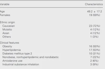
As shown in Table 1, in this cohort of 32 patients, age ranged from 32 to 76 years, average 49.2 years. Ethnic origin was as follows: 72% Caucasians, 12% Mulattoes and 12% Asians. Fifty-nine percent of pa-tients were females, 50% obese, 53% hyper-lipidemic and 31% diabetic type 2. Other clinical features were the continuous use of amiodarone and the daily inhalation of in-dustrial substances for at least six months.
Serum alanine aminotransferase was in-creased in 30 patients (93.5%), and the high-est value was 4.8 times the upper normal limit (average 2.42 ± 1.12). Serum iron con-centration was normal (50-150 µg/dl) in 93.5%, average 99.4 ± 31.3 g/dl. Transferrin iron saturation was within the normal range (20-40%) in 68.7%, average 33.1 ± 12.7%. Ferritin level was normal (females: 25-250 µg/dl; males: 50-500 µg/dl) in 78.1% of women, average 219.8 ± 163.8 µg/dl and in 76.9% of men, average 444.8 ± 305.9 µg/dl.
Table 1. Clinical and demographic characteristics of the 32 patients in the present study.
Variable Characteristics
Age 49.2 ± 17.2
Females 19 (59%)
Ethnic origin
Caucasian 23 (72%)
Mulatto 4 (12%)
Asian 4 (12%)
Others 1 (3%)
Clinical features
Obesity 16 (50%)
Hyperlipidemia 17 (53%)
Diabetes mellitus type 2 10 (31%)
Nonobese, nonhyperlipidemic and nondiabetic 7 (22%)
Amiodarone use 2 (6%)
Industrial substance inhalation 3 (9%)
The histopathological study revealed ste-atosis grade 2 in 5 cases (15.6%), grade 3 in 20 (62.5%) and grade 4 in 7 (21.9%). Necroinflammatory injury was grade 1 in 13 cases (40.6%), grade 2 in 5 (6.3%), grade 3 in 12 (37.5%), and grade 4 in 2 (15.6%). Ten patients (31.2%) had architectural damage grades 1, 2 and 3; the remaining two patients were already cirrhotic, with grade 4 damage (6.3%).
Liver biopsies submitted to Perls’ stain-ing revealed siderosis in only three patients, all of whom were males. The first one, an obese and hyperlipidemic Caucasian, had siderosis grade 2 in both lobular hepatocytes and Kupffer cells. The second patient was a diabetic Asian and presented siderosis grade 1 in lobular hepatocytes and grade 3 in Kupf-fer cells. The third one, a Caucasian without any associated condition, presented grade 1 siderosis homogeneously distributed in the periportal region, lobular hepatocytes and
Kupffer cells.
Of the 31 patients whose DNA was ob-tained, one was C282Y , seven H63D +/-and one H63D +/+. There were no C282Y +/+ or compound C282Y/H63D heterozygotes. The allelic frequencies found in this cohort of patients for C282Y and H63D were 1.6 and 14.1%, respectively. Among the three patients with liver siderosis, only one (an obese and hyperlipidemic Caucasian) had an
HFE mutation, H63D +/-. The patient who
had the H63D +/+ mutation was an obese, hyperlipidemic female with hyperferritine-mia, but with no hepatic iron deposition.
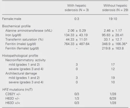
The biochemical, histopathological and genetic profile of NASH patients was com-pared according to the presence or absence of siderosis in liver tissue (Table 2). Patients with hepatic iron overload had significantly higher levels of serum ferritin (P < 0.05); however, there was no statistically signifi-cant correlation of hepatic iron overload with necroinflammatory injury or with HFE
mu-tation frequencies.
Discussion
In our cohort of 32 patients there was no evidence of the association between NASH and hepatic iron overload suggested in re-cent publications (7,14,15,18). Siderin pig-ment in liver tissue was not significant in frequency or intensity by Perls’ staining. The HFE gene mutation frequencies were
compatible with the characteristics of the local general population (19).
Hyperferritinemia was found in less than a quarter of our patients, in contrast to the majority of the patients reported by the French group from Rennes (18,20,21). According to the cited authors, the phenomena of hyper-ferritinemia, normal transferrin saturation and hepatic siderosis were predominant in patients with NASH and insulin resistance syndrome. Insulin resistance syndrome was defined by the presence of one or more of the following features: obesity, hyperlipidemia
Table 2. Biochemical, histopathological and HFE mutational results of patients with and without hepatic siderosis.
With hepatic Without hepatic siderosis (N = 3) siderosis (N = 29)
Female:male 0:3 19:10
Biochemical profile
Alanine aminotransferase (xNL) 2.06 ± 0.29 2.46 ± 1.17 Iron (µg/dl) 134.33 ± 43.19 95.83 ± 28.41 Transferrin saturation (%) 44.33 ± 11.01 33.1 ± 12.7 Ferritin (male) (µg/dl) 764.33 ± 487.64 348.9 ± 166.36* Ferritin (female) (µg/dl) - 219.8 ± 163.8
Histopathological profile Necroinflammatory activity
mild (grades 1 and 2) 3 17
severe (grades 3 and 4) 0 12
Architectural damage
mild (grades 1 and 2) 3 19
severe (grades 3 and 4) 0 10
HFE mutations (m/T)
C282Y +/- 0/3 1/28
H63D +/- 1/3 6/28
H63D +/+ 0/3 1/28
and abnormal glucose metabolism. In the present study, however, only 9 (36%) of the 25 patients with insulin resistance syndrome had hyperferritinemia but no hepatic iron overload. In contrast, three patients with he-patic iron overload had significantly higher ferritin levels and two of them had insulin resistance syndrome. This divergence could be partially attributed to the difference be-tween the databases: the patients reported by the cited investigators were selected among subjects with hepatic iron overload, while ours were from the population routinely at-tended at a Gastroenterology Outpatient Clinic. Since hepatic steatosis, insulin resis-tance syndrome and iron overload can occur independently or in combination, the hypo-thesis that postulates insulin resistance as the pivotal abnormality leading to iron over-load and subsequently to steatohepatitis (20) is acceptable for the subgroup affected by all of these conditions. This narrows down our population to only two subjects.
The predominant profile of our NASH patients agrees with that reported by Younossi et al. (22) for patients from Cleveland, OH, USA. These authors did not observe signifi-cant iron accumulation in cases with nonal-coholic fatty liver, including those with NASH. They carefully excluded alcoholism, by defining 20 g as the upper limit of accept-able daily ethanol consumption by review-ing the charts and followreview-ing up patients when necessary. Our criteria were similarly strict about this aspect, a fact that could explain the scarcity of iron deposition also in our cohort, since alcohol induces secondary iron overload.
The absence of Africans and the same proportion of Mulattoes and Asians (12%) with NASH was conspicuously different from the ethnic distribution of the local popula-tion, described as 6.7, 28.4 and 0.5%, re-spectively, according to Brazilian govern-ment statistics in 1999 and referring to the southeast region of the country (http:// www1.ibge.gov.br/english/estatistica/
populacao/condicaodevida/indicadoresminimos/ tabela1.shtm#a112). The interpretation of the ethnic profile of Brazilians is more diffi-cult because of the intense pluriracial mixing which has been taking place in the last five centuries, involving natives, Africans, Cau-casians and others (23). To our knowledge, patients of African origin having NASH have not been reported in the literature, and the lower proportion of Mulattoes suggests that an ethnically related protective effect might need to be investigated. The higher propor-tion of Asians in our NASH populapropor-tion could be attributed to the alcohol intolerance ge-netically determined among them (24), fa-cilitating the determination of alcohol con-sumption. All Asians were of Japanese ori-gin and all of them had glucose intolerance. Interestingly, it is known that western accul-turation increases the frequency of diabetes among the Japanese (25). Since NASH pa-tients in Japan are not excessively diabetic (26), an eventual major susceptibility to de-veloping liver alterations secondary to a glu-cose intolerance condition might be further studied in this group.
Three patients, two of whom were hyper-cholesterolemic, had been continuously ex-posed to fumes while working in a steel factory. They were neither obese nor dia-betic, nor did they have any hepatic iron overload. Even though the occupational eti-ology was not established in this study, Cotrim et al. (27) have already demonstrated in Brazil that chronic exposure to volatile petrochemical products can lead to NASH. Others have also recognized the association between environmental toxicity and NASH (8,28).
that this kind of surgery leads to an increase in permeability of the remaining bowel (29) and bacterial translocation (30). These phe-nomena could be related to the absorption of endotoxins and bacterial lipopolysaccharides, increasing TNF-α levels and finally leading to steatohepatitis (31).
The allelic frequencies of C282Y (1.6%) and H63D (14.5%) observed among Brazil-ian NASH patients were compatible with those described for the local population: C282Y allelic frequency ranges from 1.1 to 1.4% and H63D allelic frequency from 6.4 to 20.3% according to the population groups analyzed (19). Only one of the three subjects with hepatic iron deposition carried an HFE
mutation (H63D +/-). Moreover, it is known
that hereditary hemochromatosis and HFE
mutations are almost absent among Japanese people (26), although they were well repre-sented in our NASH series. These findings provide evidence that HFE mutations are not
necessarily involved in NASH pathogenesis, although they seem to play an important role
in Australian and North American Cauca-sians (8,14,15).
For those populations with high allelic frequencies of HFE mutations, secondary
hepatic iron overload might determine oxi-dative stress and inflammation. In Brazil, however, NASH patients comprise a hetero-geneous group with many associated condi-tions such as hyperinsulinism, environmen-tal hepatotoxin exposure and drugs - but not hepatic iron overload, and their disease sus-ceptibility could be related to genetic and environmental features other than HFE
mu-tations.
Acknowledgments
The authors would like to thank Ms. Lusane Leão Baía for statistical analysis of the results. Special thanks are due to Dr. Cláudia Tani, Dr. Roseneli D’Este and Ms. Sylvia Assumpção for their contributions to this study.
References
1. Ludwig J, Viggiano TR, McGill DB & Ott BJ (1980). Nonalcoholic steatohepatitis. Mayo Clinic Experiences with a hitherto unnamed disease. Mayo Clinic Proceedings, 55: 434-438.
2. James OF & Day CP (1998). Nonalcoholic steatohepatitis (NASH): a disease of emerging identity and importance. Journal of Hepatolo-gy, 29: 495-501.
3. Itoh S, Yougel T & Kawagoe K (1987). Comparison between nonal-coholic steatohepatitis and alnonal-coholic hepatitis. American Journal of Gastroenterology, 82: 650-654.
4. Diehl AM, Goodman Z & Ishak KG (1988). Alcohol-like disease in nonalcoholics. A clinical and histologic comparison with alcohol-induced liver injury. Gastroenterology, 95: 1056-1062.
5. Powell EE, Cooksley WGE, Hanson R, Searle J, Halliday JW & Powell LW (1990). The natural history of nonalcoholic steato-hepatitis: a follow-up study of forty-two patients for up to 21 years.
Hepatology, 11: 74-80.
6. Lee RG (1989). Nonalcoholic steatohepatitis: a study of 49 patients.
Human Pathology, 20: 594-598.
7. Bacon BR, Farakvash MJ, Janney CG & Neuschwander-Tetri BA (1994). Nonalcoholic steatohepatitis: an expanded clinical entity.
Gastroenterology, 107: 1103-1109.
8. Lonardo A, Bellini M, Tondelli E, Frazzoni M, Grisendi A, Pulvirenti M & Della-Casa G (1995). Nonoalcoholic steatohepatitis and the “bright liver syndrome”: should a recently expanded clinical entity be
fur-ther expanded? (Letter). American Journal of Gastroenterology, 90: 2072-2074.
9. Propst A, Propst T, Judmaier G & Vogel W (1995). Prognosis in nonalcoholic steatohepatitis. Gastroenterology, 108: 1607. 10. James O & Day C (1999). Nonalcoholic steatohepatitis: another
disease of affluence. Lancet, 353: 1634-1636.
11. Feder JN, Gnirke A, Thomas W et al. (1996). A novel MHC class I-like gene is mutated in patients with hereditary haemochromatosis.
Nature Genetics, 13: 399-408.
12. Adams PC (1999). Population screening for hemochromatosis. Hep-atology, 29: 1324-1327.
13. Gochee PA, Powell LW, Cullen DJ, Sart DD, Rossi E & Olynyk JK (2002). A population-based study of the biochemical and clinical expression of the H63D hemochromatosis mutation. Gastroenterol-ogy, 122: 646-651.
14. George DK, Goldwurm S, MacDonald GA, Cowley LL, Walker NI, Ward PJ, Jazwinska EC & Powell LW (1998). Increased hepatic iron concentration in nonalcoholic steatohepatitis is associated with increased fibrosis. Gastroenterology, 114: 311-318.
15. Bonkovsky HL, Jawaid Q, Tortorelli K, Leclair P, Cobb J, Lambrecht RW & Banner BF (1999). Nonalcoholic steatohepatitis and iron: increased prevalence of mutations of the HFE gene in nonalcoholic steatohepatitis. Journal of Hepatology, 31: 421-429.
Nonalcoholic steatohepatitis with iron: part of insulin resistance-associated hepatic iron overload? (Letter). Journal of Hepatology, 33: 1025-1026.
17. Datz C, Lalloz MRA, Vogel W et al. (1997). Predominance of the HLA-H Cys282Tyr mutation in Austrian patients with genetic hae-mochromatosis. Journal of Hepatology, 27: 773-779.
18. Mendler M, Turlin B, Moirand R, Jouanolle A, Sapey T, Guyader D, Le Gall J, Brissot P, David V & Deugnier Y (1999). Insulin resistance-associated hepatic iron overload. Gastroenterology, 117: 1155-1163. 19. Agostinho MF, Arruda VR, Basseres DS, Bordin S, Soares MCP, Menezes RC, Costa FF & Saad STO (1999). Mutation analysis of the HFE gene in Brazilian populations. Blood Cells, Molecules, and Diseases, 25: 324-327.
20. Guillygomarc’h A, Mendler MH, Moirand R, Jouanolle AM, David V & Deugnier Y (2000). HFE mutations in insulin resistance-associ-ated hepatic iron overload (Letter). Journal of Hepatology, 33: 515-516.
21. Moirand R, Mendler MH, Guillygomarc’h A, Brissot P & Deugnier Y (2000). Nonalcoholic steatohepatitis with iron: part of insulin resis-tance-associated hepatic iron overload? (Letter). Journal of Hepatol-ogy, 33: 1024-1026.
22. Younossi ZM, Gramlich T, Bacon BR, Matteoni CA, Boparai N, O’Neill RO & McCullough AJ (1999). Hepatic iron and nonalcoholic fatty liver disease. Hepatology, 30: 847-850.
23. Alves-Silva J,da Silva-Santos M,Guimarães PE, Ferreira AC, Bandelt HJ, Pena SD & Prado VF (2000). TheancestryofBrazilianmtDNA lineages.American Journal of Human Genetics, 67: 444-461.
24. Lumeng L & Crabb DW (1994). Genetic aspects and risk factors in alcoholism and alcoholic liver disease. Gastroenterology, 107: 572-578.
25. Franco LJ (1996). Diabetes in Japanese-Brazilians - influence of the acculturation process. Diabetes Research and Clinical Practice, 34 (Suppl): S51-S57.
26. Ueno T, Sugawara H, Sujaku K, Hashimoto O, Tsuji R, Tamaki S, Torimura T, Inuzuka S, Sata M & Tanikawa K (1997). Therapeutic effects of restricted diet and exercise in obese patients with fatty liver. Journal of Hepatology, 27: 103-107.
27. Cotrim HP, Andrade ZA, Parana R, Portugal ML, Lyra LG & Freitas LAR (1999). Nonalcoholic steatohepatitis: a toxic liver disease in industrial workers. Liver, 19: 299-304.
28. Neuschwander-Tetri BA & Bacon BB (1996). Nonalcoholic steato-hepatitis. Medical Clinics of North America, 80: 1147-1166. 29. Vazquez CM, Molina MT & Ilundain A (1988). Distal small bowel
resection increases mucosal permeability in the large intestine.
Digestion, 40: 168-172.
30. Schimpl G, Feierl G, Linni K, Uitz K, Ozbey H & Hollwarth ME (1999). Bacterial translocation in short-bowel syndrome in rats. European Journal of Pediatric Surgery, 9: 224-227.