he importance of recognizing antiphospholipid syndrome in
vascular medicine
A importância de reconhecer a síndrome antifosfolípide na medicina vascular
Andreas Funke1
*
, Adriana Danowski2, Danieli Castro Oliveira de Andrade3, Jozelia Rêgo4, Roger Abramino Levy5
Abstract
Antiphospholipid syndrome (APS) is a systemic autoimmune disease characterized by recurrent arterial or venous thrombosis and/or gestational morbidity and by the presence of antiphospholipid antibodies. It can also cause other vascular manifestations such as microangiopathy, chronic arteriopathy and catastrophic APS (CAPS). Certain laboratory tests for the syndrome (for example, the lupus anticoagulant test) can be afected by the use of anticoagulant agents, making diagnosis more diicult. he pathophysiology of APS is complex, and several mechanisms of pathogenesis related to coagulation, endothelium, and platelets are discussed in this article. We conclude by discussing treatment of APS according to the presence and type of clinical manifestations, use of direct oral anticoagulants (DOAs), and perioperative management of patients with APS.
Keywords: antiphospholipid syndrome; lupus anticoagulant; anticardiolipin antibodies; thrombosis; autoimmunity.
Resumo
A síndrome antifosfolipíde (SAF) é uma doença autoimune sistêmica caracterizada por trombose arterial ou venosa recorrente e/ou morbidade gestacional e pela presença dos anticorpos antifosfolipídeos, podendo apresentar outras manifestações vasculares, como microangiopatia, arteriopatia crônica e SAF catastróica. Determinados testes laboratoriais para a síndrome (por exemplo, o anticoagulante lúpico) podem sofrer interferência do uso de medicações anticoagulantes, diicultando o diagnóstico. A isiopatologia da SAF é complexa, sendo enumerados no texto diversos mecanismos patogênicos relacionados à coagulação, ao endotélio e às plaquetas. Por im, discutimos o tratamento da SAF de acordo com a presença e o tipo de manifestações clínicas, o uso dos anticoagulantes orais diretos e o manejo perioperatório de pacientes com SAF.
Palavras-chave: síndrome antifosfolípide; anticoagulante do lúpus; anticorpos anticardiolipina; trombose; autoimunidade.
1Universidade Federal do Paraná – UFPR, Hospital de Clínicas, Curitiba, PR, Brazil. 2Hospital Federal dos Servidores do Estado – HFSE, Rio de Janeiro, RJ, Brazil. 3Universidade de São Paulo – USP, Faculdade de Medicina, São Paulo, SP, Brazil. 4Universidade Federal de Goiás – UFG, Faculdade de Medicina, Goiânia, GO, Brazil. 5Universidade do Estado do Rio de Janeiro – UERJ, Rio de Janeiro, RJ, Brazil.
Financial support: None.
Conlicts of interest: No conlicts of interest declared concerning the publication of this article. Submitted: December 23, 2016. Accepted: April 24, 2017.
INTRODUCTION
Antiphospholipid syndrome (APS) is a systemic
autoimmune disease characterized by recurrent arterial
or venous thrombosis and/or gestational morbidity
and persistent presence of what have become known
as “antiphospholipid antibodies”
1(aPL), which can be
detected by laboratory tests for lupus anticoagulant
(LA), anticardiolipin (aCL) IgG and IgM, and
anti-β2-glycoprotein I (anti-β2-GPI) IgG and IgM.
Antiphospholipid syndrome can occur in combination
with other autoimmune diseases, most commonly
systemic lupus erythematosus (SLE), and it can also
be found in isolation (primary APS).
1,2CLINICAL MANIFESTATIONS
Antiphospholipid syndrome can affect any organ or
system. It most often manifests through deep venous
thrombosis (DVT). However, its manifestations are
not limited to the venous bed and it can also manifest
through arterial thrombosis (with or without subjacent
atherosclerosis), resulting, for example, in an ischemic
stroke, or thrombotic microangiopathy, as is seen in
nephropathy of APS.
3Vascular disease associated
with accentuated intimal hyperplasia and arterial
stenosis have also been attributed to APS.
3,4Finally,
cases of accelerated atherosclerosis have also been
described in patients with aPL or APS.
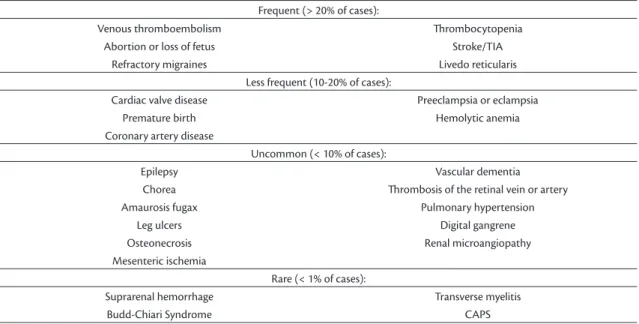
5-7Table 1 lists
the frequencies of the various different manifestations
of APS, according to analysis of a large European
cohort (1,000 patients).
8,9Rarely, a devastating condition with rapid onset
known as catastrophic antiphospholipid syndrome
(CAPS) may be observed. This is characterized by
multiple vascular occlusions (in more than three
organs or systems) over a short period of time
(less than 1 week). More than half of CAPS cases occur
in the presence of identiiable trigger factors, such
as bacterial or viral infections, surgical procedures,
withdrawal of anticoagulant treatment, obstetric
complications, neoplasms, or concomitant SLE.
Catastrophic antiphospholipid syndrome is linked
with elevated mortality rates, varying from 36.8% to
over 50% of cases, depending on the publication.
10,11DIAGNOSIS
The preliminary classification criteria, which
were published in 1999 (Sapporo) and updated in
2006 (Sydney),
12deine APS as the presence of at
least one clinical criterion and one laboratory test
criterion (listed in
Table 2). The objective of these
criteria is to deine the syndrome’s characteristics
for the purposes of etiologic and therapeutic studies.
They are also frequently used for precise diagnosis of
APS in clinical practice, but should not be considered
absolute prerequisites for prescribing or withholding
treatment.
Only laboratory test results and clinical manifestations
that are most speciic to APS were included in the
classiication criteria. Other clinical and laboratory
findings that are also associated with APS are
Table 1. Clinical manifestations of APS.
Frequent (> 20% of cases):
Venous thromboembolism hrombocytopenia
Abortion or loss of fetus Stroke/TIA
Refractory migraines Livedo reticularis
Less frequent (10-20% of cases):
Cardiac valve disease Preeclampsia or eclampsia
Premature birth Hemolytic anemia
Coronary artery disease
Uncommon (< 10% of cases):
Epilepsy Vascular dementia
Chorea hrombosis of the retinal vein or artery
Amaurosis fugax Pulmonary hypertension
Leg ulcers Digital gangrene
Osteonecrosis Renal microangiopathy
Mesenteric ischemia
Rare (< 1% of cases):
Suprarenal hemorrhage Transverse myelitis
Budd-Chiari Syndrome CAPS
denominated “non-criterion” manifestations or
tests. Non-criterion manifestations of APS include
cardiac valve disease, livedo reticularis, skin ulcers,
thrombocytopenia, nephropathy of APS, and neurological
manifestations related to aPL.
12,13Non-criterion
Laboratory tests include the antibodies aCL IgA,
anti-β2-GPI IgA, anti-phosphatidylserine (aPS),
anti-phosphatidylethanolamine (aPE), anti-prothrombin
alone (aPT-A), anti-phosphatidylserine-prothrombin
complex (aPS/PT), and anti-domain I of β2-glycoprotein I.
12,14Efect of anticoagulant therapy on laboratory
investigation of APS
In practice, it can sometimes be necessary to
conduct laboratory investigations of APS for patients
who are already being treated with parenteral or oral
anticoagulant drugs. In this situation it is not expected
that there will be a signiicant inluence on the results
of ELISA aCL and anti-β2-GPI tests, but there can be
dificulties with correctly diagnosing LA.
Guidelines published by the International Society
on Thrombosis and Haemostasis (ISTH)
15recommend
that LA should be tested 1 to 2 weeks after withdrawal
of treatment with vitamin K antagonists (VKA) or
when the international normalized ratio (INR) value
is less than 1.5. Treatment can be bridged using low
molecular weight heparin (LMWH) when VKA
are withdrawn, taking care to be sure that the last
administration of heparin occurred more than 12 hours
before the sample for LA testing is collected. Another
possibility is to test for LA during treatment with
VKA, at a point at which INR is between 1.5 and 3.0,
diluting the patient’s plasma sample at a proportion
of 1:1 with pooled normal plasma. In this case, low
levels of LA may not be detected, since the sample
is diluted by half.
One point that should be stressed is that the growing
popularity of the new direct oral anticoagulants (DOAs),
such as rivaroxaban and dabigatran etexilate, for
treatment of patients with venous thromboembolism
can lead to false-positive results in the usual tests for
LA (dilute Russell’s viper venom time [dRVVT] and
activated partial thromboplastin time [APTT]) caused
by therapeutic concentrations of these drugs.
16,17Speciically in the case of rivaroxaban, this effect
can be avoided by using Taipan snake venom time
or ecarin clotting time tests to diagnose LA,
18or by
waiting at least 24 hours after the last dose of this
medication before testing for LA using the usual
techniques.
19PATHOPHYSIOLOGY OF APS
Presence of aPL, and especially presence of
LA mediated by anti-β2-GPI antibodies, confers
an increased risk of thrombosis and/or gestational
morbidity. Experimental infusion of autoantibodies
from patients with APS potentiated formation of
thrombi in mice with induced vascular injuries.
However, the same effect was not seen in the absence
of prior vascular injury, suggesting that APS occurs
over two distinct phases (the double-hit hypothesis):
(1) existence of a latent prothrombotic state, induced
by circulating autoantibodies; (2) endothelial injury
or activation, triggering the thrombotic event. Recent
studies indicating elevated plasma levels of circulating
microparticles derived from platelets and endothelial
cells in patients with APS or aPL provide conirmation
Table 2. Updated criteria (Sydney) for classiication of APS.
Clinical criteria
1. Venous, arterial, or small vessel thrombosis* conirmed objectively (imaging or histopathology**) 2. Gestational morbidity
(a) one or more unexplained occurrence of death of a morphologically normal fetus at or after the 10th week of gestation, conirmed by ultrasound or examination of the fetus; or
(b) one or more premature deliveries (before the 34th week of gestation) of a morphologically normal neonate, caused by eclampsia, severe pre-eclampsia or placental insuiciency; or
(c) three or more consecutive unexplained spontaneous abortions before the 10th week of pregnancy, after ruling
out maternal anatomic or hormonal causes and paternal and maternal chromosomal causes.
Laboratory criteria
(a) lupus anticoagulant (LA) detected according to ISTH§ guidelines on two or more separate occasions at least
12 weeks apart; or
(b) anticardiolipin antibody (aCL) IgG or IgM detected in serum or plasma with medium to high titers (> 40 GPL or MPL units or > 99th percentile) on two or more separate occasions at least 12 weeks apart, measured using
standardized ELISA; or
(c) anti-β2-glycoprotein I antibodies (anti-β2-GPI) detected in serum or plasma on two or more separate occasions at least 12 weeks apart, measured using standardized ELISA
*Does not include supericial venous thrombosis; **Without signiicant inlammation of the vascular wall (vasculitis); §International
of the concept of a continuous prothrombotic state
in these patients.
20,21Mechanisms potentially involved in the pathogenesis
of APS include
22:
(i)
Increased immunogenicity of domain I of plasma
β2-glycoprotein I, caused by oxidative stress;
(ii) Conformational change to the β2-glycoprotein
I molecule when it binds to cell membrane
anionic phospholipids, enabling the binding of
autoantibodies and the subsequent pathogenic
effects;
(iii) Interference of these autoantibodies with the
nitric oxide endothelial synthase function;
(iv) Increased expression of thromboplastin in
endothelial cells and monocytes, induced by
aPL;
(v) Uncontrolled activation of factor XI and
consequent predisposition towards venous and
arterial thrombosis;
(vi) Potentiation of platelet activation by interaction
between β2-glycoprotein I, platelet receptors,
and anti-β2-GPI antibodies;
(vii) Interference by antigen/antibody β2-GPI/anti-β2-GPI
complexes in the protective function of annexin
A5 against activation of coagulation;
(viii) Antibody-mediated activation of the complement
(C3 and C5), resulting in fetal loss and/or
thrombosis;
(ix) Increased expression of toll-like receptors 7
and 8 (TLR7 and TLR8); and
(x)
In patients with APS nephropathy, aPL activation
of the mammalian target of rapamycin complex
(mTORC) pathway, inducing accentuated intimal
hyperplasia and vascular disease.
23TREATING APS – GESTATIONAL MORBIDITY
Treatment for patients with obstetric APS includes
non-pharmacological measures such as management
by a multidisciplinary team at a high-risk pregnancy
clinic, rigorous clinical and laboratory control, and
ultrasound monitoring of fetal growth and uteroplacental
circulation.
24,25Patients with a history of APS with
exclusively obstetric manifestations can be treated,
in the event of future pregnancies, with low-dose
aspirin (LDA) (75-100 mg/day) plus unfractionated
heparin (UFH) or LMWH at prophylactic doses
until 6 weeks after delivery.
24However, pregnant
women with APS and a history of thrombotic events
need antithrombotic therapy throughout the entire
pregnancy and immediate postpartum period, with
a combination of LDA and full anticoagulant doses
of heparin (UFH or LMWH).
24,26Once the pregnancy-puerperium cycle is complete,
patients who have exhibited gestational APS remain
at increased risk of thrombotic events, even when
there is no prior history of thrombosis, and in these
circumstances indeinite primary prophylaxis with
LDA is recommended, in addition to controlling the
classic risk factors for thrombosis.
26TREATING APS – THROMBOTIC
MANIFESTATIONS
Stratiication of risk of thrombosis and
hemorrhage
All treatment decisions for patients with APS
should take account of the risk of thrombosis
recurrence and risks resulting from the anticoagulant
treatment (for example, hemorrhage).
27In general,
it is recommended that risk of thrombosis should
be stratiied by identiication of cardiovascular risk
factors (primarily: smoking, hypertension, diabetes,
dyslipidemia, and obesity), presence or absence of
associated autoimmune diseases (for example, SLE),
and deinition of whether or not there is a high-risk
aPL proile,
27which includes (1) positive LA result;
(2) triple positivity, (i.e. LA, aCL, and anti-b2-GPI
all positive in the same patient); and (3) persistently
high aCL levels in patients with SLE. Factors that can
increase the risk of hemorrhagic complications from
anticoagulant treatment include concomitant use of
aspirin (especially if > 100 mg/day), age > 75 years,
prior severe hemorrhage, polypharmacy, cancer, and
cerebral white matter abnormalities (leukoaraiosis).
27Secondary thromboprophylaxis
Patients who have had one or more thrombotic
events and meet the classiication criteria for APS are
at increased risk of recurrence of this type of event
and long-term anticoagulant therapy is therefore
indicated. In cases with a prior history of venous
thrombosis, VKA are indicated with a target INR of
2.0 to 3.0. For patients with APS who have had arterial
thrombosis, the prevalent recommendation,
1,26,27based
on specialists’ opinions, is long-term treatment with
VKA with a target INR of >3.0 (intensive regime),
or INR of 2.0 to 3.0 in combination with platelet
antiaggregation (LDA). Treatment for patients who
have had thrombotic events but do not meet the
results, but at low levels, and/or in whom aPL is not
persistent, should be treated in the same way as if
these tests had been negative.
27In cases of APS conirmed by the classiication
criteria (
Table 2
) and with arterial or venous thrombosis,
the duration of secondary thromboprophylaxis is
indeinite. However, for patients who have a irst
venous thrombosis event with an identiied transient
trigger factor and a low risk aPL proile (isolated,
non-persistent positive result with low aCL or
anti-β2-GPI titers – i.e. insuficient criteria to conirm
APS), withdrawing anticoagulant therapy can be
considered after 3 to 6 months, possibly supporting this
decision with Doppler ultrasonography examination
and D-dimer testing in order to rule out signiicant
residual venous thrombosis
.
Primary thromboprophylaxis
Long-term primary thromboprophylaxis in patients
positive for aPL but with no previous thrombosis is a
controversial subject. A prospective study did not detect
any beneit from using LDA,
28but a meta-analysis
of 11 studies did detect a protective action from this
intervention against thrombotic events (arterial, but not
venous), and although this protection was eliminated
when the analysis was restricted to prospective and
higher-quality studies,
1,29another meta-analysis that
used individual patient data from ive cohorts also
found evidence of protection.
30The following recommendations made by a task-force
that was convened during the 13
thInternational
Congress on Antiphospholipid Antibodies
27remain
valid for management of patients with aPL but without
antecedents of thrombosis: (1) assessment and control
of associated cardiovascular risk factors; (2) use of
LMWH or equivalent during periods of increased risk
of thromboembolic events (surgery, immobilization,
puerperium); (3) prescription of an antiplatelet drug
(LDA) for higher-risk patients, such as patients with
concomitant prothrombotic risk factors and patients
with a high-risk aPL proile (see above); and (4) use
of an antiplatelet drug (LDA) and hydroxychloroquine
for patients with SLE who are positive for LA or
have persistently positive hig
h-level aCL test results.
Direct oral anticoagulants, non-vitamin K
antagonists
Secondary, long-term thromboprophylaxis with
warfarin or other VKA involves several inconvenient
aspects, such as the need for regular monitoring of
INR, multiple drug and dietary interactions and, in
some cases, dificulty with achieving or maintaining
adequate and stable levels of anticoagulation.
Another problem mentioned in the literature is the
variable interaction between LA and the reagents
used to evaluate prothrombin time (PT), which
sometimes results in prolonged baseline values and
reduced reliability of INR values for anticoagulation
intensity monitoring.
16,31Direct oral anticoagulants, including a direct
thrombin inhibitor (dabigatran etexilate), and two
direct factor Xa inhibitors (rivaroxaban and apixaban)
are commercially available in Brazil, and have been
approved for, among other indications, DVT and/or
venous thromboembolism prophylaxis. The eficacy
and safety of their use for these indications have been
conirmed in at least three systematic reviews.
32-34The advantages offered by these medications over
VKA are as follows: administration in ixed doses,
usually without the need for laboratory monitoring
of their anticoagulant action; absence of signiicant
interactions with alcohol or food; and fewer signiicant
drug interactions.
16,17In view of the inconveniences related to use of VKA
and the advantages offered by DOAs, it is natural that
there is interest in employing the newer drugs to treat
APS as well. It has been estimated that around 10% of
patients with DVT have APS,
35from which it can be
deduced that there were probably patients with APS
enrolled on studies of DOAs. However, considering
that these studies did not systematically document
aPL status, their results cannot be generalized to
patients with APS.
36Furthermore, the intervention
with which DOAs were compared in those studies was
warfarin with a target INR of 2.5 (2.0 – 3.0), which
is generally appropriate for secondary prophylaxis
of venous thrombosis, but may offer an insuficient
level of protection for patients with APS, particularly
those with arterial thrombosis. There are currently,
to the knowledge of the authors of the present
article, at least two clinical trials underway assessing
rivaroxaban for patients with APS: the Rivaroxaban
In Antiphospholipid Syndrome (RAPS) study,
36which has been recruiting patients with APS and prior
history of venous thrombosis, and the Rivaroxaban
in Thrombotic Antiphospholipid Syndrome (TRAPS)
study,
37in which the study population comprises
patients with APS and venous, arterial or microvascular
thrombosis, and a triple-positive aPL proile. When the
results of these studies become available, they may
elucidate the role that DOAs have to play in treatment
of patients with APS.
While the results of speciic clinical trials are not
yet available, there are several reports in the literature
of cases and/or case series in which DOAs were used
on patients with APS, with contradictory results. In a
thrombosis with indication for moderate intensity
anticoagulation (INR 2 to 3), but in whom there were
dificulties achieving or maintaining the therapeutic
target, VKA were substituted with 20 mg rivaroxaban
per day, after which no new venous thromboembolism
events were observed during follow-up with a median
duration of 10 (6-24) months. Another publication
38describes treatment of 26 patients with APS and
history of venous and/or arterial thrombosis with
dabigatran or rivaroxaban. After median follow-up
of 19 (8-29) months, treatment was discontinued in
just four cases (recurrent thrombosis in one patient,
hemorrhagic complications in two patients, and
recurrent migraines in one patient). However, we
also found reports in the literature describing highly
unfavorable experiences with use of DOAs in patients
with APS. Schaefer et al.
39reported three cases (two
of primary APS with arterial events and one of APS
and SLE with venous events) that exhibited further
thrombotic events after warfarin was substituted with
a DOA (dabigatran or rivaroxaban). Win and Rodgers
also described three cases of dificult to treat APS
that exhibited further thrombotic or ischemic events
while on rivaroxaban or dabigatran.
40In another case
series, reported by Signorelli et al.,
41eight patients
with APS and prior histories of venous and/or arterial
thromboses (three of whom were triple positive for
aPL) had additional thrombotic events while on
treatment with rivaroxaban; seven were put back
onto treatment with VKA and did not have further
thrombosis relapses thereafter.
A task force on antiphospholipid syndrome
treatment trends met during the 14th International
Congress on Antiphospholipid Antibodies
42and their
recommendations state that VKA are still the foundation
of anticoagulation in patients with thrombotic APS,
although DOAs can be considered in patients with
recurrent or initial venous thromboembolism that
occurs in the absence of or with subtherapeutic levels
of anticoagulation and only if there is known allergy
to or intolerance of VKA or inadequate anticoagulant
control. According to the same recommendations,
there are no data to support use of DOAs in patients
with APS and recurrent venous thromboembolism
that occurs despite therapeutic anticoagulation levels
or who exhibit arterial thrombosis caused by APS.
Additionally, these recommendations highlight
the crucial role of patient compliance with DOA
treatment, since the anticoagulant effect is normally
not monitored, with the consequence that poor patient
compliance with VKA treatment is not a reason for
prescribing DOAs. Finally, they also caution that
there are no antidotes for DOAs that could be used
in the event of severe hemorrhagic complications or
urgent need for surgery.
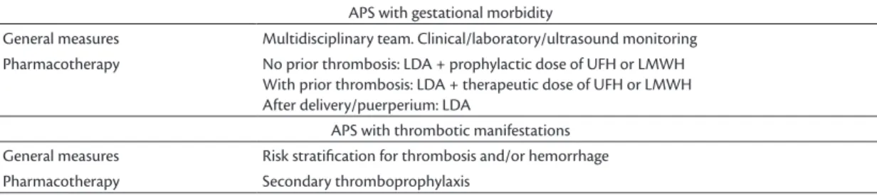
A summary of the treatment of APS according to
the type of manifestation (gestational or thrombotic)
can be seen in Table 3
. The primary and secondary
thromboprophylaxis in APS are summarized in Table 4
.
Treatment of catastrophic APS (CAPS)
The severity of CAPS demands immediate and
vigorous action. Reversible precipitating factors
(for example, infections) should be identiied and
treated. Analyses of case series and case reports
10,11,43-45indicates that survival rates are higher with combined
triple therapy with full anticoagulation with heparin
and corticoid therapy in high doses combined with
plasmapheresis and/or intravenous immunoglobulin
(IgIV), preferring the latter if there is an active
infection. If both are used (plasmapheresis and IgIV),
the plasmapheresis sequence should be concluded
before starting administration of IgIV. If SLE or
other underlying autoimmune disease are present
in addition to CAPS, additional treatment with
cyclophosphamide or another immunosuppressant
may be indicated. Use of rituximab has been suggested
for treatment of recurrent refractory CAPS, when
there is microangiopathic hemolytic anemia or
when anticoagulation is contraindicated because of
concomitant hemorrhagic complications.
11Table 3. Summary of treatment for APS.
APS with gestational morbidity
General measures Multidisciplinary team. Clinical/laboratory/ultrasound monitoring
Pharmacotherapy No prior thrombosis: LDA + prophylactic dose of UFH or LMWH
With prior thrombosis: LDA + therapeutic dose of UFH or LMWH After delivery/puerperium: LDA
APS with thrombotic manifestations
General measures Risk stratiication for thrombosis and/or hemorrhage
Perioperative management of patients with
aPL or APS
46Surgical procedures can trigger thrombotic events
47and even CAPS
11in patients with aPL or APS and
adequate preoperative planning of prophylaxis is
therefore necessary. The medical team (clinical
team, surgeon, anesthetist) should, by consensus,
deine the approach that will be taken in terms of
the timing of preoperative suspension of VKA,
using LMWH as a treatment bridge, application of
intermittent compression devices, early postoperative
mobilization, postoperative reintroduction of VKA,
and laboratory monitoring strategy.
46This approach
should take into consideration any history of thrombotic
or hemorrhagic events, whether a clinically relevant
aPL proile exists (titers > 40 GPL or MPL units,
persistence > 12 weeks, and detection of LA according to
the ISTH guidelines) and presence of other risk factors
for thrombosis or hemorrhage.
46,48Although 20 to 40%
of patients with APS have thrombocytopenia, most
often above 70,000 platelets/mm
3, this rarely results
in signiicant hemorrhage and does not reduce the
risk of thrombosis.
49In the absence of anticoagulant
treatment, an elevated APTT (above the reference
value) may indicate presence of LA and a minimally
prolonged PT (above the upper limit of normality)
may indicate presence of antiprothrombin antibodies.
More accentuated PT abnormalities (INR > 2.0)
require the investigation of other causes including
the LA-associated hypoprothrombinemia.
46,50Prevention of perioperative thrombosis in
patients with APS includes general measures,
such as (i) minimizing intravascular manipulation for
access and monitoring; (ii) reducing venous stasis,
avoiding use of tourniquets for drawing blood samples,
and reducing the frequency of inlation of cuffs to take
blood pressure; (iii) thromboprophylaxis, combining
mechanical modalities (graduated compression
stockings or intermittent pneumatic compression
devices) and pharmacological measures.
46Perioperative
pharmacological thromboprophylaxis in patients with
APS on oral anticoagulation with VKA is provided
parenterally using LMWH (enoxaparin) or UFH to
provide a therapeutic bridge, returning to VKA as
soon as possible postoperatively (UFH or LMWH
should only be withdrawn when INR is on target).
46,51Occurrence of hemorrhagic complications does not
eliminate the risk of thromboembolism, and in these
situations intensive use of mechanical methods is
recommended and, as soon as possible, resumption
of pharmacological method
s of thromboprophylaxis.
46FINAL COMMENTS
Antiphospholipid Syndrome is very important in
vascular medicine because it is a recognized cause of
recurrent venous and/or arterial thrombosis. Diagnosis
is dependent on recognition of its clinical thrombotic
and/or gestational manifestations and on requesting
and interpreting the appropriate laboratory tests for
the syndrome. The results of tests for LA can be
affected by use of parenteral or oral anticoagulant
medications and it is recommended that particular
care is taken with choosing the time of sample
collection and with interpreting the results in these
circumstances. Increasingly, additional pathogenic
APS mechanisms are being revealed, which could
facilitate development of new treatment options
that do not involve anticoagulants. An appropriate
approach to treatment of these patients should take
account of their history of thrombotic events and/or
prior gestational morbidity and other associated
cardiovascular risk factors. Although DOAs offer
several beneits over use of warfarin or other VKA
for treatment and prophylaxis of venous thrombosis in
Table 4. hromboprophylaxis in APS.
Secondary thromboprophylaxis (conirmed APS)
Venous thrombosis Anticoagulation with VKA, INR 2.0 to 3.0
Arterial thrombosis
Anticoagulation with VKA, INR > 3.0 or
Anticoagulation with VKA, INR 2.0 to 3.0 + LDA Primary thromboprophylaxis (positive for aPL, clinical criteria not present)
General hromboprophylaxis with LMWH or equivalent during high-risk
periods (surgery, immobilization, puerperium) With associated risk factors (cardiovascular risk, high-risk aPL
proile) LDA
the general population, conirmation of their eficacy
for APS is still awaiting the results of speciic trials.
Perioperative management of patients with APS
demands special precautions related to prophylaxis
against both thromboembolic manifestations and
hemorrhagic com
plications.
REFERENCES
1. Merashli M, Noureldine MHA, Uthman I, Khamashta M. Antiphospholipid syndrome: an update. Eur J Clin Invest. 2015;45(6):653-62. PMid:25851448. http://dx.doi.org/10.1111/ eci.12449.
2. Gómez-Puerta JA, Cervera R. Diagnosis and classification of the antiphospholipid syndrome. J Autoimmun. 2014;48-49:20-5. PMid:24461539. http://dx.doi.org/10.1016/j.jaut.2014.01.006.
3. Pons-Estel GJ, Cervera R. Renal involvement in antiphospholipid syndrome. Curr Rheumatol Rep. 2014;16(2):397. PMid:24357443. http://dx.doi.org/10.1007/s11926-013-0397-0.
4. Christodoulou C, Sangle S, D’Cruz DP. Vasculopathy and arterial stenotic lesions in the antiphospholipid syndrome. Rheumatology. 2007;46(6):907-10. PMid:17403711. http://dx.doi.org/10.1093/ rheumatology/kem040.
5. Belizna CC, Richard V, Primard E, et al. Early atheroma in primary and secondary antiphospholipid syndrome: an intrinsic finding. Semin Arthritis Rheum. 2008;37(6):373-80. PMid:17977581. http:// dx.doi.org/10.1016/j.semarthrit.2007.08.002.
6. Ames PRJ, Margarita A, Alves JD. Antiphospholipid antibodies and atherosclerosis: insights from systemic lupus erythematosus and primary antiphospholipid syndrome. Clin Rev Allergy Immunol. 2009;37(1):29-35. PMid:18987784. http://dx.doi.org/10.1007/ s12016-008-8099-5.
7. Denas G, Jose SP, Bracco A, Zoppellaro G, Pengo V. Antiphospholipid syndrome and the heart: a case series and literature review. Autoimmun Rev. 2015;14(3):214-22. PMid:25461836. http:// dx.doi.org/10.1016/j.autrev.2014.11.003.
8. Cervera R, Piette J-C, Font J, et al. Antiphospholipid syndrome: clinical and immunologic manifestations and patterns of disease expression in a cohort of 1,000 patients. Arthritis Rheum. 2002;46(4):1019-27. PMid:11953980. http://dx.doi.org/10.1002/ art.10187.
9. Ruiz-Irastorza G, Crowther M, Branch W, Khamashta MA. Antiphospholipid syndrome. Lancet. 2010;376(9751):1498-509. PMid:20822807. http://dx.doi.org/10.1016/S0140-6736(10)60709-X.
10. Asherson RA, Cervera R, de Groot PG, et al. Catastrophic antiphospholipid syndrome: international consensus statement on classification criteria and treatment guidelines. Lupus. 2003;12(7):530-4. PMid:12892393. http://dx.doi.org/10.1191/0961203303lu394oa.
11. Cervera R, Rodríguez-Pintó I, Colafrancesco S, et al. 14th International Congress on Antiphospholipid Antibodies Task Force Report on Catastrophic Antiphospholipid Syndrome. Autoimmun Rev. 2014;13(7):699-707. PMid:24657970. http://dx.doi.org/10.1016/j. autrev.2014.03.002.
12. Miyakis S, Lockshin MD, Atsumi T, et al. International consensus statement on an update of the classification criteria for definite antiphospholipid syndrome (APS). J Thromb Haemost. 2006;4(2):295-306. PMid:16420554. http://dx.doi.org/10.1111/j.1538-7836.2006.01753.x.
13. Meroni PL, Chighizola CB, Rovelli F, Gerosa M. Antiphospholipid syndrome in 2014: more clinical manifestations, novel pathogenic players and emerging biomarkers. Arthritis Res Ther. 2014;16(2):209. PMid:25166960. http://dx.doi.org/10.1186/ar4549.
14. Rodríguez-García V, Ioannou Y, Fernández-Nebro A, Isenberg DA, Giles IP. Examining the prevalence of non-criteria anti-phospholipid antibodies in patients with anti-phospholipid syndrome: a systematic review. Rheumatology. 2015;54(11):2042-50. PMid:26152548. http://dx.doi.org/10.1093/rheumatology/kev226.
15. Pengo V, Tripodi A, Reber G, et al. Update of the guidelines for lupus anticoagulant detection. Subcommittee on Lupus Anticoagulant/ Antiphospholipid Antibody of the Scientific and Standardisation Committee of the International Society on Thrombosis and Haemostasis. J Thromb Haemost. 2009;7(10):1737-40. PMid:19624461. http://dx.doi.org/10.1111/j.1538-7836.2009.03555.x.
16. Savino S, Breen K, Hunt BJ. Rivaroxaban use in patients with antiphospholipid syndrome and previous venous thromboembolism. Blood Coagul Fibrinolysis. 2015;26(4):476-7. PMid:25923780. http:// dx.doi.org/10.1097/MBC.0000000000000247.
17. Arachchillage DJ, Cohen H. Use of new oral anticoagulants in antiphospholipid syndrome. Curr Rheumatol Rep. 2013;15(6):331. PMid:23649961. http://dx.doi.org/10.1007/s11926-013-0331-5.
18. Van Os GMA, de Laat B, Kamphuisen PW, Meijers JCM, de Groot PG. Detection of lupus anticoagulant in the presence of rivaroxaban using Taipan snake venom time. J Thromb Haemost JTH. 2011;9(8):1657-9. PMid:21668740. http://dx.doi. org/10.1111/j.1538-7836.2011.04395.x.
19. Góralczyk T, Iwaniec T, Wypasek E, Undas A. False-positive lupus anticoagulant in patients receiving rivaroxaban: 24 h since the last dose are needed to exclude antiphospholipid syndrome. Blood Coagul Fibrinolysis. 2015;26(4):473-5. PMid:25402189. http:// dx.doi.org/10.1097/MBC.0000000000000235.
20. Chaturvedi S, Cockrell E, Espinola R, et al. Circulating microparticles in patients with antiphospholipid antibodies: characterization and associations. Thromb Res. 2015;135(1):102-8. PMid:25467081. http://dx.doi.org/10.1016/j.thromres.2014.11.011.
21. Breen KA, Sanchez K, Kirkman N, et al. Endothelial and platelet microparticles in patients with antiphospholipid antibodies. Thromb Res. 2015;135(2):368-74. PMid:25496997. http://dx.doi. org/10.1016/j.thromres.2014.11.027.
22. Giannakopoulos B, Krilis SA. The pathogenesis of the antiphospholipid syndrome. N Engl J Med. 2013;368(11):1033-44. PMid:23484830. http://dx.doi.org/10.1056/NEJMra1112830.
23. Canaud G, Bienaimé F, Tabarin F, et al. Inhibition of the mTORC pathway in the antiphospholipid syndrome. N Engl J Med. 2014;371(4):303-12. PMid:25054716. http://dx.doi.org/10.1056/ NEJMoa1312890.
24. Jesus GRR, dos Santos FC, Oliveira CS, Mendes-Silva W, Jesus NR, Levy RA. Management of obstetric antiphospholipid syndrome. Curr Rheumatol Rep. 2012;14(1):79-86. PMid:22105547. http:// dx.doi.org/10.1007/s11926-011-0218-2.
25. Galarza-Maldonado C, Kourilovitch MR, Pérez-Fernández OM, et al. Obstetric antiphospholipid syndrome. Autoimmun Rev. 2012;11(4):288-95. PMid:22001418. http://dx.doi.org/10.1016/j. autrev.2011.10.006.
27. Ruiz-Irastorza G, Cuadrado MJ, Ruiz-Arruza I, et al. Evidence-based recommendations for the prevention and long-term management of thrombosis in antiphospholipid antibody-positive patients: report of a task force at the 13th International Congress on antiphospholipid antibodies. Lupus. 2011;20(2):206-18. PMid:21303837. http:// dx.doi.org/10.1177/0961203310395803.
28. Erkan D, Harrison MJ, Levy R, et al. Aspirin for primary thrombosis prevention in the antiphospholipid syndrome: a randomized, double-blind, placebo-controlled trial in asymptomatic antiphospholipid antibody-positive individuals. Arthritis Rheum. 2007;56(7):2382-91. PMid:17599766. http://dx.doi.org/10.1002/art.22663.
29. Arnaud L, Mathian A, Ruffatti A, et al. Efficacy of aspirin for the primary prevention of thrombosis in patients with antiphospholipid antibodies: an international and collaborative meta-analysis. Autoimmun Rev. 2014;13(3):281-91. PMid:24189281. http:// dx.doi.org/10.1016/j.autrev.2013.10.014.
30. Arnaud L, Mathian A, Devilliers H, et al. Patient-level analysis of five international cohorts further confirms the efficacy of aspirin for the primary prevention of thrombosis in patients with antiphospholipid antibodies. Autoimmun Rev. 2015;14(3):192-200. PMid:25461472. http://dx.doi.org/10.1016/j.autrev.2014.10.019.
31. Rosborough TK, Jacobsen JM, Shepherd MF. Factor X and factor II activity levels do not always agree in warfarin-treated lupus anticoagulant patients. Blood Coagul Fibrinolysis. 2010;21(3):242-4. PMid:20182349. http://dx.doi.org/10.1097/MBC.0b013e32833581a3.
32. Tahir F, Riaz H, Riaz T, et al. The new oral anti-coagulants and the phase 3 clinical trials: a systematic review of the literature. Thromb J. 2013;11(1):18. PMid:24007323. http://dx.doi. org/10.1186/1477-9560-11-18.
33. Kakkos SK, Kirkilesis GI, Tsolakis IA. Editor’s Choice - efficacy and safety of the new oral anticoagulants dabigatran, rivaroxaban, apixaban, and edoxaban in the treatment and secondary prevention of venous thromboembolism: a systematic review and meta-analysis of phase III trials. Eur J Vasc Endovasc Surg. 2014;48(5):565-75. PMid:24951377. http://dx.doi.org/10.1016/j.ejvs.2014.05.001.
34. Cohen AT, Hamilton M, Bird A, et al. Comparison of the non-VKA oral anticoagulants apixaban, dabigatran, and rivaroxaban in the extended treatment and prevention of venous thromboembolism: systematic review and network meta-analysis. PLoS One. 2016;11(8):e0160064. PMid:27487187. http://dx.doi.org/10.1371/ journal.pone.0160064.
35. Andreoli L, Chighizola CB, Banzato A, Pons-Estel GJ, Jesus GR, Erkan D. Estimated frequency of antiphospholipid antibodies in patients with pregnancy morbidity, stroke, myocardial infarction, and deep vein thrombosis: a critical review of the literature. Arthritis Care Res. 2013;65(11):1869-73. PMid:23861221. http:// dx.doi.org/10.1002/acr.22066.
36. Cohen H, Doré CJ, Clawson S, et al. Rivaroxaban in antiphospholipid syndrome (RAPS) protocol: a prospective, randomized controlled phase II/III clinical trial of rivaroxaban versus warfarin in patients with thrombotic antiphospholipid syndrome, with or without SLE. Lupus. 2015;24(10):1087-94. PMid:25940537. http://dx.doi. org/10.1177/0961203315581207.
37. ClinicalTrials.gov [site na Internet]. Rivaroxaban in Thrombotic Antiphospholipid Syndrome (TRAPS). USA; 2014. [citado 2015 nov 7]. https://clinicaltrials.gov/ct2/show/record/NCT02157272
38. Noel N, Dutasta F, Costedoat-Chalumeau N, et al. Safety and efficacy of oral direct inhibitors of thrombin and factor Xa in antiphospholipid syndrome. Autoimmun Rev. 2015;14(8):680-5. PMid:25864630. http://dx.doi.org/10.1016/j.autrev.2015.03.007.
39. Schaefer JK, McBane RD, Black DF, Williams LN, Moder KG, Wysokinski WE. Failure of dabigatran and rivaroxaban to prevent thromboembolism in antiphospholipid syndrome: a case series of three patients. Thromb Haemost. 2014;112(5):947-50. PMid:25118790. http://dx.doi.org/10.1160/TH14-03-0272.
40. Win K, Rodgers GM. New oral anticoagulants may not be effective to prevent venous thromboembolism in patients with antiphospholipid syndrome. Am J Hematol. 2014;89(10):1017. PMid:25043836. http://dx.doi.org/10.1002/ajh.23797.
41. Signorelli F, Nogueira F, Domingues V, Mariz HA, Levy RA. Thrombotic events in patients with antiphospholipid syndrome treated with rivaroxaban: a series of eight cases. Clin Rheumatol. 2016;35(3):801-5. PMid:26219490.
42. Erkan D, Aguiar CL, Andrade D, et al. 14th International Congress on Antiphospholipid Antibodies: task force report on antiphospholipid syndrome treatment trends. Autoimmun Rev. 2014;13(6):685-96. PMid:24468415. http://dx.doi.org/10.1016/j.autrev.2014.01.053.
43. Rodriguez-Pintó I, Espinosa G, Cervera R. Catastrophic antiphospholipid syndrome: The current management approach. Best Pract Res Clin Rheumatol. 2016;30(2):239-49. PMid:27886797. http://dx.doi. org/10.1016/j.berh.2016.07.004.
44. Asherson RA, Cervera R, Piette JC, et al. Catastrophic antiphospholipid syndrome: clinical and laboratory features of 50 patients. Medicine. 1998;77(3):195-207. PMid:9653431. http://dx.doi. org/10.1097/00005792-199805000-00005.
45. Asherson RA, Cervera R, Piette JC, et al. Catastrophic antiphospholipid syndrome: clues to the pathogenesis from a series of 80 patients. Medicine. 2001;80(6):355-77. PMid:11704713. http://dx.doi. org/10.1097/00005792-200111000-00002.
46. Saunders KH, Erkan D, Lockshin MD. Perioperative management of antiphospholipid antibody-positive patients. Curr Rheumatol Rep. 2014;16(7):426. PMid:24828479. http://dx.doi.org/10.1007/ s11926-014-0426-7.
47. Erkan D, Yazici Y, Peterson MG, Sammaritano L, Lockshin MD. A cross-sectional study of clinical thrombotic risk factors and preventive treatments in antiphospholipid syndrome. Rheumatology. 2002;41(8):924-9. PMid:12154210. http://dx.doi.org/10.1093/ rheumatology/41.8.924.
48. Akkara Veetil BM, Bongartz T. Perioperative care for patients with rheumatic diseases. Nat Rev Rheumatol. 2011;8(1):32-41. PMid:22083219. http://dx.doi.org/10.1038/nrrheum.2011.171.
49. Arkfeld DG, Weitz IC. Immune thrombocytopenia in patients with connective tissue disorders and the antiphospholipid antibody syndrome. Hematol Oncol Clin North Am. 2009;23(6):1239-49. PMid:19932431. http://dx.doi.org/10.1016/j.hoc.2009.08.010.
50. Mazodier K, Arnaud L, Mathian A, et al. Lupus anticoagulant-hypoprothrombinemia syndrome: report of 8 cases and review of the literature. Medicine. 2012;91(5):251-60. PMid:22932789. http://dx.doi.org/10.1097/MD.0b013e31826b971f.
*
Correspondence
Andreas Funke Universidade Federal do Paraná – UFPR, Hospital de Clínicas Rua General Carneiro, 181, Uniclin, 12º andar – Alto da Glória CEP 80060-900 - Curitiba (PR), Brazil Tel.: +55 (41) 3360-1098 E-mail: [email protected]
Author information
AF - Board-certiied in Rheumatology by Universidade Federal do Paraná (UFPR), Rheumatologist and Chief of Ambulatório de SAF e Vasculites, Hospital de Clínicas, UFPR; Member of Comissão de Vasculopatias da Sociedade Brasileira de Reumatologia (SBR). AD - MSc in Internal Medicine from Universidade Federal do Rio de Janeiro (UFRJ); Rheumatologist and Chief of Clínica de Lupus, Hospital Federal dos Servidores do Estado do Rio de Janeiro; Member of Comissão de Vasculopatias da Sociedade Brasileira de Reumatologia (SBR). DCOA - Post-doctoral fellow, Universidade de Cornell/HSS; Assistant Professor, Disciplina de Reumatologia, FMUSP; Member of Comissão de Vasculopatias da Sociedade Brasileira de Reumatologia (SBR). JR - PhD in Health Sciences from Universidade Federal de Goiás (UFG); Adjunct Professor of Rheumatology at Faculdade de Medicina; Member of Comissão de Vasculopatias da Sociedade Brasileira de Reumatologia (SBR). RAL - PhD in Biological Sciences from Universidade do Estado do Rio de Janeiro (UERJ); Associate Professor at UERJ; Member of Comissão de Vasculopatias da Sociedade Brasileira de Reumatologia (SBR).
Author contributions