The expression of TGF
β
1 mRNA in the early stage
of the midpalatal suture cartilage expansion
Emilia Teruko Kobayashi*, Yasuaki Shibata**, Vanessa Cristina Veltrini***, Rosely Suguino****, Fabricio Monteiro de Castro Machado*****, Maria Gisette Arias Provenzano******, Tatiane Ferronato*******, Yuzo Kato********
Introduction: The application of orthodontic expansion force induces bone formation at the midpalatal suture because of cell proliferation and differentiation. Expansion forces may stimulate the production of osteoinductive cytokines, such as transforming growth factor β1 (TGFβ1), in the progenitor cells. Objectives: This study determined the role of TGFβ1 in the early stage of midpalatal suture cartilage expansion. Methods: A orthodontic appliance was placed between the right and left upper molars of 4-week-old rats. The initial expansion force was 50 g. Animals in the control and experimental groups were sacrified on days 0, 2, and 5 and 6 µmm thick sections were prepared for an in situ hybridization technique. Results: Two days after the application of force, prechondroblastic and undifferentiated mesenchymal cells distributed along the inner side of the cartilaginous tissue had high levels of TGFβ1 transcription. On day 5, the TGFβ1 transcription was found in osteocytes and osteoblastic cells on the surface of newly formed bone. Immunohistochemistry using Osteocalcin-Pro (OC-Pro) confirmed osteoblastic activity. Conclusions: Results suggest that the expansion of midpalatal su-ture cartilage induces differentiation of osteochondroprogenitor cells into osteoblasts after stimulation by cytokine production.
Abstract
Keywords: Transforming growth factor ß1. Proliferation. Differentiation. Osteoblasts. "In-situ" Hibridization.
* PhD in Orthodontics and Dentofacial Orthopedics and Associate Professor, Discipline of Pediatric Denistry I and II, Maringá University Center (CESUMAR). ** PhD in Pathology and Associate Professor, Division of Oral Pathology and Bone Metabolism, Nagasaki University Graduate School of Biomedical Science,
Japan.
*** PhD in Oral Pathology (FO-USP). Professor of Pathology at State University of Maringá (UEM) and Universitary Center of Maringá (CESUMAR). **** PhD Student in Orthodontics (UNESP). Associate Professor, Discipline of Pediatric Dentistry I and II, CESUMAR.
***** MSc in Orthodontics and Associate Professor, Discipline of Pediatric Dentistry I and II, CESUMAR.
****** MSc in Pedodontics and Specialist in Orthodontics and Dentofacial Orthopedics and Associate Professor, Discipline of Pediatric Dentistry I and II, State University of Maringá.
******* Specialization Student, Discipline of Orthodontics, State University of Londrina.
introduction
The midpalatal suture cartilage of growing rats is composed of layers of precartilaginous cells located in the central part of the suture, and of mature cartilaginous cells layers on both sides of the precartilaginous layers. The precar-tilaginous cells layers are filled with prechon-droblastic and undifferentiated mesenchymal cells with a high capacity to proliferate and differentiate into chondrocytes and osteoblasts.
Bone formation at the midpalatal suture cartilage initiates from the outer side of the cartilaginous tissue by means of endochondral ossification. However, when an orthodontic expansion force is applied to the suture, new bone formation is initiated on the inner side of the cartilaginous tissue by means of intramem-branous ossification.7,18 This process involves the proliferation of undifferentiated mesen-chymal cells and their differentiation into os-teoblasts.
Kobayashi et al7 described the early cell re-sponse caused by the induction of orthodontic forces, which increase the expression of prolif-erating cell nuclear antigen (PCNA), a specific cell proliferation marker, and many other pro-teins of the bone matrix in the inner side of the cartilaginous tissue. Their results showed that mechanical stress is an important mediator of proliferation and differentiation of osteochon-droprogenitor cells into osteoblasts.
However, no studies have definitively ex-plained the molecular mechanism of cell re-sponse mediated by orthodontic expansion forces that leads to proliferation and differen-tiation of the progenitor cells into osteoblasts.
Both in vivo11,12,14 and in vitro4,8,9 studies have demonstrated the participation of trans-forming growth factor β1 (TGFβ1), a cytokine that belongs to the TGFβ superfamily, in bone formation.
This study was performed using an in situ hybridization technique to evaluate the tran-scription level of TGFβ1, a cytokine with high
osteogenic capacity, after an orthodontic ex-pansion force was applied to the midpalatal suture cartilage of growing rats.
MAtEriALS And MEtHodS Expansion of the midpalatal suture
Four-week-old male Wistar rats (Charles River Corporation, Kanagawa, Japan) weigh-ing 67-83g were housed at the animal labora-tory and fed a standard pellet chow (Oriental Yeast, Tokyo, Japan) and water ad libitum. All experimental procedures were approved by the Animal Welfare Committee of Nagasaki Uni-versity, Japan.
An orthodontic expansion appliance (0.014 inch Co-Cr wire, green Elgiloy Semi-Resilient wire; Rocky Mountain Morita Corporation, Denver, CO, USA) was placed between the maxillary right and left molars, as described by Kobayashi et al.7
A strain gauge (Tomy International Co., To-kyo, Japan) was used to adjust the initial ex-pansion force to 50 g. The animals in the con-trol and experimental groups were sacrified on days 0, 2, and 5. Each group was composed of 3 animals.
tissue preparation for immunohistochemistry
The maxillary bone was surgically removed and fixed by immersion in 4% paraformal-dehyde overnight at 4°C. After fixation, the maxilla was demineralized in 10% ethylenedi-aminetetraacetic acid (EDTA) for 10 days at 4°C, and then dehydrated using an increasing ethanol series. The specimens was embedded in paraffin, cut into 6 µmm thick serial fron-tal sections at the mesial root of the maxillary first molar, and mounted on 3-aminopropyl-triethoxysilane coated slides.
tissue preparation for in situ hybridization
immunohisto-chemical staining. All solutions were free of RNase due to the addition of 0.1% diethyl py-rocarbonate (DEPC) to H2O.
Preparation of crnA digoxigenin-labeled probes for in situ hybridization
The plasmid containing TGFβ1 cDNA was transferred into Escherichia coli to amplify cDNA. TGFβ1 cDNA was cut at the BamHI/HindIII site, subcloned into Bluescript KS+ vector, and then used as a model for cRNA production. Single strand RNA antisense (complementary) and sense (non complementary) digoxigenin-labeled probes were prepared according to the instructions sup-plied with the DIG-RNA labeling kit (Boehringer Mannheim, Germany). Transcriptions were per-formed using T3 or T7 RNA polymerase. Labeling with digoxigenin was confirmed using a hybrid-ization filter. Each probe reacted only with a cor-responding RNA reverse strand.
in situ hybridization
In situ hybridization was performed accord-ing to the method described by Nakase et al.13 After blocking the alkaline phosphatase activ-ity with acid, the sections were incubated with RNA DIG-UTP (1.5 mg/ml) label probes at 55° C overnight, and then washed extensively and treated for RNase. The DIG-labeled probes were detected using an anti-DIG antibody conjugated with alkaline phosphatase and 5-bromo-4-chloro-3-indolyl phosphate as a substrate and developed using a DIG nucleic acid detection kit (Boehring-er Mannheim).
Controls were: (a) hybridization with sense (mRNA) probe; (b) hybridization with non probe.
immunohistochemical and histochemical staining
Immunohistochemistry was performed us-ing the peroxidase-anti-peroxidase method as described by Sakai et al.16 Briefly, the sections were pretreated with first antibody. Rabbit
an-tisera against rat Cathepsin K (CK)3 and rat Os-teocalcin-Pro peptide (OC-pro)2 were diluted at 1:200 and 1:100 and kept in blocking buffer overnight at 4°C.
On the following day, the sections were washed and incubated with the second antibody (goat anti-rabbit IgG).
The immunoreactivity sites were visualized us-ing peroxidase-anti-peroxidase and reacted with 3,3 diaminobenzidine to produce a brown benzi-dine staining precipitation.17 For Proliferating Cell Nuclear Antigen (PCNA) detection, specimens were kept overnight at 4oC in mouse monoclonal antibody (clone PC10, DAKO, Tokyo, Japan) at 1:50 dilution as the first antibody.
The sections were stained with streptavidin-biotin peroxidase (Histofine ABC kit-Nichirei Co. Ltd., Tokyo) according to the manufacturer’s instructions. Negative control immunoreactivity was evaluated using normal rabbit serum (1:100 dilution) or normal mouse IgG (100 mg/ml). The histochemical tests for hematoxylin and eosin were performed using the method described by Lyon.10
rESuLtS
Histological changes during
midpalatal suture cartilage expansion
On day 0, the central area of the suture carti-lage was filled with a cartilaginous cell layer com-posed of undifferentiated mesenchymal at the center, and prechondroblastic cells. Around this area, the cells exhibited features of mature chon-droblasts and/or chondrocytes (Fig 1A).
On day 2, the mature cartilaginous cell layers were displaced laterally, and the central part of the suture still had immature prechondroblastic and mesenchymal cells. In addition, a cell cluster was observed at the border of prechondroblastic and chondroblastic cells (Fig 1B).
A
A
B
B
C
C
in situ hybridization and immunohistochemistry
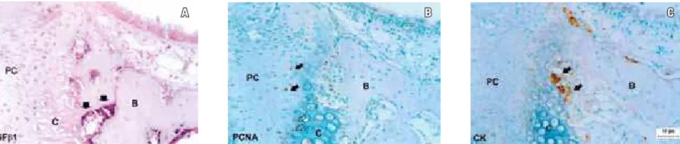
In this study, in situ hybridization tech-nique was carried out using cRNA-DIG-labeled probes to evaluate the expression of TGFβ1 mRNA localized in the midpalatal suture car-tilage. On day 0 (Fig 2A), a positive TGFβ1 mRNA (a) transcription level was detected in the mature osteoblasts located in the periphery of trabecular bone, laterally to the layer of cells compatible with chondroblasts, as shown by ar-rows in Figure 2A.
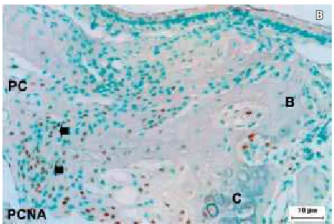
Positive PCNA immunoreactivity (Fig 2B) was found in the prechondroblastic cells in the central part of the suture and in the mature and hypertrophying cartilage cells located in the pe-riphery of the suture. Intense cathepsin (CK) (Fig 2C) immunoreactivity was observed in the outer side of the cartilaginous cell layers, in the
transition to bone tissue.
On day 2 (Fig 3A), an intense positive TGFβ1 mRNA (a) transcription was seen at the border of prechondroblastic and chondroblastic cells.
Strong PCNA (Fig 3B) immunoreactivity was seen in the same area. The positive immunoreactiv-ity pattern for CK found in the control group (day 0) was also seen in the outer side of cartilaginous tissue, which was indicative of osteoclastic activity.
During the following days (Fig 4A), an intense specific signal for TGFβ1 mRNA (a) was observed in osteocytes (open arrowheads) and osteoblasts (filled arrowheads) inside and on the surface of newly formed bone (day 5).
Osteoblastic activity was confirmed by im-munohistochemistry using Osteocalcin-Pro (OC-Pro) (Fig 4B). The pattern of osteoclastic activity (Fig 4C) was the same found on day 0 (control group).
FIGURE 1 - Sequence of histological changes in midpalatal suture after application of expansion force. Con: control; PC: precartilaginous cells; c: cartilaginous cells; B: bone; NB: newly formed bone. Bars A, B and C = 50 µm.
FIGURE 2 - Expression of TGFß1 mRNA (A), PCNA (B) and CK (C) on day 0. TGFß1: transforming growth factor ß1; PCNA: proliferating cell nuclear antigen; CK: cathepsin K. Bars A, B and C = 10 µm.
A B
A B C
⇓⇓
diScuSSion
On day 0, positive PCNA immunoreactivity was expressed in the prechondroblastic cells lo-cated in the central area of the midpalatal suture cartilage and in some mature and hypertrophy-ing cartilage cells, which was indicative of their proliferative activity. PCNA is a protein found in the cell nucleus that acts as a DNA polymerase delta cofactor during the DNA synthesis stage.1 It is used to determine the level of proliferative activity. At this stage, proliferative activity may be associated with normal cross-sectional develop-ment of the palate.7
On day 2, the expression of PCNA immu-noreactivity increased substantially in the bor-der of the prechondroblastic and chondroblas-tic layers after the orthodonchondroblas-tic expansion force was applied.
Previously to our study7, positive
immu-noreactivity for osteocalcin (OCN), a specific marker for osteoblasts, and alkaline phospha-tase (ALPase) activity were found in the same stage and area, which suggests that osteochon-droprogenitor cells differentiate into osteoblasts in response to the expansion force. Accordingly, high TGFβ1 mRNA transcription levels were expressed in the same region on day 2, as well as in mature osteoblasts on day 0.
TGFβ1 expression associated with newly formed bone has been investigated by many authors. Noda et al14 reported the occurrence of bone formation after TGFβ1 injection in the calvarium of newborn rats.
In addition, the role of TGFβ1 in osteoblastic differentiation from undifferentiated mesenchy-mal cells was been investigated by Joyce et al,5 who reported that TGFβ1 induces differentia-tion of mesenchymal-like cells into osteoblasts
FIGURE 3 - Expression of TGFβ1 mRNA (A) and PCNA (B) on day 2. Bars A and B = 10 µm.
Submitted: September 2008 Revised and accepted: April 2009 by stimulating proliferation and extracellular
matrix protein production.
TGFβ1 may mediate osteogenesis because of its chemotactic effect on the osteoblastic pre-cursor cells as it recruits those cells to the region to start the process of bone formation.15
In the late stage of the treatment (day 5), new bone formation continued and developed a columnar bone structure that grew from the center of the suture.
Positive immunoreactivity for Osteocalcin-Pro (OC-Osteocalcin-Pro), a specific osteoblastic marker, confirmed osteoblastic activity on the surface of the newly formed bone in this region.
TGF1 transcription was detected in osteo-cytes and osteoblasts on the surface of newly formed bone, which suggests that those cyto-kines participate in the regulation of the differ-entiation of mesenchymal cells into osteoblasts. At all experimental time points (day 0, 2, 5), CK immunoreactivity was expressed exclu-sively in the outer side of the cartilaginous cell layers, following the normal pathway of calci-fied cartilage matrix absorption by osteoclastic cells. This protease is involved in the degrada-tion of type I and type II collagen and osteo-nectinby osteoclasts.3
However, there was no osteoclastic activity in the inner side of the cartilaginous tissue, al-though there were blood vessels that promoted the migration of precursor osteoclasts to this re-gion. Osteoclastic activity may change due to the
high TGFβ1 expression, which can inhibit the differentiation of precursor osteoclasts and also the absorptive activity of mature osteoclasts.6
concLuSionS
The results of this study suggest that: The expansion of the midpalatal suture in-creases TGFβ1 transcription in the cells in the border of precartilaginous and cartilaginous cell layers and in osteocytes and osteoblasts on the surface of newly formed bone.
The expression of TGFβ1, osteocalcin (OCN), and alkaline phosphatase (ALPase) in the border of the precartilaginous and cartilagi-nous cell layers on day 2, was an indicative of the beginning of osteochondroprogenitor cells differentiation into osteoblasts.
New bone formation by means of intramem-branous ossification was induced in the inner side of the cartilaginous layers.
The absence of osteoclastic activity in the inner side of the expanded cartilaginous tissue may be associated with the high level of TGFβ1 transcription.
AcKnowLEdgMEnt
1. Bravo R, Frank R, Blundell PA, MacDonald-Bravo H. Cyclin/PCNA is the auxiliary protein of DNA polymerase-delta. Nature. 1987 Apr 2-8;326(6112):515-7.
2. Hashimoto F, Kobayashi Y, Kamiya T, Kobayashi K, Kato Y, Sakai H. Antigenicity of pro-osteocalcin in hard tissue: the authenticity to visualize osteocalcin-producing cells. J Bone Miner Metab. 1997 Sep;15(3):122-31.
3. Hou WS, Li Z, Gordon RE, Chan K, Klein MJ, Levy R, et
al. Cathepsin k is a critical protease in synovial
ibroblast-mediated collagen degradation. Am J Pathol. 2001 Dec;159(6):2167-77.
4. Janssens K, Ten Dijke P, Janssens S, Van HW. Transforming growth factor beta 1 to the bone. Endocr Rev. 2005 Oct;26(6):743-4.
5. Joyce ME, Roberts AB, Spom MB, Bolander ME. Transforming growth factor-beta and the initiation of chondrogenesis and osteogenesis in the rat femur. J Cell Biol. 1990 Jun;110(6):2195-207.
6. Karst M, Gorny G, Galvin RJ, Oursler MJ. Roles of stromal cell RANKL, OPG, and M-CSF expression in biphasic TGF-beta regulation of osteoclast differentiation. J Cell Physiol. 2004 Jul;200(1):99-106.
7. Kobayashi ET, Hashimoto F, Kobayashi Y, Sakai E, Miyazaki Y, Kamiya T, et al. Force-induced rapid changes in cell fate at midpalatal suture cartilage of growing rats. J Dent Res. 1999 Sep;78(9):1495-504.
8. Lee JY, Kim KH, Shin SY, Rhyu IC, Lee YM, Park YJ, et al. Enhanced bone formation by TGF1 releasing collagen/ chitosan microgranules. J Biomed Mater Res A. 2006 Mar 1;76(3):530-9.
9. Lieb E, Vogel T, Milz S, Dauner M, Schulz MB. Effects of Transforming Growth Factor 1 on bone-like tissue formation in three-dimensional cell culture II: osteoblastic differentiation. Tissue Eng. 2004 Sep-Oct;10(9-10):1414-25.
rEfErEncES
10. Lyon H. Hematoxylin-eosin: an example of a common histological staining method. In: Celis JE. Cell biology: a laboratory handbook. 2nd ed. San Diego: Academic Press; 1998. p. 232-7.
11. Mackie EJ, Trechsel U. Stimulation of bone formation in vivo by transforming growth factor: remodeling of woven bone and lack of inhibition by indomethacin. Bone. 1990;11(4):295-300. 12. Marcelli C, Yates AJ, Mundy GR. In vivo effects of human
recombinant transforming growth factor on bone turnover in normal mice. J Bone Miner Res. 1990 Oct;5(10):1087-96. 13. Nakase T, Takaoka K, Hirakawa K, Hirota S, Takemura T,
Onoue H, et al. Alterations in the expression of osteonectin, osteopontin and osteocalcin mRNAs during the development of skeletal tissues in vivo. Bone Miner. 1994 Aug;26(2):109-22. 14. Noda M, Camilliere JJ. In vivo stimulation of bone formation
by transforming growth factor-beta. Endocrinology. 1989 Jun;124(6):2991-4.
15. Pfeilschifter J, Wolf O, Naumann A, Minne HW, Mundy GR, Zielgler R. Chemotactic response of osteoblastic-like cells to transforming growth factor beta. J Bone Miner Res. 1990 Aug. 5(8):825-30.
16. Sakai H, Saku T, Kato Y, Yamamoto K. Quantitation and immunohistochemical localization of cathepsins E and D in rat tissues and blood cells. Biochim Biophys Acta. 1989 May 31;991(2):367-75.
17. Sternberger LA, Hardy PH Jr, Cuculis JJ, Meyer HG. The unlabeled antibody enzyme method of immunohistochemistry. Preparation and properties of soluble antigen-antibody
complex (horseradish peroxidase) and its use in identiication
of spirochetes. J Histochem Cytochem. 1970 May;18(5):315-33. 18. Takahashi I, Mizoguchi I, Nakamura M, Sasano Y, Saitoh
S, Kagayama M, et al. Effects of expansive force on the differentiation of midpalatal suture cartilage in rats. Bone. 1996 Apr;18(4):341-8.
contact address
Emilia Teruko Kobayashi