Immunohistochemical analysis of the neural structures
of the posterior cruciate ligament in osteoarthritis
patients submitted to total knee arthroplasty: an
analysis of thirty-four cases
Glaucus Cajaty Martins,I,IIGilberto Camanho,IMara Ibis RodriguesIII
IFaculdade de Medicina da Universidade de Sa˜o Paulo, Instituto de Ortopedia (IOT/FMUSP), Department of Orthopedics and Traumatology, Sa˜o Paulo/SP, Brazil.IIHospital Federal de Ipanema, Department of Orthopedics and Traumatology, Rio de Janeiro/RJ, Brazil.IIIFaculdade de Medicina da Universidade do Estado do Rio de Janeiro (UERJ), Department of Histology, Rio de Janeiro/RJ, Brazil.
OBJECTIVES:Many authors recommend posterior cruciate ligament-retaining arthroplasty with the intention to maintain the proprioception properties of this ligament. Preservation of the neuroreceptors and nervous fibers may be essential for retaining the proprioception function of the posterior cruciate ligament. The present study was thus developed to evaluate the presence of neural structures in the posterior cruciate ligament resected during posterior stabilized arthroplasty in osteoarthritis patients. In particular, clinical, radiographic and histological parameters were correlated with the presence or absence of neural structures in the posterior cruciate ligament.
METHODS:In total, 34 posterior cruciate ligament specimens were stained with hematoxylin-eosin and Gomori trichrome. An immunohistochemical analysis using antibodies against the S100 protein and neurofilaments was also performed. The presence of neural structures was correlated with parameters such as tibiofemoral angulation, histological degeneration of the posterior cruciate ligament, Ahlba¨ck radiological classification, age, gender and the histologic pattern of the synovial neurovascular bundle around the posterior cruciate ligament.
RESULTS:In total, 67.5% of the cases presented neural structures in the posterior cruciate ligament. In 65% of the cases, the neurovascular bundle was degenerated. Nervous structures were more commonly detected in varus knees than in valgus knees (77% versus 50%). Additionally, severe histologic degeneration of the
posterior cruciate ligament was related to neurovascular bundle degeneration.
CONCLUSIONS:Severe posterior cruciate ligament degeneration was related to neurovascular bundle compromise. Neural structures were more commonly detected in varus knees. Intrinsic neural structures were detected in the majority of the posterior cruciate ligaments of patients submitted to knee arthroplasty for osteoarthritis.
KEYWORDS: Posterior Cruciate Ligament; Total Knee Arthroplasty; Osteoarthritis; Neuroreceptors;
Immunohistochemical Analysis.
Martins GC, Camanho GL, Rodrigues MI. Immunohistochemical analysis of the neural structures of the posterior cruciate ligament in osteoarthritis patients submitted to total knee arthroplasty: an analysis of thirty-four cases. Clinics. 2015;70(2):81-86.
Received for publication onSeptember 12, 2014;First review completed onOctober 14, 2014;Accepted for publication onNovember 19, 2014 E-mail: [email protected]
& INTRODUCTION
Total knee arthroplasty has been demonstrated to be extremely efficient in the treatment of advanced osteoar-throsis of this joint (1).
Currently, there are two main arthroplasty techniques, which involve either preservation or replacement of the posterior cruciate ligament (PCL). Both techniques have yielded consistent functional results (1).
The choice of the most suitable implant has generated controversy. Advocates of the use of a prosthesis to preserve the PCL allege that this would ensure an increased range of motion, better stair-climbing performance and enhanced proprioception (2,3).
An innovative study performed by Kennedy et al. (4) in 1982 revealed the presence of nerve fibers and mechan-oreceptors in anterior and posterior cruciate ligaments, menisci and capsular structures, indicating that these structures may be related to proprioception. These authors
Copyrightß2015CLINICS– This is an Open Access article distributed under
the terms of the Creative Commons Attribution Non-Commercial License (http:// creativecommons.org/licenses/by-nc/3.0/) which permits unrestricted non-commercial use, distribution, and reproduction in any medium, provided the original work is properly cited.
No potential conflict of interest was reported.
also described a nerve bundle in the synovium surrounding the PCL that is supposedly continuous with the tibial nerve and that carries nerve impulses.
In subsequent studies, Franchi et al. (5) and Del Valle et al. (6) recognized Ruffini and Pacini receptors, Golgi endings and nerve fiber terminals in the PCL of osteoarthritis patients submitted to prosthetic knee replacement.
To achieve adequate biomechanical performance, a knee prosthesis needs PCL preservation to ensure sufficient conservation of the anatomical and functional structures (7,8). Similarly, it is necessary for the nerve endings and neuroreceptors to remain present and functional to ensure that the proprioceptive properties of the retained PCL are maintained (5).
An analysis of PCL innervation in patients with advanced arthrosis is likely to be important in clinical practice, as the presence of neuroreceptors remaining in the ligament would be a sign that this ligament should be preserved for proprioception purposes (6). Along the same line of reasoning, if we can establish a subgroup of patients with particular clinical and/or radiological parameters in whom PCL innervation is seriously impaired, a posterior-stabilized prosthesis, which would require the ligament to be sacrificed, could be considered. These data may provide additional evidence that may be used to determine the most suitable type of implant for a particular patient.
& MATERIALS AND METHODS
A cross-sectional study in which 31 patients (34 liga-ments) were evaluated was conducted. Of these patients, 22 were female and 9 were male. The patients’ age ranged from 53 to 87 years and they were submitted to arthroplasty with PCL replacement at our service between June 2009 and June 2011.
This research complied with the Declaration of Helsinki and was approved by the Ethics Committee of Universidade de Sa˜o Paulo (USP).
Only cases of primary osteoarthritis were included in this series; patients with rheumatic diseases and patients who underwent osteosynthesis surgery, osteotomy or previous arthrotomy at the knee level and posttraumatic sequelae were excluded.
The patients who participated in this survey signed an informed consent form declaring their awareness of the methodology and objectives of this study.
The PCL was sectioned at its femoral origin during surgery and its tibial bone insertion was maintained. This ligament was removed after tibial sectioning was per-formed.
The PCL was preserved in 10% formaldehyde and was subsequently placed in a paraffin mold such that the ligament fibers were oriented along the longitudinal axis. The paraffin blocks were then sectioned and stained with hematoxylin-eosin.
For Gomori trichrome staining, the paraffin mold was soaked in this reagent. Gomori trichrome staining is an excellent technique for ligament analysis, as this reagent stains whole or new collagen fibers green, aged collagen fibers red and cell nuclei purplish-blue (hematoxylin) (9).
For immunohistochemical analysis, paraffin sections measuring approximately 10mm were processed in
strepta-vidin-biotin-peroxidase and incubated with anti-S100 pro-tein antibodies. In the axonal neurofilament experiments,
the process was similar; however, the processed mold was instead placed in contact with anti-neurofilament antibo-dies.
The S100 protein is found in the Schwann cells and myelinated fibers present in mechanoreceptors. The anti-neurofilament antibody labels the cytoskeleton (axon).
Histological degeneration of the collagen fibers of the ligament was analyzed using optical microscopy (OM) and was classified as follows (7):
a) Mild: defined by impairment of fewer than 20% of the collagen fibers based on an examination of 10 fields at 1006 magnification. The predominant
findings were disorganized collagen bundles in certain areas (fields of the slide) and an increase in slack connective tissue between the bundles. b) Moderate: defined by impairment of at least 20% but
less than 50% of the collagen fibers. Based on microscopic examination, the predominant altera-tions were an inflammatory reaction; hypercellular-ity; increased vascularization and disorganized, frayed or dense collagen bundles in several places. c) Severe: defined by the involvement of more than 50%
of the collagen fibers in the bundles based on the evaluation of 10 fields at 1006magnification. The
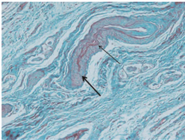
predominant alterations observed by OM were the presence of cysts, collagen degeneration (fibrinoid/ mucoid/myxoid) and intratendon fatty infiltration, with the possibility of total loss of the structure and integrity of the collagen fiber bundles (coalescent fields) (Figure 1).
The neural immunomarking of specific corpuscles and of neurofilaments was qualitatively assessed and classified as the presence or absence of marking.
The mechanoreceptors were classified into four types according to the classification established by Freeman and Wyke (10) (Figure 2):
Type I (Ruffini): encapsulated ovoid or globular corpus-cles with slow adaptation function.
Type II (Pacini): elongated conical corpuscles with fast adaptation function.
Type III (Golgi): spindle-shaped corpuscles with fast adaptation function.
Figure 1 -Gomori trichrome stain (406magnification). Severe
Type IV: non-corpuscular endings that can be free (pain) or efferent and amyelinic (vasomotricity).
The Ahlba¨ck classification was used to classify the radiological pattern of arthrosis into five types (11).
Measurement of the tibiofemoral angulation involved knee radiographs taken with orthostatic support encom-passing the distal femoral and proximal tibial diaphysis. A goniometer was used to measure the tibiofemoral angle, which consisted of the intersection of the femoral and tibial anatomical axes. The following angles were considered: varus (less than or equal to 4
˚
of the tibiofemoral valgus), neutral (from 5˚
to 9˚
of the valgus) and valgus (above 10˚
). The radiological parameters (the Ahlba¨ck classification and the measurement of the tibiofemoral axis) were evaluated together by two specialist surgeons with more than 10 years of experience in arthroplasty surgery. In case of discordance, a third colleague with the same training assisted in determining the end result.The patients were divided into two age groups: 70 years and over and under 70 years.
Based on macroscopic analysis at the time of surgery, the anterior cruciate ligament (ACL) was considered either present or absent. No attempt was made to classify the ACL as normal or undergoing macroscopic degeneration, as this would generate a bias in the analysis due to extreme subjectivity.
The neurovascular bundle (NVB) was classified as preserved, degenerated or absent. In the degenerated cases, the following characteristics were observed by microscopy: a thickened tunica intima in the vessels, degeneration in the elastic laminae of the vascular wall and nerve atrophy demonstrated by perineural retraction (Figure 3). In the cases classified as absent, the NVB was not visualized. This absence of visualization can be interpreted in two ways: either the NVB was not included in the histological section, or it was not present. The four absent cases were excluded from the statistical analysis due to these two distinct possibilities.
The presence of neural structures in the PCL was evaluated and correlated with the tibiofemoral angulation (varus-valgus), the degree of histological degeneration of the collagen fibers, the macroscopic state of the ACL, Ahlba¨ck’s radiological classification, age, sex and the histological state of the NVB contained in the synovium that surrounds the PCL. The histological pattern of the
vascular bundle (preserved or degenerated) was also correlated with previously described parameters.
Statistical analysis
The chi-square test was used for analysis of parametric data and the Kruskal-Wallis test and Mann-Whitney test were used for analysis of non-parametric data. Values of p,0.05 were considered significant. Fisher’s exact test was used for analysis of categorical data.
& RESULTS
Regarding the grade of histological degeneration, there were 23 severe cases (67.5%), 9 moderate cases (26.5%) and 2 mild cases (6%).
According to Ahlba¨ck’s radiological classification of arthrosis, there were seven grade I (20.5%), five grade II (14.5%), 14 grade III (41%), six grade IV (18%) and two grade V (6%) cases.
There were six cases of genu valgum (17.5%), 2 neutral cases (6%) and 26 cases of genu varum (76.5%).
The ACL was present in 64.7% of cases (22 cases) and absent in 35.3% of cases (12 cases).
The NVB contained in the synovium of the PCL was identified in 30 cases (88.5%), whereas it appeared degen-erated in 22 cases (65%) (Figure 3) and preserved in eight cases (23.5%). Additionally, in 4 cases, the NVB could not be identified in the histological sections (11.5%).
Immunomarking for neural structures was positive in 23 of the cases (67.5%). Specific mechanoreceptors were identified in 10 ligaments, with 9 type II cases (Pacini) and six type IV cases predominating. Moreover, neurofilaments were identified in 14 ligaments.
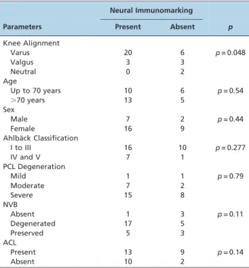
Neural immunomarking was more frequent in the genu varum (77%) than in the genu valgum (50%); this difference was statistically significant, as demonstrated by the chi-square test (p= 0.048) (Table 1).
Immunomarking positivity was observed even at increas-ing levels of the Ahlba¨ck classification (p= 0.277).
A significant correlation was demonstrated between severe histological degeneration of the PCL and degenera-tion of the NVB (Mann-Whitney test, p= 0.015) (Table 2). Considering the 30 cases in which the NVB was identified, in the event of severe degeneration of the PCL, the NVB was
Figure 2 - Immunomarking for S100 (2006 magnification).
Presence of a type II mechanoreceptor (Pacinian corpuscle) in the upper left field (red arrow) and a type IV mechanoreceptor (perivascular terminations) in the center of the image (black arrow).
Figure 3 - Gomori trichrome stain (406 magnification).
degenerated 85.7% of the time (18/21 cases). In the cases with mild or moderate histological impairment of the PCL, the NVB was degenerated 44% of the time (4/9 cases).
There was no significant relationship between the immunomarking of the neural structures or the pattern of degeneration of the NVB and the other parameters studied (Tables 1 and 2).
& DISCUSSION
The functional integrity of the PCL is essential for the functionality of the knee prosthesis in which this ligament is preserved. The preservation of this structure, at least in theory, could benefit proprioception (6).
In 1982, Kennedy et al. (4) performed experiments that were based on a study by Gardiner (12) and demonstrated that the tibial nerve is the source of the nerve fibers that innervate the synovium that surrounds the cruciate liga-ments. Using the silver nitrate impregnation technique, these authors detected the presence of superficial axons and Golgi receptors in the PCL synovium.
In 1984, Schultz et al. (13) used a similar technique and also described the presence of several unmyelinated axons in the peripheral fat (synovium) that coats the cruciate ligaments, in addition to significant degeneration of the collagen fibers of these ligaments. In 22 cruciate ligaments, they observed only 1 mechanoreceptor.
Studies conducted by Franchi et al. (5) and Dell Valle et al. (6) revealed that neural structures and mechanoreceptors were present in the PCL of patients with arthrosis submitted to knee arthroplasty, even in the presence of degeneration of the ligament’s collagen fibers. In fact, Franchi et al. (5) observed a decrease in the number of mechanoreceptors in the arthritic knee. Both groups of investigators, however, failed to determine either the histological grade of PCL
degeneration or the radiological grade of the associated arthrosis.
Proprioception is the ability to detect movement and positioning in the space of a particular joint and is performed by neural receptors located within the joint capsule in the ligaments, muscles and skin (14).
In studies conducted by Barret et al. (2) and Skinner (15), the authors found that proprioception diminishes with age and that proprioception in cases of knee osteoarthritis differs from that in control individuals of the same age. Disorganization of the collagen fibers of the PCL suppo-sedly leads to greater lassitude of this ligament. This process could raise the threshold of response in the nerve fibers and mechanoreceptors located along the axis of the pathologi-cally slackened ligament. Moreover, the function of the afferent neural structures present in the PCL could be impaired, either due to a decrease in their number or due to their degeneration.
Several studies (15-17) have not described proprioception differences between patients who received a PCL replace-ment prosthesis and those with preservation of this ligament.
Regarding mechanoreceptors, Pacinian corpuscles pre-dominated in the present study, in accordance with studies by Franchi et al. (5) and Katonis et al. (18), whereas no Golgi receptors were found. These results are consistent with the work of Dell Valle et al. (6), who did not observe this type of mechanoreceptor in humans.
In our study, in most cases (67.5%), neural structures were present in the PCL, even in the presence of severe degeneration. These data are in accordance with the work of Nelissen and Hogendoorn (8), who examined a series of 11 knee arthrosis patients and observed eight cases of severe degeneration of the PCL. In seven of these cases, neural structures were present in the ligament.
Table 1 -Correlation between the studied parameters and the immunomarking of the neural structures of the PCL.
Neural Immunomarking
Parameters Present Absent p
Knee Alignment
Varus 20 6 p= 0.048
Valgus 3 3
Neutral 0 2
Age
Up to 70 years 10 6 p= 0.54
.70 years 13 5
Sex
Male 7 2 p= 0.44
Female 16 9
Ahlba¨ck Classification
I to III 16 10 p= 0.277
IV and V 7 1
PCL Degeneration
Mild 1 1 p= 0.79
Moderate 7 2
Severe 15 8
NVB
Absent 1 3 p= 0.11
Degenerated 17 5
Preserved 5 3
ACL
Present 13 9 p= 0.14
Absent 10 2
Table 2 -Correlation between the studied parameters and the state of conservation of the PCL’s subsynovial NVB.
NVB
Parameters Preserved Degenerated p
Knee Alignment
Varus 6 18 p= 0.68
Valgus 2 4
Neutral 0 0
Age
Up to 70 years 2 10 p= 0.31
.70 years 6 12
Sex
Male 1 8 p= 0.20
Female 7 14
Ahlba¨ck Classification
I to III 7 15 p= 0.391
IV and IV 1 7
PCL Degeneration
Mild 1 0 p= 0.015
Moderate 4 4
Severe 3 18
ACL
Present 5 13 p= 0.86
In the current study, mechanoreceptors and neurofila-ments were mostly present in the individuals with advanced grades of arthrosis, as determined using Ahlba¨ck’s radiological classification (IV and V). In contrast, in the study by Nelissen and Hogendoorn (8), in which only Ahlba¨ck type IV and V cases were considered, neural structures were identified in all but one case.
Positive immunomarking was significantly more likely to be observed in cases of genu varum (77%) than in cases of genu valgum (50%) in the present study. Although statistical significance could not be determined due to the small size of the study group, all six cases of genu valgum presented severe histological degeneration of the PCL. Such findings support the practice of sacrificing the PCL in cases of genu valgum, which has been reported by certain authors (19). Cases of genu valgum are usually more technically demanding than those of genu varum (19). In particular, ligament release is more complex and sacrificing the PCL facilitates better ligament balance in the knee. Moreover, if the proprioceptive capacity of this ligament is more severely impaired in cases of valgus, a posterior-stabilized prosthesis in which the PCL ligament is discarded would be more suitable based on this hypothesis.
In our study, 30/34 of PCL specimens (88%), it was possible to identify the subsynovial NVB described pre-viously by Kennedy et al. (4) and Schultz et al. (13). In 22 cases (64.7%), degeneration of this bundle could be observed by microscopy. Our data are consistent with those of Stubbs et al. (20), who examined 50 PCLs from arthrosis patients submitted to arthroplasty and found arteriosclero-sis and perineural fibroarteriosclero-sis in 78% and 50% of the ligaments studied, respectively, after histological analysis.
Degenerative alterations of the NVB could lead to impaired function and decreased transmission of impulses captured in the knee. This degeneration of the subsynovial NVB should be studied in more depth, as it could be, at least in theory, one of the factors responsible for the propriocep-tion deficit of arthritic patients.
A significant statistical correlation (p= 0.015) was identi-fied between the grade of PCL degeneration and the state of the NVB in the current study. The cases of severe histological degeneration of the PCL were related to degenerated NVBs 80% of the time and to impaired bundles 44% of the time in cases of mild or moderate degeneration. These data are important because if we can identify patients with severe PCL degeneration, aside from altered biome-chanical properties, this may be an indicator of deficient proprioception in the ligament. Under this potential set of circumstances, PCL replacement arthroplasty would, in theory, be an indication to consider.
The good results obtained with prostheses with preserva-tion of the PCL, despite advanced degenerapreserva-tion in many cases, could be due to the design of the implants; the ligament plays a secondary role when applying an axial load to the prosthesis (20). In addition, after arthroplasty, the collateral ligaments and the capsule are responsible for knee proprioception, which helps to explain the similar results obtained using prostheses with and without sacrifice of the PCL (20).
This study has certain limitations. The first is that the immunomarking analysis was merely qualitative; thus, the density of the ligament innervation was not quantified. Another weak point is the use of Ahlba¨ck’s radiological classification, which has limited reproducibility (21); however,
it is the most widely used classification system among orthopedic surgeons. The method by which the grade of histological PCL degeneration was evaluated was also subjective, although this method was based on previous studies (7,20,22) and conducted by a histologist with extensive experience in scientific studies. One strong point of the study is that it was performed in patients submitted to surgery in whom neural structures were identified using one of the most widely accepted current techniques: immunomarking for S100 and neurofilaments. Furthermore, we demonstrated the relationship between severe degeneration of the PCL and impairment of the NVB. We also established that neural structures are more commonly present in cases of varus deformity than in cases of valgus deformity. These original results pave the way for the development of new questions. Additionally, the results should inspire surveys geared toward the identification of epidemiological, clinical and radiological parameters that may be related to the histological and functional states of the PCL and its applications in surgical practice.
The topic of articular mechanoreceptors is far from being merely academic, as it has served as one of the foundations of prestigious authors (1,6) recommendation of PCL preserva-tion in knee arthroplasty. Addipreserva-tionally, articular mechanor-eceptors have been the subject of recent surveys on other joints, such as the shoulder (15) and intervertebral disc (23).
& CONCLUSION
1) Intrinsic neural structures were detected in the majority of the PCLs of patients submitted to knee arthroplasty for osteoarthritis, even in the presence of severe structural degeneration of the ligament’s collagen fibers. 2) There was an association between severe PCL degeneration and NVB compromise. 3) Neural structures were more frequently observed in varus knees than in valgus knees.
& AUTHOR CONTRIBUTIONS
Martins GC conceived the study, harvested the ligaments, the data and wrote the manuscript. Camanho GL analyzed the data and revised the manuscript. Rodrigues MI performed the histological analysis.
& REFERENCES
1. Jacobs WC, Clement DJ, Wymenga AB. Retention versus removal of the posterior cruciate ligament in total knee replacement: a systematic literature review within the Cochrane framework. Acta Orthop. 2005;76(6):757-68, http://dx.doi.org/10.1080/17453670510045345. 2. Barrett DS, Cobb AG, Bentley G. Joint proprioception in normal,
osteoarthritic and replaced knees. J Bone Joint Surg Br. 1991;73(1):53-6. 3. Andriacchi TP, Galante JO. Retention of the posterior cruciate in total
knee arthroplasty. J Arthroplasty. 1988;3[Suppl]:S13-9.
4. Kennedy JC, Alexander IJ, Hayes KC. Nerve supply of the human knee and its functional importance. Am J Sports Med. 1982;10(6):329-35, http://dx.doi.org/10.1177/036354658201000601.
5. Franchi A, Zaccherotti G, Aglietti P. Neural system of the human posterior cruciate ligament in osteoarthritis. J Arthroplasty. 1995;10(5): 679-82, http://dx.doi.org/10.1016/S0883-5403(05)80215-3.
6. Del Valle ME, Harwin SF, Maestro A, Murcia A, Vega JA. Immunohisto-chemical analysis of mechanoreceptors in the human posterior cruciate ligament: a demonstration of its proprioceptive role and clinical relevance. J Arthroplasty. 1998;13(8):916-22, http://dx.doi.org/10.1016/S0883-5403 (98)90199-1.
7. Kleinbart FA, Bryk E, Evangelista J, Scott WN, Vigorita VJ. Histologic comparison of posterior cruciate ligaments from arthritic and age-matched knee specimens. J Arthroplasty. 1996;11(6):726-31, http://dx. doi.org/10.1016/S0883-5403(96)80012-X.
9. Prophet E, Mills B, Arrington JB, Sobin L. Laboratory methods in histotechnology.Washington: Armed Forces Institute of Pathology; 1994. 10. Freeman MA, Wyke B. The innervation of the knee joint. An anatomical
and histological study in the cat. J Anat. 1967;101(Pt 3):505-32. 11. Ahlba¨ck S. Osteoarthrosis of the knee. A radiographic investigation. Acta
Radiol Diagn (Stockh). 1968:Suppl 277:7-72.
12. Gardner E. The innervation of the knee joint. Anat Rec. 1948;101(1):109-30, http://dx.doi.org/10.1002/ar.1091010111.
13. Schultz RA, Miller DC, Kerr CS, Micheli L. Mechanoreceptors in human cruciate ligaments. A histological study. J Bone Joint Surg Am. 1984;66 (7):1072-6.
14. Ejnisman B, Faloppa F, Carrera EF, Andreoli CV, Alves MTS, Odashiro A, et al. Estudo imunohistoquı´mico dos mecanorrecptores do ligamento glenoumeral inferior em cada´veres humanos. Rev Bras Ortop. 2002; 37(7):289-98.
15. Skinner HB. Pathokinesiology and total joint arthroplasty. Clin Orthop Relat Res. 1993;(288):78-86.
16. Lattanzio PJ, Chess DG, Mac Dermid JC. Effect of the posterior cruciate ligament in knee-joint proprioception in total knee arthroplasty. J Arthroplasty. 1998;13(5):580-5, http://dx.doi.org/10.1016/S0883-5403 (98)90059-6.
17. Jacobs WC, Clement DJ, Wymenga AB. Retention versus removal of the posterior cruciate ligament in total knee replacement: a systematic
literature review within the Cochrane framework. Acta Orthop. 2005; 76(6):757-68, http://dx.doi.org/10.1080/17453670510045345.
18. Katonis P, Papoutsidakis A, Aligizakis A, Tzanakakis G, Kontakis GM, Papagelopoulos PJ. Mechanoreceptors of the posterior cruciate ligament. J Int Med Res. 2008;36(3):387-93.
19. Sah AP, Scott RD. How to balance the posterior cruciate ligament in a cruciate retaining total knee arthroplasty. Tech Knee Surg 2010;9(1):43, http://dx.doi.org/10.1097/BTK.0b013e3181d16672.
20. Stubbs G, Dahlstrom J, Papantoniou P, Cherian M. Correlation between macroscopic changes of arthrosis and the posterior cruciate ligament histology in the osteoarthritic knee. ANZ J Surg. 2005;75(12):1036-40.
21. Galli M, De Santis V, Tafuro L. Reliability of the Ahlba¨ck classification of knee osteoarthritis. Osteoarthritis Cartilage. 2003;11(8):580-4, http://dx. doi.org/10.1016/S1063-4584(03)00095-5.
22. Allain J, Goutallier D, Voisin MC. Macroscopic and histological assess-ments of the cruciate ligaassess-ments in arthrosis of the knee. Acta Orthop Scand. 2001;72(3):266-9, http://dx.doi.org/10.1080/00016470152846592. 23. Oliveira VM, Puertas EB, Alves MTS, Yamashita HK. Estudo