www.rbo.org.br/
issn/$–see front matter © 2013 Sociedade Brasileira de Ortopedia e Traumatologia. Published by Elsevier Editora Ltda. All rights reserved. doi: 10.1016/j.rboe.2012.04.004
*Corresponding author: R. Caracas, 418, CEP: 18046-718, Sorocaba, SP, Brazil. Fax: (+55 15) 3233-4171
E-mail: [email protected] A RT I C L E I N F O
Article history:
Received on January 15, 2012 Approved on April 12, 2012
Keywords: Femur
Posterior Cruciate Ligament Orthopaedic Procedures
a b s t r a c t
Purpose: To provide an anatomical and morphometric basis for the femoral insertions of the posterior cruciate ligament (PCL) in order to aid in the creation of anatomical femoral tunnels in ligament surgical reconstruction. Study design: laboratory controlled study. Material and methods: The macroscopic details of the femoral insertions of the PCL’s anterolateral (AL) and posteromedial (PM) bundles were analyzed in 24 cadaver knees. The specimens were photographed with a digital camera and the images obtained were studied using the software ImageJ. The bundles’ insertion areas were measured in square millimeters, and the length of the structures and the distances between significant points were measured in millimeters. Results: The PCL’s femoral insertion average total area was 87.29 ± 31.42 mm². The mean insertion’s areas of the AL and PM bundles were, respectively, 47.13 ± 19.14 and 40.67 ± 16.19 mm². In 95.8% of the examined knees was verified the presence of the medial intercondylar ridge and in 83.3% of the knees was noted the medial bifurcated ridge. The average length of the medial intercondylar ridge was 20.54 ± 2.26 mm and the medial bifurcated ridge’s average length was 7.62 ± 2.35 mm. Conclusions: The AL had a femoral insertion area larger than the PM bundle; these bundles’ insertion areas were lower than those previously described in the literature. There were important individual variations related to the area of the bundles in the samples, suggesting that there should be an individual recommendation for anatomical reconstructions of the PCL with single or double femoral tunnels.
© 2013 Sociedade Brasileira de Ortopedia e Traumatologia. Published by Elsevier Editora Ltda. All rights reserved.
Original Article
Anatomical study and morphometric analyses on the femoral
insertions of the posterior cruciate ligament
Julio Cesar Gali,
1,*Heetor Campora de Sousa Oliveira,
2Adriano Bordini Camargo,
3Carlos Rodrigo Barbosa Martins,
3Phelipe Augusto Cintra da Silva,
3Edie Benedito Caetano
4 1PhD in Orthopedics and Traumatology from the School of Medicine, Universidade de São Paulo (USP); Voluntary Attending Physician in theOrthopedics and Traumatology Service, Faculdade de Ciências Médicas e da Saúde (FCMS) de Sorocaba, Pontifícia Universidade Católica de São Paulo (PUC-SP), Sorocaba, SP, Brazil.
2Resident Physician in the Orthopedics and Traumatology Service, FCMS de Sorocaba, PUC-SP (2011), Sorocaba, SP, Brazil.
3Third-year Resident Physician in the Orthopedics and Traumatology Service, FCMS de Sorocaba, PUC-SP, Sorocaba, SP, Brazil. 4Full Professor and Head of the Orthopedics and Traumatology Service, FCMS de Sorocaba, PUC-SP, Sorocaba, SP, Brazil.
Introduction
The posterior cruciate ligament (PCL) originates in the lateral face of the medial femoral condyle (MFC) and heads posteriorly, laterally and distally to become attached in the posterior intercondylar area of the tibia, extending for a few millimeters on the adjacent posterior tibial surface.1,2
It is composed of two functional bundles: the anterolateral (AL) and the posteromedial (PM). The AL bundle becomes tense at close to 90° of flexion, while the PM becomes tense at full extension.2 It is considered to be the main primary restrictor of
posterior translation of the knee and the secondary restrictor of varus, valgus and external rotation movements of this joint.3
Lesions of this ligament can occur separately, but a growing number of them have been recognized as forming part of combined lesions.4
Regarding the natural history of posterior cruciate injuries, Dejour et al.5 followed up 45 patients, for periods ranging from
four to 44 years. They concluded that kinematic disorders of the knee resulting from loss of the PCL led to femorotibial or generalized osteoarthrosis 25 years after the injury, on average.
Torg et al.6 evaluated 47 patients over follow-ups of one
to 37 years and inferred that PCL injuries in association with other forms of instability presented higher incidence of degenerative alterations on radiographs and patellofemoral diseases on clinical examination.
Surgical treatment for these injuries is frequently recommended for active patients, in order to achieve joint stability and prevent secondary arthrosis. Success in the surgical reconstructions depends on accurate reproduction of the anatomy of the PCL.7
The aim of this study was to describe the format, location and measurements of the femoral insertions of the PCL in order to assist surgeons in performing anatomical reconstruction of this ligament.
Materials and methods
This anatomical and morphometric study on the femoral insertions of the PCL was conducted on 24 anatomical knee specimens (12 right and 12 left knees that did not form pairs) from adult individuals of unknown age. None of the knees presented any signs of arthrosis and all of them presented intact anterior and posterior cruciate ligaments.
The fresh specimens were fixed in 10% formol and were conserved in a mixture of 2.5% phenol, 2.5% formol and 1% sodium chloride. Before dissection, they were kept in liquid glycerin for 60 days.
The femur of the specimens was cut using a saw in the sagittal plane in order to precisely delimit the femoral insertion of the PCL.
We carefully removed the synovial cover of the PCL, the fibrous expansions and the meniscofemoral ligaments. Using a scalpel with a no. 11 blade, each bundle was delicately removed after marking out its limits using small dots of ink. In this manner, we were able to delineate its bone insertions in the medial wall of the MFC.
We observed the macroscopic details of the femoral insertions of the AL and PM bundles of the PCL and photographed the specimens using a Canon EOS Rebel T1i camera. All the images produced included a reference marker (Fig. 1) and were always perpendicular to the area of ligament insertion.
We used the ImageJ software for measuring the insertion area of the bundles, in square millimeters, and for measuring the lengths of the structures and the distances between the significant points, in millimeters.
Fig. 2 - Macroscopic view of the lateral face of the medial femoral condyle of a right knee, with the linear representation of the medial intercondyle ridge (a-b) and the medial bifurcated ridge (c-d), with reference marker. Fig. 1 - Macroscopic appearance of the femoral insertions of the anterolateral bundle (a) and posteromedial bundle (b) of the posterior cruciate ligament, on the lateral face of the medial femoral condyle of a left knee, with reference marker. Also note the area of insertion of the posterior meniscofemoral ligament (c).
Results
The insertion of the PM bundle was distal and lateral to the insertion of the AL bundle. Macroscopically, the femoral insertions of the PCL presented variation both in the geometrical shape and in the topography. In 18 knees (75%), we found that the insertion shape was oval, while in six knees (25%), a semicircular insertion shape was found. The topography of the insertion was concave in 17 knees (70.8%) and planar in the other seven (29.2%).
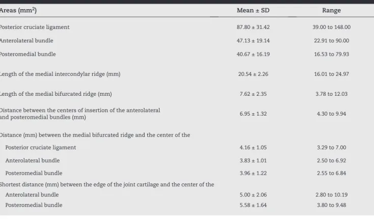
Table 1 presents the measurements on the insertion areas of the AL and PM bundles, the lengths of the medial intercondylar and medial bifurcated ridges, the distances between the centers of insertion of the AL and PM bundles, the distances between the medial bifurcated ridge and the centers of insertion of the AL and PM bundles, and the shortest distance between the edge of the joint cartilage and the centers of insertion of the AL and PM bundles, with their respective standard deviations and ranges between the minimum and maximum values.
Discussion
Surgical reconstruction of the PCL requires clear knowledge of the femoral anatomy and reproducible methods for locating the tunnel entrances.8
The aim of our study was to provide an anatomical and morphometric basis for the femoral insertions of the PCL in order to guide surgeons in placing grafts in anatomical femoral tunnels, in reconstructions on this ligament.
Regarding descriptions of the anatomy, we found in the literature that the format of the femoral insertion of the PCL is relatively planar and in a half-moon shape.7,9 According
to Dargel et al.,10 this insertion has an oval shape, with the
largest diameter in the anteroposterior orientation, with the knee extended.
Lopes et al.11 reported that the femoral insertion of the PCL
was semicircular in 15 (75%) of the knees studied and oval in the other five (25%). They also stated that the femoral insertion of the PCL was concave in 19 knees (95%) and planar in only one (5%). In the study by Cury et al.,12 the insertion shape was
a quarter of an ellipse in all the patients.
Our results were identical to those of these authors regarding the shape of the insertion, although we found a greater proportion of planar insertions (29.2%).
Also in relation to descriptive anatomy, Lopes et al.11
reported that the medial intercondylar ridge was present in 18 of the 20 knees evaluated (90%) and that the medial bifurcated ridge was present in eight knees (40%). The mean length of the first of these was 14.24 mm and of the second, 5.8 mm.
Regarding these parameters, our results were similar only in relation to the presence of the medial intercondylar ridge. We found the medial bifurcated ridge in a much smaller proportion of the specimens (83.3%) and mean lengths of the medial intercondylar and medial bifurcated ridges in our sample were also greater (20.54 and 7.62 mm, respectively).
In relation to the areas of the femoral insertions of the AL and PM bundles of the PCL, Takahashi et al.13 evaluated 32
knees by means of photographs with a measurement scale, with subsequent computer analysis. They found that the insertion area of the AL bundle was 58.0 ± 25.4 mm2 and that
the insertion area of the PM bundle was 64.6 ± 24.7 mm2.
Areas (mm2) Mean ± SD Range
Posterior cruciate ligament 87.80 ± 31.42 39.00 to 148.00
Anterolateral bundle 47.13 ± 19.14 22.91 to 90.00
Posteromedial bundle 40.67 ± 16.19 16.53 to 79.93
Length of the medial intercondylar ridge (mm) 20.54 ± 2.26 16.01 to 24.97
Length of the medial bifurcated ridge (mm) 7.62 ± 2.35 3.78 to 12.03
Distance between the centers of insertion of the anterolateral
and posteromedial bundles (mm) 6.95 ± 1.32 4.30 to 9.94
Distance (mm) between the medial bifurcated ridge and the center of the
Posterior cruciate ligament 4.16 ± 1.05 3.29 to 7.00
Anterolateral bundle 3.83 ± 1.01 2.50 to 6.92
Posteromedial bundle 3.96 ± 1.22 2.55 to 6.84
Shortest distance (mm) between the edge of the joint cartilage and the center of the
Anterolateral bundle 5.00 ± 2.06 2.80 to 10.19
Posteromedial bundle 5.58 ± 1.64 3.80 to 9.48
Dargel et al.10 marked the femoral insertion of the PCL using
radiopaque barium sulfate emulsion in 30 pairs of knees that had been fixed in formalin. Following this, the specimens were radiographed, digitized and examined using image processing software. It was found that the femoral insertion area of the PCL was 133.8 ± 34.53 mm2 in the left knees and 147.1 ± 33.81
mm2 in the right knees.
Lopes et al.11 photographed 20 knees using a digital camera
equipped with three-dimensional laser, and the images obtained were studied using specific software. They found that the mean femoral insertion areas of the PCL were 118.0 ± 23.95 mm2 for the AL bundle and 90.0 ± 16.13 mm2 for the PM bundle.
The total insertion area of the PCL was 209.0 ± 33.82 mm2.
Cury et al.12 evaluated the total insertion area of the PCL in
20 knees from cadavers by means of the AutoCAD software and found a mean of 153.5 mm2.
In our evaluation, like that of Lopes et al.,11 the insertion
area of the AL bundle was greater than that of the PM bundle, which was contrary to the findings of Takahashi et al.13
We also found, between our cases and those of the other authors, that there was a large standard deviation for the mean insertion area, ranging from 18% to 44% of the total area. This probably demonstrates that there are notable differences between individual measurements in the populations studied.
The mean for the total insertion area of the PCL in our sample was much smaller than that of the other authors. In approximate numbers, our area was 71% of the mean total insertion area of Takahashi et al.,13 66% of the mean area found
by Dargel et al.10 in left knees, and only 42% of the mean area
found by Lopes et al.11
The latter authors attributed the divergences in the values encountered to the different measurement methods used and the variations in sex and ethnicity of the populations studied. In addition, in their measurements, they included all the peripheral fibers of the PCL.11
Our numerical results were smaller probably because in our study, we solely delimited the bone sites for femoral insertion of the PCL bundles. It was possible to identify these by means of careful dissection. We did not include the fibrous expansions that are inserted at the periphery of the bone insertion sites, because these do not form part of the ligament body, strictly speaking.
Concordant with the objective of our study, we determined the diameter that would be needed for a femoral bone tunnel filled with a graft, to cover the entire insertion of the PCL. Thus, since the total area is equal to pi multiplied by the radius squared (Δ = π x r2), we calculated that for the mean total
femoral insertion area of the PCL of our sample, a diameter of 10.57 mm would be needed; for the lower standard deviation, a tunnel of 8.47 mm in diameter would be needed and for the upper standard deviation, a tunnel of 12.32 mm in diameter. These dimensions are bigger than those of the single tunnels that are generally used.
We therefore disagree with Morgan et al.,14 who reported
that reconstruction with a single band should not occupy the entire insertion area of the PCL. The notable individual variations possibly do not allow a single reconstruction technique to be applied. Perhaps there should be individualized
indications for anatomical reconstructions (of insertion sites) with single bundles and double bundles.
This study has two limitations: our sample of 24 knees might be considered to be small, given the great anatomical variation found; and also, the photographic analysis that we did may have had imperfections, given that it did not provide adequate information regarding the depth of the insertion.
Conclusions
The insertion area of the anterolateral bundle of the PCL is larger than that of the posteromedial bundle and the insertion areas of these bundles were smaller than has previously been described in the literature.
The insertions of the PCL have bone limits: the medial intercondylar ridge, which is present in almost all cases; and the medial bifurcated ridge, which is seen less frequently. Both of these may provide reference points for drilling the femoral tunnels for PLC reconstruction and for assessing their positioning, by means of radiographs or computed tomography scans after the operation.
There were notable individual variations in the sample analyzed and, for this reason, the indications for anatomical reconstruction of the PCL using a single or double tunnel should be individualized.
Conflicts of interest
The authors declare that there was no conflict of interests in conducting this study.
R E F E R E N C E S
1. Amis AA, Gupte CM, Bull AM, Edwards A. Anatomy of the posterior cruciate ligament and the meniscofemoral ligaments. Knee Surg Sports Traumatol Arthrosc. 2006;14(3):257-63.
2. Girgis FG, Marshall JL, Al Monajem ARS. The cruciate ligaments of the knee joint. Anatomical, functional, and experimental analysis. Clin Orthop Relat Res. 1975;(106):216-31.
3. Grood ES, Stowers SF, Noyes FR. Limits of movement in the human knee. Effect of sectioning the posterior cruciate ligament and posterolateral structures. J Bone Joint Surg Am. 1988;70(1):88-97.
4. Clancy WG, Sutherland TB. Combined posterior cruciate ligament injuries. Clin Sports Med. 1994;13(3):629-47. 5. Dejour H, Walch G, Peyrot J, Eberhard P. The natural history of
rupture of the posterior cruciate ligament. Rev Chir Orthop Reparatrice Appar Mot. 1988;74(1):35-43.
6. Torg JS, Barton TM, Pavlov H, Stine R. Natural history of the posterior cruciate ligament-deficient knee. Clin Orthop Relat Res. 1989;(246):208-16.
8. Saddler SC, Noyes FR, Grood ES, Knochenmuss DR, Hefzy MS. Posterior cruciate ligament anatomy and length-tension behavior of PCL surface fibers. Am J Knee Surg. 1996;9(4):194-99. 9. Edwards A, Bull AMJ, Amis AA. The attachments of the fiber
bundles of the posterior cruciate ligament: an anatomic study. Arthroscopy. 2007;23(3):284-90.
10. Dargel J, Pohl P, Tzikaras P, Koebke J. Morphometric side-to-side differences in human cruciate ligament insertions. Surg Radiol Anat. 2006;28(4):398-402.
11. Lopes OV, Ferretti M, Shen W, Ekdahl M, Smolinski P, Fu FH. Topography of the femoral attachment of the posterior cruciate ligament. J. Bone Joint Surg Am. 2008;90(2):249-55.
12. Cury RPL, Severino NR, Camargo OPA, Aihara T, Batista Neto LV, Goarayeb DN. Estudo anatomico da insercao femoral do ligamento cruzado posterior. Rev Bras Ortop. 2011;46(5):591-5. 13. Takahashi M, Matsubara T, Doi M, Suzuki D, Nagano A.
Anatomical study of the femoral and tibial insertions of the anterolateral and posteromedial bundles of human posterior cruciate ligament. Knee Surg Sports Traumatol Arthrosc. 2006;14(11):1055-9.