Auditory characteristics of individuals
with temporomandibular dysfunctions
and dentofacial deformities
Tatiane Totta1, Giselda Santiago2, Eduardo Sanches Gonçales3, Sandra de Oliveira Saes4, Giédre Berretin-Felix5
Objective:To investigate whether there is any relationship between otological as well as vestibular symptoms, audiological findings and type of temporomandibular disorder (articular, muscular and mixed); and to check the distribution of the temporomandibular disorders (TMD) dysfunction degree in the research population. Methods: A retrospective study involving 30 patients of both sexes, aged between 18 and 49 years old, diagnosed with TMD and dentofacial deformities, who were subject to clinical evaluation (muscle palpation, auscultation of temporomandibular joint during mandibular motion and measurement of jaw movement), audiological testing (pure tone audiometry and immittance testing) and two questionnaires, one on otological and vestibular symptoms and the other on TMD anamnesis. Based on both the anamnesis questionnaire and the clinical assessment, the subjects were divided according to the type and degree of TMD dysfunction (mild, moderate and severe), and compared regarding the occurrence of auditory signs and symptoms, vestibular symptoms and audiological findings according to TMD type. Results: The anamnesis questionnaire demonstrated higher prevalence (83.33%) of severe TMD. Subjects with mixed TMD had more complaints about hypoacusis than those with muscular TMD (p < 0.05). The results showed no change in either audiological and immittance testing for all assessed individuals. Conclusion: Otological symptoms are present in subjects with TMD and dentofacial deformities, regardless of the classification of TMD (articular, muscular or mixed). Those with mixed TMD may have higher incidence of complaints about hypoacusis than subjects with muscular TMD. Further studies are needed to investigate the relationship between otological symptoms and the different types of TMD.
Keywords:Temporomandibular joint disorders. Hearing disorders. Audiometry. Malocclusion.
How to cite this article: Totta T, Santiago G, Gonçales ES, Saes SO, Berre-tin-Felix G. Auditory characteristics of individuals with temporomandibular dysfunctions and dentofacial deformities. Dental Press J Orthod. 2013 Sept-Oct;18(5):70-7.
» The authors report no commercial, proprietary or inancial interest in the prod-ucts or companies described in this article.
Contact address: Giédre Berretin-Felix Alameda Dr. Otávio Pinheiro Brizolla, 9-75 Vila Universitária, Bauru/SP - Brazil CEP: 17012-901 – E-mail: [email protected] 1 Postgraduate student in Rehabilitation Sciences, Craniofacial Anomalies
Rehabilitation Hospital – University of São Paulo (HRAC-USP). 2 MSc in Rehabilitation Sciences, HRAC-USP.
3 Associate Professor, Oral and Maxillofacial Surgery and Traumatology, Bauru School of Dentistry, University of São Paulo (FOB-USP). 4 Professor, Speech Therapy, Sacred Heart University (USC).
5 Associate Professor, department of Speech Therapy, Bauru School of Dentistry, University of São Paulo (FOB-USP).
Submitted: November 17, 2009 - Revised and accepted: April 27, 2011
Objetivo: investigar se há relação entre os sintomas otológicos, vestibulares, achados audiológicos e o tipo de disfunção temporomandibular (articular, muscular e misto), e verificar a distribuição do grau de disfunção da DTM nessa população. Métodos:
estudo retrospectivo, envolvendo 30 pacientes com deformidades dentofaciais diagnosticados com DTM, de ambos os sexos, entre 18 e 49 anos de idade, submetidos a avaliação clínica (palpação muscular, ausculta da articulação temporomandibular durante os movimentos mandibulares e mensuração da movimentação da mandíbula), exame audiológico (audiometria tonal limiar e imitanciometria) e a dois questionários, sendo um sobre sintomas otológicos e vestibulares e outro anamnético da DTM. A partir do questionário anamnético e da avaliação clínica, os sujeitos foram divididos conforme o tipo e o grau da disfunção da DTM (leve, moderado e severo), e comparados quanto à ocorrência dos sinais e sintomas auditivos, vestibulares e achados audiológicos, de acordo com o tipo de DTM. Resultados:
houve maior prevalência (83,33%) de DTM severa de acordo com questionário anamnético. Sujeitos com DTM mista apresentaram mais queixas de hipoacusia do que aqueles com DTM muscular (p < 0,05). Os resultados evidenciaram ausência de alterações nos exames audiológico e imitanciométrico para todos os indivíduos avaliados. Conclusão: sintomas auditivos estão presentes nos sujeitos com DTM e deformidades dentofaciais, independentemente da classificação da DTM (articular, muscular ou mista), e aqueles com DTM mista podem apresentar maior ocorrência de queixa de hipoacusia do que sujeitos com DTM muscular. Estudos futuros são necessários para investigar a relação entre a sintomatologia auditiva e os diversos tipos de DTM.
INTRODUCTION
Temporomandibular disorders (TMD) represent a set of clinical signs and multifactorial etiology symp-toms characterized by pain in the temporomandibular joint (TMJ) and/or the tissue surrounding it, functional limitations in the jaw or crackling during TMJ
move-ments.13 The usual classiication, according to The
American Academy of Orofacial Pain criteria,18 divides
TMDs into groups according to anatomical etiology, respectively: Articular disorder, including the articular surface, the intra-articular disc or the articular bone; muscular disorder, involving the masticatory muscles surrounding the TMJ; or mixed disorder when there
are signals of articular and muscular TMD.12
There are several factors involved in the etiology of TMD, such as disturbances of occlusion as well as max-illary and mandibular bone bases, degenerative disorder, traumatic factors, muscle disorders such as hyperactivity or hypoactivity, stress and emotional problems as well as functional changes and harmful habits that generate
per-sistent overload in the TMJ or in the muscle.1,12 Although
the literature is discordant with regard to the real inluence of malocclusion on the occurrence of TMD, a review
car-ried out by McNamara et al15 related speciic diagnostic
groups of TMD to occlusal factors, such as skeletal ante-rior open bite, overjet greater than 6 to 7 mm, diference between centric relation and maximal habitual intercuspa-tion greater than 4 mm, unilateral crossbite, and absence of ive or more posterior dental elements. Regarding Angle’s
malocclusions, Thilander et al27 correlated Classes I, II and
III to the prevalence of TMD and found higher prevalence of TMD in the group with Class III. However, the litera-ture systematic review analysis of longitudinal studies
con-ducted by Mohlin et al16 did not ind signiicant clinical
associations between diferent malocclusions, orthodontic treatment and signs and symptoms of TMD.
Auditory complaints and symptoms such as otalgia, tinnitus, hypoacusis, ear fullness and vertigo are oten correlated to the presence of TMDs.9,11,19,21,24,26,28,30
Several studies have been conducted in order to un-derstand the etiology of auditory symptoms in subjects
with TMD, irstly described by Costen.6 The author
sug-gested that poor positioning of the mandibular condyle, caused by loss of posterior tooth support, could cause blockage of the Eustachian tube, symptoms of otalgia,
tinnitus and vertigo. Later, Myrhaug17 stated that both
stress and compression of the structures that conduct
sound lead to increase in impedance, otentimes causing ear fullness of loating feature associated with tinnitus.
However, Penkner et al20 demonstrated that masticatory
muscle spasms do not afect the function of the sot palate tensor muscle and the Eustachian tube.
More recently, other studies found an association between pain on palpation of the mandibular condyle
and the presence of otalgia in TMD subjects.8,11,24
Ad-ditionally, high prevalence of tinnitus and vertigo9,28,30
symptoms as well as ear fullness,8,14,25 was observed in
the research population. These conditions may be
relat-ed both to TMD signs, the presence of muscle tension,29
and pain on palpation in the masticatory muscles.14
With regard to hearing thresholds in subjects with TMD, the literature indicates lowering of airway
thresholds in the frequencies of 6000 Hz and 8000 Hz.25
However, no changes in audiological testing and
in-creased incidence of normal immittance were found.9
Although many studies have described auditory symptoms present in TMD patients, few studies have correlated the type of dysfunction (articular, muscle or mixed) to auditory signs and symptoms. Among them,
Tuz et al28 have not found any prevalent occurrence
of otalgia symptoms, tinnitus, vertigo or hearing loss among subjects classiied as presenting articular, mus-cular and mixed TMD. On the other hand, the authors found higher prevalence of complaints in this popula-tion compared to the control group.
Hence, considering that skeletal malocclusion can be a contributing factor to TMD, this study aims to char-acterize the hearing function of individuals with TMD and dentofacial deformities according to each one of the TMD groups: articular, muscular or mixed (articular and muscular) and to investigate the distribution of the TMD dysfunction degree (mild, moderate or severe) in the studied population.
MATERIAL AND METHODS
The research subjects comprised 30 young adults of both sexes, clinically diagnosed with temporoman-dibular disorders by means of implementation of the Research Diagnostic Criteria for Temporomandibular
Disorders (RDC/TMD) Axis I,7 and who presented
head and neck, noise exposure and early hearing-loss.
This study was conducted at Universidade do Sagrado
Co-ração/USC (Sacred Heart University) Bauru, Brazil, and was approved by the University Ethics Committee for Research on Human Beings (Protocol No. 077/2003).
Individuals were selected based on analysis of medical records of patients treated at the Oral and Maxillofacial Surgery Clinic of the College of Den-tistry at the aforementioned university. These sub-jects had undergone a dental clinical evaluation and answered two questionnaires. One questionnaire
was the Anamnesis Index (AI)4,5 based on a
modi-fication of the Helkimo anamnesis index,10 and
pre-viously used by Conti et al4 at a significance level
of 5%; while the other investigated the history of otological and vestibular symptoms.
Clinical evaluation was performed by the same ex-aminer, properly trained and calibrated, and consid-ered the following: measurement of both maximum mouth opening and laterality, presence of mandibu-lar deviation or deflection, TMJ(s) palpation, analysis of joint noises and bilateral palpation of the temporal muscles, masseter, posterior digastric muscle, medial pterygoid muscle, sternocleidomastoid and upper tra-pezius. Based on the aspects investigated in the inter-view and clinical evaluation, the TMD patients were diagnosed and classified according to The American
Academy of Orofacial Pain criteria.18
• GI: Group with articular TMD – presence
of disc displacement, limitation of maximum mouth opening (less than or equal to 35 mm), mandibular deflection or deviation, spontane-ous pain and/or upon TMJ palpation; cases with signs of myogenic TMD were excluded.
• GII: Group with muscular TMD – report of pain
in the masticatory muscles during functional ex-amination and/or muscle palpation; cases with arthrogenous TMD signs were excluded.
• GIII: Group with mixed TMD – signs of mus -cular and arti-cular TMD.
Thus, the GI group (articular TMD) comprised 10 subjects, 7 with Angle Class II malocclusion and 3 with Angle Class III malocclusion. The GII group (muscular TMD) comprised 10 subjects, 4 with Class II and 6 with Class III. And finally, the GIII group (mixed TMD) comprised 10 subjects, 6 with Class II and 4 with Class III.
In the anamnesis index, the following aspects were investigated: Difficulty in mouth opening and jaw laterality, discomfort or muscle pain when chewing, presence of pain and/or muscle fatigue, perception of noise in the TMJ, headaches, neck or shoulder pain, pain in the ears (otalgia) or near them, use of one side of the mouth for chewing, facial pain in the morning and whether the subject considered his bite as nor-mal.5,12,26 Three possible answers were offered: “yes”, “no” and “sometimes”; each “yes” corresponding to 2 points, “sometimes” represented 1 point and “no” represented 0. The items related to frequent head-aches, earaches or close to it, and the presence of TMJ noises had a total of 3 points when they were
bilat-erally or intensively referred.4 The sum of points
al-lowed the classification of subjects into no-TMD (0 to 3 points), mild TMD (4-8 points), moderate TMD
(9-14 points) and severe TMD (15 to 23 points).4
For specific investigation on ontological and ves-tibular symptomatology, the subjects were inter-viewed by a speech therapist who considered the presence of the following symptoms: vertigo and/ or dizziness, tinnitus, ear fullness, earache and hy-poacusis. They were also submitted to hearing eval-uation at the Center of Health Education (CEPS), Bauru, Brazil, at properly equipped and acoustically treated rooms. The subjects underwent otoscopic examination, pure tone audiometry, logoaudiom-etry and immittance testing, using the GSI-60 audi-ometer and AZ-7 imittanciaudi-ometer.
Pure tone audiometry determines the least
quan-tity of audible acoustic energy (hearing threshold),23
through the analysis of hearing thresholds for air and bone conduction of each frequency, using pure tones that are generated by two monaural headphones. The subject is instructed to raise his hand to the evaluator every time he listens to the tone stimulus. The evaluator reduces the signal strength tone, de-creasingly, in values ranging from 10 to 10 dB(HL) until the hearing threshold is found. There must be confirmation of response in 50% of the 4 stimula-tion trials and the maximum hearing threshold value
of 20 dB(HL)24 should be considered as normal.
Data analysis was performed by means of descrip-tive statistics, parametric and nonparametric. To com-pare the occurrence of auditory and vestibular symp-toms according to the three different groups of TMD, the analysis of variance (ANOVA) was applied, as a classification criterion adopting a significance level
of p < 0.05. To compare the TMD groups
regard-ing the severity of TMJ disorder, the nonparametric
Kruskal-Wallis test was applied, considering p < 0.05.
Friedman test was used to investigate the
tympano-metric results, considering p < 0.01.
RESULTS
The study comprised 30 subjects of both sexes, 24 women and 6 men, aged 18-49 years (mean 27.3 ± 7.05 years), divided into 3 groups: GI (10 subjects with ar-ticular TMD), GII (10 subjects with muscular TMD) and GIII (10 subjects with mixed TMD).
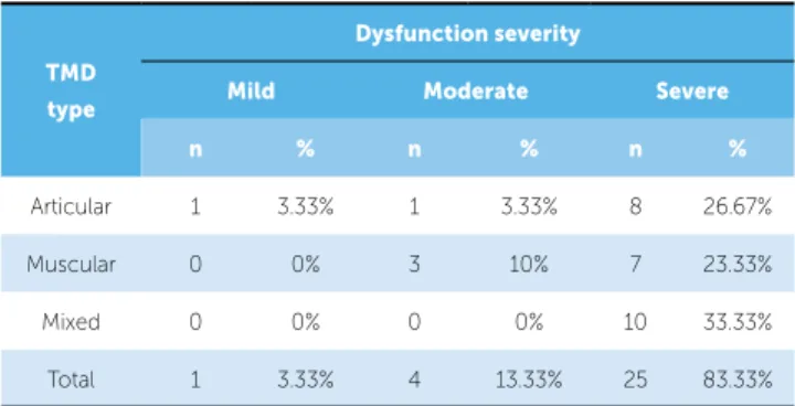
With regard to the anamnesis index, it was veriied higher prevalence of subjects presenting severe symp-toms of dysfunction for the three TMD groups stud-ied, as shown in Table 1. However, no statistically
sig-niicant diference (p=0.069) was found among them.
Additionally, symptoms of moderate dysfunction occurred in one subject of GI group (10%) and in three subjects of GII group (30%). Mild dysfunction was observed for one subject of GI group (10%), only.
Individuals of articular (GI), muscular (GII) and mixed (GIII) TMD groups were questioned regarding the audiological and vestibular symptoms (Fig 1), and tinnitus and vertigo were reported by most subjects. lowest intensity, in which the subject can
discrimi-nate 50% of the words dictated to him. The Speech Recognition Index (SRI) test parameters were also evaluated: the subject must repeat a list of 25 mono-syllabic words (for each ear), in the same intensity, 40 dB above the tonal threshold mean of the
follow-ing frequencies: 500, 1000 and 2000 kHz.23
Immittance testing represents an indirect test of tubal function, capable of evaluating both static and dynamic air pressure in the middle ear. This exam measures the ability of the tympanic membrane to reflect a sound introduced into the outer acoustic meatus, in response to gradual changes of pressure in the same space, thus, verifying the permeability of the tympanic-ossicular system with the passage of
a sound wave within normal limits.23
The criteria used to define changes in the audio-logical tests were the following: Pure tone air-con-duction thresholds (in the frequencies of 250 Hz, 500 Hz, 1000 Hz, 2000 Hz, 3000 Hz, 4000 Hz, 6000 Hz and 8000 Hz) and pure tone bone-con-duction (in the frequencies of 500 Hz, 1000 Hz, 2000 Hz and 4000 Hz) greater than 20 dB(HL); air-bone GAP greater than 10 dB(HL); Speech Rec-ognition Index (SRI, monosyllables) lower than 92%, SRT with values above 20 dB; tympanometric curve deviated from -100 daPa; contralateral acous-tic reflexes exceeding 115 dB(HL) (absent); differ-ence between the pure tone air-conduction thresh-old and reflex threshthresh-old less than 60 dB(HL), which
would be suggestive of recruitment.9
TMD type
Dysfunction severity
Mild Moderate Severe
n % n % n %
Articular 1 3.33% 1 3.33% 8 26.67%
Muscular 0 0% 3 10% 7 23.33%
Mixed 0 0% 0 0% 10 33.33%
Total 1 3.33% 4 13.33% 25 83.33%
Table 1 - Distribution of subjects in accordance with Helkimo Anamnesis In-dex for each group.
Figure 1 - Distribution of subjects with audiological and vestibular symptoms according to the three different groups of TMD.
8
6 6 6 6
4 4
2 2
7 7 7 7 7 7
5 5 5 5
3 3
1 1
0
Vertigo and/ or dizziness
Ear Fullness Tinnitus
Articular Muscular Mixed
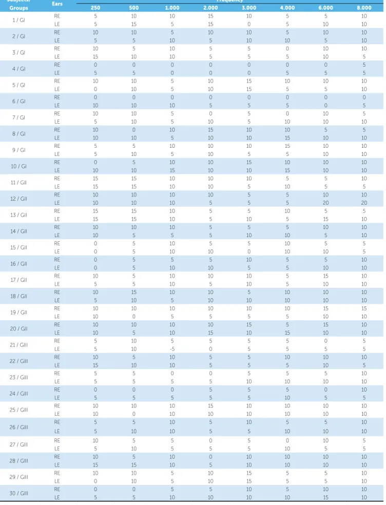
Table 2 - Distribution of pure tone air conduction thresholds for all frequencies of each individual from the GI (articular), GII (muscular) and GIII (articular and muscular) groups for the right (RE) and left ears (LE).
Subjects/
Groups Ears
Frequency
250 500 1.000 2.000 3.000 4.000 6.000 8.000
1 / GI RE 5 10 10 15 10 5 5 10
LE 5 15 5 15 0 5 10 10
2 / GI RE 10 10 5 10 10 5 10 10
LE 5 5 10 5 10 10 5 10
3 / GI RE 10 5 10 5 5 0 10 10
LE 15 10 10 5 5 5 10 5
4 / GI RE 0 0 0 0 0 0 0 5
LE 5 5 0 0 0 5 5 5
5 / GI RE 10 10 5 10 15 10 10 10
LE 0 10 5 10 15 5 5 10
6 / GI RE 0 0 0 0 0 0 0 0
LE 10 10 10 5 5 5 0 5
7 / GI RE 10 10 5 0 5 0 10 5
LE 5 10 5 10 5 10 10 10
8 / GI RE 10 0 10 15 10 10 5 5
LE 10 10 5 10 10 15 10 10
9 / GI RE 5 5 10 10 10 15 10 10
LE 5 10 5 10 5 5 10 10
10 / GI RE 0 5 10 10 15 10 10 10
LE 10 10 15 10 10 15 10 10
11 / GII RE 15 15 10 10 10 5 5 10
LE 15 15 10 10 5 10 5 5
12 / GII RE 10 10 10 10 5 5 10 10
LE 10 10 10 5 5 5 20 20
13 / GII RE 15 15 10 5 5 10 5 5
LE 15 15 10 5 10 5 15 10
14 / GII RE 10 10 10 5 5 5 10 10
LE 10 5 5 5 10 10 5 10
15 / GII RE 0 5 10 5 5 10 5 5
LE 0 5 10 10 0 10 10 5
16 / GII RE 0 5 5 5 10 5 5 10
LE 0 5 10 10 5 5 10 10
17 / GII RE 10 5 10 10 10 5 15 10
LE 5 5 10 5 10 5 10 10
18 / GII RE 10 15 10 10 5 10 10 10
LE 5 10 5 10 10 10 10 10
19 / GII RE 10 10 10 10 10 10 15 15
LE 10 0 5 5 5 5 10 10
20 / GII RE 10 10 10 10 15 5 15 10
LE 10 5 10 15 10 15 10 10
21 / GIII RE 5 10 5 5 5 5 0 5
LE 5 10 -5 0 5 5 5 5
22 / GIII RE 10 5 10 5 5 10 10 10
LE 15 10 10 5 5 5 10 5
23 / GIII RE 5 5 0 0 5 5 5 10
LE 5 5 5 5 10 10 10 10
24 / GIII RE 0 0 0 5 5 5 0 10
LE 5 5 5 5 5 10 5 5
25 / GIII RE 10 10 10 15 10 10 10 10
LE 10 0 10 10 10 10 10 10
26 / GIII RE 5 5 10 5 10 5 5 10
LE 5 10 10 5 5 10 10 10
27 / GIII RE 10 5 5 0 5 0 10 5
LE 5 10 5 5 5 10 5 5
28 / GIII RE 10 5 10 0 10 10 10 10
LE 15 15 10 5 10 10 10 10
29 / GIII RE 10 10 5 10 15 5 5 10
LE 0 10 5 10 15 5 5 10
30 / GIII RE 0 0 5 5 10 5 10 10
Furthermore, otalgia and hypoacusis were less fre-quently reported for individuals with muscular TMD. Statistical analysis showed that subjects with mixed TMD have more complaints about hypoacusis when
compared to subjects with muscular TMD (p=0.0581).
In regard to pure tone audiometry, the subjects of the three TMD groups showed, for both ears, hear-ing thresholds ranghear-ing between 5 and 20 dB(HL), for all air-conduction frequencies tested (Table 2).
As for pure tone bone-conduction thresholds, all subjects presented normal compatible values and showed air-bone GAP lower than 10 dB(HL) for all frequencies tested.
In logoaudiometry, the Speech Recognition Index found for all individuals showed values between 96% and 100% of accuracy, which is consistent with the ref-erences of normality. Similarly, normal results in SRT (between 0 and 20 dB(HL)) were observed for the en-tire sample. The immittance testing showed signiicant
prevalence (p=0.0001) of type A curve, representing
consistent results with normal values.
DISCUSSION
In this study, patients were divided into groups ac-cording to TMD classiication (articular, muscular and mixed) and dysfunction severity (mild, moderate and severe), aiming at elucidating the relationship between audiological / vestibular signs and symptoms and the diferent types of TMD.
In this sample there was higher incidence of TMD in females (80%) compared to males (20%), and this prevalence is very close to the values reported in the lit-erature, since many studies have shown that TMD signs and symptoms are more common in women aged be-tween 20 to 39 years old.2,17,20,21,26
Twenty four women were evaluated in this study, 15 (50%) of which aged between 20 and 29 years old, who were diagnosed with TMD. This result corroborates
those obtained by Williamson29 and Felício.8
There is also the hypothesis that degradation of cartilage and articular bone, due to menopause and
increase in estrogen and prolactin,17 may be related to
the presence of TMD symptoms in women aged 40-49 years. In this sample, such symptoms were observed in 2 (6.67%) women within this age range.
With regard to the degree of dysfunction, 25 subjects (88.33%) of the sample, among the diferent groups, showed severe TMD symptoms. The study carried out
by Penkner et al20 found higher prevalence of
moder-ate dysfunction, followed by severe dysfunction, among
evaluated subjects with TMD. Keersmaekers et al11 also
measured the symptoms in TMD, by means of applying
the Pain and Dysfunction Index,10 and found signiicant
higher prevalence of severe dysfunction in individuals with TMD concomitant with otalgia, when compared to subjects without otalgia. On the other hand, Silveira
et al26 found higher severity rate of mild TMD, followed
by absent, moderate and severe TMD, respectively, in a sample of patients treated in an otorhinolaryngology outpatient unit. To carry out this evaluation, the authors used the same anamnesis questionnaire of the present
research. Conti et al5 investigated the association
be-tween malocclusion (Class I and Class II), orthodontic treatment, prevalence and severity of TMD signs and symptoms in 200 adolescents. They found that 34% of them had mild symptoms, according to the anamnesis index applied, while 3.5% had moderate symptoms. The lack of agreement in the literature regarding the degree of TMD prevalence may be related to the dif-ferent conditions presented by the subjects of the cited studies, and it is important to consider that this study investigated individuals with dentofacial deformities.
As for the auditory and vestibular symptoms, it was found that more than half of the subjects in each studied group had symptoms of vertigo and/or dizziness,
tinni-tus and ear fullness. Pereira et al21 also observed 65% of
prevalence of tinnitus, otalgia and ear fullness in patients
with TMD, as well as Parker and Chole19 found
signii-cant prevalence of tinnitus and vertigo in patients with
TMD. A study carried out by Tuz et al28 also subdivided
the subjects according to the types of TMD and found no predilection of audiological symptoms of otalgia, tinni-tus, vertigo and hearing loss among groups with muscu-lar, articular or mixed TMD. However, these symptoms were greatly prevalent in the control group of subjects
without TMD. Nevertheless, Felício et al9 found a
re-lationship between subjects with severe pain in muscles and in TMJ, and the propensity for otalgia and tinnitus.
According to the literature, the complaint of ear fullness may be due to muscle changes in patients with TMD, such as the lateral pterygoid muscle spasm, lead-ing to hypertonia of the tensor tympani muscle, caus-ing changes in the cycle of Eustachian tube opencaus-ing and leading to reduction in ventilation of the middle
hypothesis by means of electromyographic evaluation. Tinnitus sensation and other otologic symptoms, such as otalgia and hypoacusis, have been linked to the pres-ence of TMD(s) in several studies, with four possible models that explain the etiology of otologic symptoms and signs in TMD(s): Embryological, muscular, bone
communication and neural network.22
In the present study, subjects with articular symp-toms associated or not with muscular complaints had higher incidence of otalgia and hypoacusis, although hearing loss had been declined by audiological testing.
These findings agree with Ciacanglini et al,3 who also
found significant association between the severity of arthropathy and the perception of audiological symp-toms. Such complaints may be justified by the close relationship between TMJ, tympanic cavity and the
Eustachian tube,2 in which the influence of changes
in the contraction of the stapedius and cheekbone muscles on the ossicular chain of the middle ear and tympanic membrane, caused by the traction of the articular disc, could impact hearing perception, with otalgia related to central exciting effects and
neuro-muscular mechanisms involved in the TMD.22
The present study identified no changes in
audio-logical tests, corroborating the literature.8,20 In
addi-tion it also found proper tubal funcaddi-tion (type A curve)
for all subjects, also discovered in other studies.8,20
However, it disagrees with a similar study25 that found
lower pure tone air-conduction thresholds in 6000 Hz and 8000 Hz frequencies in TMD patients.
It is worth considering the limitations of this study: the heterogeneity of dentofacial deformities present in each studied group as well as the lack of control group with dentofacial balance. Further studies shall be conducted in this direction, attempting to better understand the rela-tionships between malocclusion, temporomandibular dys-function and audiological/vestibular signs and symptoms.
CONCLUSION
This study found that audiological symptoms are present in subjects with temporomandibular disorders and dentofacial deformities, regardless of TMD classi-ication (articular, muscular or mixed). Subjects with mixed TMD may have higher incidence of complaints of hypoacusis than subjects with muscular TMD, how-ever, with neither hearing loss, diagnosed by audiologi-cal testing, nor tympanometric alterations being ob-served in the research population.
1. Bianchini EMG. Articulação temporomandibular e fonoaudiologia. In: Ferreira LP, Bei-Lopes DM, Limongi SCO, organizadores. Tratado de fonoaudiologia. São Paulo: Roca; 2009. cap. 57, p. 532-44.
2. Bubon MS. Documented instance of restored conductive hearing loss.
Funct Orthod. 1995;12(1):26-9.
3. Ciacanglini R, Loreti P, Radaelli G. Ear, nose, and throat symptoms in
patients with TMD: the association of symptoms according to severity of arthropathy. J Orofacial Pain. 1994;8(3):293-7.
4. Conti PC, Ferreira PM, Pegoraro LF, Conti JV, Salvador MC. A
cross-sectional study of prevalence and etiology of signs and symptoms of temporomandibular disorders in high school and university students. J Orofac Pain. 1996;10(3):254-62.
5. Conti A, Freitas M, Conti P, Henriques J, Janson G. Relationship between
signs and symptoms of temporomandibular disorders and orthodontic treatment: a cross-sectional study. Angle Orthod. 2003;73(4):411-7.
6. Costen JB. Syndrome of ear and sinus simptons dependent upon
disturbed functions of the temporomandibular joint. Ann Otol Rhinol Laryngol. 1934;43:1-15.
7. Dworking SF, LeResche L, editors. Research diagnostic criteria for
temporomandibular disorders: review, criteria, examinations and speciications, critique. J Craniofamandib Disord. 1992;6:301-55.
8. Felício CM, Oliveira JAA, Nunes LJ, Jeronymo LFG, Jeronymo RRF.
Alterações auditivas relacionadas ao zumbido nos distúrbios otológicos e da articulação têmporo-mandibular. Rev Bras Otorrinolaringol. 1999;65(2):141-6.
9. Felício CM, Faria TG, Silva MAMR, Aquino AMCM, Junqueira CA.
Temporomandibular disorder: relationship between otologic and orofacial symptoms. Rev Bras Otorrinolaringol. 2004;70(6):786-93. 10. Helkimo M. Studies on function and dysfunction of the masticatory
system. II. Index for anamnestic and clinical dysfunction and occlusal state. Svensk Tandläk T. 1974;67(2):101-21.
11. Keersmaekers K, De Boever JA, Van den Bergue L. Otalgia in patients with temporomandibular joint disorders. J Prosthet Dent. 1996;75(1):72-6. 12. Kogawa EM. Avaliação da discriminação interoclusal para
microespessuras e da força máxima de mordida em pacientes
portadores de disfunções temporomandibulares [dissertação]. Bauru (SP): Universidade de São Paulo; 2005.
13. Koh H, Robinson PG. Occlusal adjustment for treating and preventing temporomandibular joint disorders. J Oral Rehabil. 2004;31(4):287-92. 14. Kuttila S, Kuttila M, Le Bell Y, Alanen P, Jouko S. Aural symptoms and
signs of temporomandibular disorder in association with treatment need and visits to a physician. Laryngoscope. 1999;109(10):1669-73. 15. McNamara JA, Seligman DA, Okeson JP. Occlusion, Orthodontic
treatment, and temporomandibular disorders: a review. J Orofacial Pain. 1995;9(1):73-90.
REFERENCES
16. Mohlin B, Axelsson S, Paulin G, Pietilä T, Brattström V, Hansen K, et al. TMD in relation to malocclusion and orthodontic treatment. Angle Orthod. 2007;77(3):542-8.
17. Myrhaug H. The incidence of ear symptoms in cases of malocclusion and temporomandibular joint disturbances. Br J Oral Surg. 1964; 2(1):28-32.
18. Okeson JP. Dor orofacial: guia de avaliação, diagnóstico e tratamento. The American Academy of Orofacial Pain. Chicago: Quintessence; 1998. 19. Parker WS, Chole RA. Tinnitus, vertigo and temporomandibular disorders.
Am J Orthod Dentofac Orthop. 1995 fev;107(2):153-158. 20. Penkner K, Köle W, Kainz J, Schied G, Lorenzoni M. The function
of tensor veli palatine muscles in patients with aural symptoms and temporomandibular disorder. An EMG study. J Oral Rehabilitation. 2000;27(4):344-8.
21. Pereira KNF, Andrade LLS, Costa MLG, Portal TF. Sinais e sintomas de pacientes com disfunção temporomandibular. Rev CEFAC. 2005;7(2):221-8.
22. Ramirez LM, Ballesteros LE, Sandoval GP. Topical review: temporomandibular disorders in an integral otic symptom model. Int J Audiology. 2008;47(4):215-27.
23. Redondo MC, Lopes Filho O. Testes básicos de avaliação auditiva. In: Lopes Filho O, et al. Tratado de Fonoaudiologia. 2. ed. Ribeirão Preto: Tecmedd, 2005. p. 89-110.
24. Reis AC, Hotta TH, Ferreira-Jerônymo RR, Felício CM, Ribeiro RF. Ear symptomatology and occlusal factors: a clinical report. J Prosthet Dent. 2000;83(1):21-4.
25. Rodrigues ACY, Berretin-Felix G, Jorge JC, Genaro KF. Caracterização das alterações miofuncionais orais e auditivas em indivíduos com disfunção temporomandibular. Pró-Fono. 1998;10(1):51-5.
26. Silveira AM, Feltrin PP, Zanetti RV, Mautoni MC. Prevalência de portadores de DTM em pacientes avaliados no setor de otorrinolaringologia. Rev Bras Otorrinolaringol. 2007;73(4):528-32.
27. Thilander B, Rubio G, Pena L, de Mayorga C. Prevalence of temporo-mandibular dysfunction and its association with malocclusion in children and adolescents: An epidemiologic study related to speciied stages of dental development. Angle Orthod. 2002;72(2):146-54.
28. Tuz HH, Onder EM, Kisnisci RS. Prevalence of otologic complaints in patients with temporomandibular disorder. Am J Orthod Dentofacial Orthop. 2003;123(6):620-23.
29. Williamson EH. The inter-relationship of internal derangements of the temporomandibular joint, headache e vertigo and tinnitus: a survey of 25 patients. J Craniomand Pract. 1990;8(4):301-6.