Effects of chin advancement surgery in hyoid bone and
tongue positions and in the dimension of the oropharynx
Milena Barreto de Arruda Cabral1, André Carlos de Freitas2, Telma Martins de Araújo3, Nilson Pena4,
Rivail Almeida Brandão Filho5
How to cite this article: Cabral MBA, Freitas AC, Araújo TM, Pena N, Brandão Filho RA. Effects of chin advancement surgery in hyoid bone and tongue positions and in the dimension of the oropharynx. Dental Press J Orthod. 2013 Sept-Oct;18(5):64-9.
Submitted: June 30, 2011 - Revised and accepted: December 03, 2011
» The authors report no commercial, proprietary or inancial interest in the prod-ucts or companies described in this article.
Contact address: Milena Barreto de Arruda Cabral
Av. Araújo Pinho, 62 – 7º andar – Canela – Salvador/BA – Brazil CEP: 40.110-150 – E-mail: [email protected] » The patients displayed in this article previously approved the use of their
radio-graphs.
1 MSc Student in Orthodontics and Facial Orthopedics, Bahia Federal
University (UFBA).
2 PhD, Oral and Maxillofacial Surgery. Associate Professor, UFBA. 3 PhD and Full professor, Department of Orthodontics, UFBA. 4 PhD in Dental Radiology, UNICAMP.
5 PhD Student in TMD and Associate Professor, UFBA.
Introduction: Advancement genioplasty has been increasingly indicated for the correction of anterior mandibular deficiency as it balances the patient’s profile and generates functional changes. Objective: Thus, the aim of this study was to assess the effects of advancement genioplasty in the oropharyngeal size and in the position of the hyoid bone and tongue. Methods: The sample comprised 22 lateral cephalometric radiographs of 11 individuals who had under-gone advancement genioplasty alone. Eleven of these radiographs depicted the immediate preoperative period (T0) and the other 11 the postoperative period after at least four months (T1). The radiographs were scanned and exported to the Radiocef Studio 2.0® software (Radio Memory Ltda., Belo Horizonte, MG, Brazil). The landmarks were then
identified and automatically measured by the program. Results: The results showed statistically significant differences between the horizontal position of the hyoid bone and tongue and in the oropharyngeal size. The hyoid bone and tongue were repositioned anteriorly after surgery (p = 0.01), thereby increasing the dimension of the oropharyngeal airspace (p = 0.01). Conclusions: The oropharyngeal dimension increased as the tongue base and hyoid bone were repositioned more anteriorly.
Keywords:Orthognathic surgery. Chin. Oropharynx. Hyoid bone. Tongue.
Introdução: a cirurgia de avanço de mento isolado tem sido cada vez mais indicada para a correção de deficiência an-terior da mandíbula, harmonizando o perfil e gerando alterações funcionais. Objetivo: essa pesquisa teve a finalidade de avaliar os efeitos da cirurgia de avanço de mento no tamanho da orofaringe e nas posições do osso hioide e da língua.
Métodos: a amostra constou de 22 radiografias cefalométricas de perfil de 11 indivíduos que se submeteram à cirurgia de mentoplastia de avanço isolada. Dessas radiografias, 11 retratavam o período imediatamente pré-cirúrgico (T0) e 11 o pós-cirúrgico de, pelo menos, quatro meses (T1). As radiografias foram digitalizadas e transferidas para o programa Radiocef Studio 2.0, por meio do qual foram feitas as medições entre os pontos demarcados. Resultados: verificaram--se diferenças estatisticamente significativas entre T0 e T1 quanto à posição do osso hioide e da língua no sentido ho-rizontal e ao tamanho da orofaringe. O osso hioide se apresentou posicionado mais anterior em T1 (p = 0,01), assim como a língua, aumentando o tamanho da orofaringe (p = 0,01). Conclusão: houve aumento do espaço aéreo da oro-faringe com o posicionamento mais anterior da base da língua, bem como a reposição do osso hioide anteriormente.
INTRODUCTION
Patients with dentofacial deformities increasingly seek combined orthodontic-surgical treatment. Above and beyond the esthetic issues inherent in facial defor-mity, functional conditions such as chewing, speech, articulation and nasal1 breathing can be compromised. Genioplasty is a surgical procedure widely used in con-junction with other orthognathic surgeries to correct transverse, vertical and sagittal imbalances of the lower third of the face. Nowadays, it is oten indicated to be performed alone as it is considered a relatively simple procedure with predictable results which, when prop-erly indicated, improves facial esthetics.
Chin hypoplasia or deficiency is one of the most frequent deformities found in the anterior region of the mandible.2 It is often seen in patients with both Angle Class II and Class I malocclusions with appro-priate three-dimensional positioning of the maxillo-mandibular complex. For many of these cases, genio-plasty is likely to be the treatment of choice.3
By means of genioplasty some deformities specif-ic to the lower third of the face can be corrected by changing the shape and size of the mandibular sym-physis, thereby improving soft tissue bone support and patient profile.2 When performed alone, it is a more conservative surgical procedure involving little morbidity and not requiring intermaxillary fixation nor a lengthy in-patient hospitalization.4
Osteotomy extends from the premolar region on one side of the mandible to the opposite side, below the apices of the teeth and the mandibular canal. The tongue muscles, including the geniohyoid and genio-glossus muscles, remain attached to the osteotomized bone segment in order to ensure the blood supply necessary for the enlarged chin.4,5
The geniohyoid muscle originates from the bottom of the lower genial tubercle of the mental symphysis, extending medially to the mylohyoid region and being attached to the upper margin of the body of the hyoid bone. The mylohyoid muscle originates from the medial surface of the mandible and is attached to the anterior surface of the hyoid bone through its posterior portion.12
The genioglossus muscle originates from the ge-nial tubercle of the mandible and has a portion of its fibers connected to geniohyoid muscle fibers so that the hyoid bone moves in tandem with the tongue and oral floor during chewing and breathing.11
In healthy individuals, the hyoid bone is situated at the level of the C3-C4 vertebrae and it is important to preserve the size of the upper airspace because it serves as an anchor for the tongue muscles.14 The dis-tance between the hyoid bone and the mandibular plane is of approximately 12 mm.15
The oropharynx is lined with lymphoid tissue and extends itself from the second through the fourth cervical vertebra, participating in the processes of swallowing and breathing.10
Dimensional changes in the airspace have been observed ater surgical repositioning of the mandible and maxilla6,7,8 as well as changes in the position of the hyoid bone, tongue and head.9 Previous studies have shown increases in oropharyngeal dimensions during mandibular advancement surgery and have recognized this surgical procedure as one of the most successful in the treatment of oropharyngeal space deiciency.7,16,17 Mandibular advancement pushes the geniohyoid and genioglossus muscles forward, causing an anterior movement of the tongue base and hyoid bone, conse-quently increasing posterior airspace.18
The technique of genioplasty with advancement of the genioglossus muscle in patients with man-dibular retrognathism promotes increased posterior airspace (PAS) and soft tissue advancement of the mental region.19
The aim of this article was to assess whether any changes occur in the oropharyngeal size and in the position of both the hyoid bone and tongue after chin advancement surgery.
MATERIAL AND METHODS
with the patient’s head immobilized by a cephalostat, orientated by the Frankfort horizontal plane posi-tioned parallel to the floor and the midsagittal plane perpendicular to the floor. At the time when the ra-diographs were taken patients were instructed to keep their facial muscles at rest and not to swallow.
Initially, all 22 radiographs were scanned using an HP Scanjet G4050 TMA [Transparent Materials Adapter] (HP Development Company, L.P.) with slide scanning capabilities at 200 dpi with 100% resolution. The images were exported to the Radiocef Studio 2.0®
sotware (Radio Memory Ltda., Belo Horizonte, MG, Brazil), and were analyzed with the Mixcef tool.
Subsequently, cephalometric landmarks were iden-tiied (Table 1). These landmarks determined the linear distances of the oropharyngeal space (f1-f2), which also revealed the anteroposterior changes in the tongue, vertical position of the tongue (D-PNS) and both the vertical position (H-SOPS) and horizontal position (C3-H) of the hyoid bone (Fig 1). These dis-tances were automatically measured by the sotware.
Assessment of intra and inter-examiner agree-ment was conducted by means of remarking through the Radiocef Studio 2.0® software (Radio Memory Ltda., Belo Horizonte, MG, Brazil the landmarks ) of five randomly selected radiographs by applying Lin’s concordance correlation coefficient. One of the au-thors performed the tracings twice on the same ra-diograph, with a 15-day interval between them, and another examiner also marked the landmarks on the same five radiographs.
STATISTICAL ANALYSIS
Assessment of intra and inter-examiner agreement was performed by means of applying Lin’s concor-dance correlation coeicient. Paired t test was used to assess the diferences between pre- and postoperative measurements. Pearson’s correlation was used to assess the correlation between the variables which yielded statistically signiicant diferences and the amount of chin advancement. All test results for p values lower than 0.05 were considered statistically signiicant.
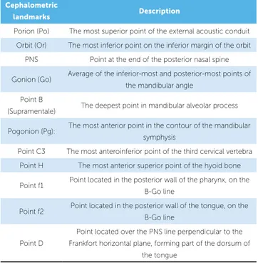
Table 1 - Cephalometric landmarks used in the study and their description.
Figure 1 - Tracings performed with Radiocef soft-ware.
Cephalometric
landmarks Description
Porion (Po) The most superior point of the external acoustic conduit
Orbit (Or) The most inferior point on the inferior margin of the orbit
PNS Point at the end of the posterior nasal spine
Gonion (Go) Average of the inferior-most and posterior-most points of the mandibular angle
Point B
(Supramentale) The deepest point in mandibular alveolar process
Pogonion (Pg): The most anterior point in the contour of the mandibular symphysis
Point C3 The most anteroinferior point of the third cervical vertebra
Point H The most anterior superior point of the hyoid bone
Point f1 Point located in the posterior wall of the pharynx, on the B-Go line
Point f2 Point located in the posterior wall of the tongue, on the B-Go line
Point D
Point located over the PNS line perpendicular to the Frankfort horizontal plane, forming part of the dorsum of
The average value of the increase in the oropharynx, though small, can have great clinical signiicance because, according to Trotman et al,23 a decrease of 1 mm in diam-eter of the upper airway of a child can decrease efective airspace by 65%, producing a critical obstruction.
Santos Júnior et al19 obtained results similar to ours in terms of oropharyngeal dimension and, conse-quently, the position of the tongue in the horizontal direction. They assessed pre- and postoperative lat-eral cephalometric radiographs of patients who had undergone genioplasty combined with nasal surgery or uvulopalatopharyngoplasty. The postoperative re-sults, which varied between 4 and 6 months, showed a 2.9 mm mean increase in oropharyngeal space.
Mandibular advancement surgery has revealed an increase in the oropharyngeal size and has been rec-ognized as one of the most successful procedures in the treatment of oropharyngeal space deiciency.7,16,17,25 According to Carlo et al,18 the principle underlying in-creases in oropharyngeal airspace is the fact that this surgery pulls the geniohyoid and genioglossus muscles forward, thereby causing an anterior movement of the tongue base and the hyoid bone and, consequently, increasing the posterior26 airspace. According to Ellis et al,5 in advancement genioplasty the tongue muscles, including geniohyoid and genioglossus muscles, must remain attached to the osteotomized bone segment in order to ensure adequate blood supply, minimizing the possibility of resorption and providing long-term stability. Preservation of the pedicle during surgery is responsible for the tongue moving anteriorly and this principle is not only embraced but advocated by other authors to this day.4,18,19,25,27
The indings of these research suggest the possibility that, with a relatively simple procedure. i.e., advance-ment genioplasty, one can expect an increase in oropha-ryngeal airspace as a result of the anterior repositioning of the base of the tongue and the hyoid bone, thereby
RESULTS
A high degree of intraexaminer agreement (0.99) was found through the correlation coefficient be-tween the measurements carried out by the research-er at two diffresearch-erent times. A high degree of correlation was also found in the inter-examiner assessment, with a coefficient of 0.98. Thus, no statistically significant difference was found for all measures assessed. In light of the results, the values of the first measurements were used for data analysis.
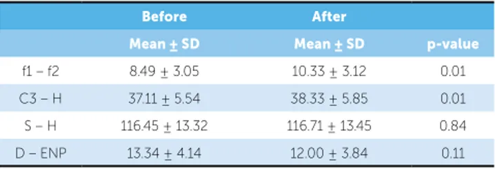
This study disclosed a statistically significant difference in the size of the oropharynx and in the positioning of the hyoid bone in the horizontal di-rection between both pre- and postoperative peri-ods (Table 2). The oropharynx increased on average 1.83 mm, pointing to a change in the position of the tongue anteriorly (p = 0.01) and the hyoid bone moved forward on average 1.59 mm (p = 0.002).
In the vertical direction, the change in the hyoid bone and tongue (p = 0.11) did not yield statistically sig-niicant results. Despite this, the hyoid bone proved to be more superior in over half of the sample (p = 0.61).
DISCUSSION
Lateral cephalometric radiographs were used be-cause airspace dimensions as well as tongue and hyoid bone position are properly reproduced in cephalo-grams where the head is in a natural position.21 Al-though radiographic cephalometry provides two-dimensional images, it has been widely used in the study of pharyngeal airspace and craniofacial mor-phology as it offers considerable advantages, such as low cost and minimal exposure to radiation.8,9,17 Moreover, according to Barbosa et al,22 lateral cepha-lometric radiography is efficient in the diagnosis of adenoid hypertrophy because it strongly correlates with nasal endoscopy, which is considered the gold standard for diagnosing this condition.
Table 2 - Means of variables at T0 and T1.
Before After
Mean ± SD Mean ± SD p-value
f1 – f2 8.49 ± 3.05 10.33 ± 3.12 0.01
C3 – H 37.11 ± 5.54 38.33 ± 5.85 0.01
S – H 116.45 ± 13.32 116.71 ± 13.45 0.84
D – ENP 13.34 ± 4.14 12.00 ± 3.84 0.11
Table 3 - Correlation between the amount of chin advancement and the
amount of oropharyngeal airspace increase and anterior repositioning of the hyoid bone.
1 Pearson’s correlation.
Variables
Chin advancement
Correlation
Coeicient p-value
Increased oropharyngeal size -0.50 0.11
rior mandibular repositioning. The authors found it to be more superiorly positioned. They argued that this change was due to increases in oral cavity space and the tendency of the tongue to occupy such space. For them, changes in the tongue occur whenever there is an increase in oral cavity size, which is not the case when advancement genioplasty is performed alone.
Santos Júnior et al19 showed a decrease of 2 mm on average in the vertical distance between the hy-oid bone and the mandibular plane. This may have occurred because in addition to advancement genio-plasty most of the sample was also subjected to uvulo-palatopharyngoplasty (UPPP), which may have con-tributed to statistically significant outcomes.
Achilleos et al9 also showed that the hyoid bone took on a higher position ater mandibular advance-ment surgery, in agreeadvance-ment with results obtained in other studies,8,16 in which its position remained un-changed in the long-term. When advancement genio-plasty was performed alone, changes in the stomato-gnathic system proved less signiicant than in man-dibular advancement surgery. Additionally, the sample size in this investigation (n = 11) may have contributed to the attainment of this result. Thus, further studies using a larger sample size are warranted.
CONCLUSIONS
Based on the results of this study, it is reasonable to conclude that advancement genioplasty alone corre-lates with a statistically signiicant increase in the oro-pharyngeal size as a result of an anterior repositioning of the base of the tongue. Changes in the position of the hyoid bone in the horizontal direction were also observed, probably due to a stretching of the suprahy-oid muscles resulting from chin advancement.
improving both esthetic and functional conditions of pa-tients with mandibular deiciency. Further studies must be conducted to assess the beneicial efect of this increase in oropharyngeal on breathing. By no means does chin advancement surgery replace mandibular advancement surgery, but it may be an alternative for some cases.
Ribeiro et al4 asserted that they failed to account for the amount of mandibular advancement when assessing changes in the same variables studied in this research, a factor that should have been taken into consideration. In this study, assessment of chin advancement alone did not disclose any correlation between the extent of the advancement and the amount of increase in oropha-ryngeal size (p = 0.11), nor between the extent of the advancement and the amount of anterior repositioning of the hyoid bone (p = 0.79) (Table 3). In other words, patients who experienced signiicant chin advance-ment did not necessarily attain considerable gains in the dimension of the oropharynx nor in anterior hy-oid bone repositioning. These results can be explained by the continuous change in the position of the hyoid bone that occurs throughout one’s lifetime, regardless of surgical interventions. These changes are age-related and difer in terms of facial pattern.28 In addition, since only the minimum period in which the surgery was performed was actually timed, the radiographs that de-picted the postoperative period might present diferent time intervals. It is expected that further studies aimed at examining the variance between groups with difer-ent levels of advancemdifer-ent will show greater accuracy in the assessment of this correlation.
ante-1. Ambrizzi DR, Franz SA, Pereira Filho VA, Gabrielli MAC, Gimenez CMM, Bertoz FA. Avaliação das queixas estético-funcionais em pacientes portadores de deformidades dentofaciais. Rev Dental Press Ortod Ortop Facial. 2002;12(5):63-70.
2. Freitas CE. Mentoplastia: um importante complemento, não uma solução.
In: Araújo A. Cirurgia Ortognática. São Paulo: Ed. Santos; 1999. cap. 12, p. 231-74.
3. Triaca A, Furrer T, Minoretti R. Chin shield osteotomy: a new genioplasty technique avoiding a deep mento-labial fold in order to increase the labial competence. Int J Oral Maxillofac Surg. 2009;38(11):1201-25.
4. Proit WR, Turvey TA, Moriarty JD. Augmentation genioplasty as an adjunct conservative orthodontic treatment. Am J Orthod. 1981;79(5):473-91. 5. Ellis III E, Dechou PC, McNamara JA Jr, Carlson DS, Liskiewicz WE.
Advancement genioplasty with and without soft tissue pedicle: an experimental investigation. J Oral Maxillofac Surg. 1984;42(10):637-45. 6. Pinto PA, Ferreira AP, Figueredo A. Perspectiva ortodôntica da cirurgia
do mento. Rev Portuguesa Estomatol Med Dent Cir Maxilofac. 2009;50(1):25-34.
7. Ribeiro C. Efeitos da cirurgia ortognática de avanço ou recuo mandibular nas posições do osso hióide, da língua e no tamanho da orofaringe [dissertação]. Salvador (BA): Universidade Federal da Bahia; 2009.
8. Turnbull NR, Battagel JM. The efects of orthognathic surgery on
pharyngeal airway dimensions and quality of sleep. Eur J Orthod. 2000;27(3):235-47.
9. Achilleos S, Krogstad O, Lyberg T. Surgical mandibular setback and changes in uvuloglossopharyngeal and head posture: a short and long-term cephamoletric study in males. Eur J Orthod. 2000;22(4):383-94. 10. Nuernberg CHG, Vilella OV. Avaliação cefalométrica da orofaringe. Rev
Odonto Ciênc. 2006;21(54):370-5.
11. Pae EK, Blasius JJ, Nanda R. Heterogeneity in vertical positioning of the hyoid bone in relation to genioglossal in men. Angle Orthod. 2004;74(3):343-48.
12. Souza RP, Pagotto SR, Paes Junior AJO, Soares AH, Rapoport A. Diagnóstico por imagem da cavidade oral. Radiol Bras. 2003;36(3):169-72.
13. Guven O, Saraçoglu U. Changes in pharyngeal airway space and hyoid bone positions after body ostectomies and sagittal split ramus osteotomies. J Cranio Surg. 2005;16(1):23-30.
14. Salles C, Campos OS, Andrade NA, Daltro C. Síndrome da apnéia e hipopnéia obstrutiva do sono: análise cefalométrica. Rev Bras Otorrinolaringol. 2005;71(3):369-72.
15. Baik UB, Suzuki M, Ikeda K, Sugawara J, Mitani H. Relationship between cephalometric characteristics and obstructive sites in obstructive sleep apnea syndrome. Angle Orthod. 2002;72(2):124-34.
REFERENCEs
16. Gonçalves JR, Buschang PH, Gonçalves DG, Wolford LM. Postsurgical stability of oropharyngeal airway changes following counter-clockwise maxillo-mandibular advancement surgery. J Oral Maxillofac Surg. 2006;64(5):755-62.
17. Pereira Filho VA, Jeremias F, Tedeschi L, Souza RF. Avaliação cefalométrica do espaço aéreo posterior em pacientes com oclusão Classe II submetidos à cirurgia ortognática. R Dental Press Ortodon Ortop Facial. 2007;12(5):119-25.
18. Bruno Carlo B, Mauro P, Silvia B, Enrico S. Modiied genioplasty and bimaxillary advancement for treating obstructive sleep apnea syndrome. J Oral Maxillofac Surg. 2008;66(9):1971-4.
19. Santos Junior JF, Abrahão M, Gregório LC, Zonato AI, Gumieiro EH. Mentoplastia para avanço do músculo genioglosso em pacientes com síndrome da apnéia-hipopnéia do sono obstrutiva e retrognatismo mandibular. Rev Bras Otorrinolaringol. 2007;73(4):480-6. 20. Heitz C, Lima LMS, Orso VA, Pinto LP, Ferreira AGM. Mentoplastia:
do planejamento à técnica cirúrgica. J Bras Ortodon Ortop Facial. 2005;10(55):29-35.
21. Malkoc S, Usumez S, Nur M, Donaghy CE. Reproducibility of airway dimensions and tongue and hyoid positions on lateral cephalograms. Am J Orthod Dentofacial Orthop. 2005;128(4):513-6.
22. Barbosa MC, Knop LAH, Lessa MM, Araújo TM. Avaliação da radiograia cefalométrica lateral como meio de diagnóstico da hipertroia de adenóide. Rev Dental Press Ortod Ortop Facial. 2009;14(4):83-91.
23. Trotman CA, McNamara JA, Dibbets JM, Van der Weele LT. Association of lip posture and the dimensions of the tonsils and sagittal airway with facial morphology. Angle Orthod. 1997;67(6):425-32.
24. Trenouth MJ, Timms DJ. Relationship of functional oropharynx to craniofacial morphology. Angle Orthod. 1999;69(5):419-23.
25. Prinsell JR. Maxillomandibular advancement surgery for obstructive sleep apnea syndrome. J Am Dent. 2002;133(11):1489-97.
26. Strauss RA, Abubaker AO. Genioplasty: a case for advancement osteotomy. J Oral Maxillofac Surg. 2000;58(7):783-87.
27. Polido WD, Bell WH. Long-term osseous and soft tissue changes after large chin advancements. J Cranio-Maxillofac Surg. 1993;21(2):54-9.
28. Pae EK, Quas C, Quas J, Garret N. Can facial type be used to predict changes in hyoid bone position with age? A perspective based on longitudinal data. Am J Orthod Dentofacial Orthop. 2008;134(6):792-7. 29. Athanasiou AE, Papadopoulos MA, Mazaheri M, Lagoudakis M.