Alberto Consolaro1,Renata Bianco Consolaro2, Leda Francischone3
How to cite this article: Consolaro A, Consolaro RB, Francischone L. Clari-ications, guidelines and questions about the dental bleaching “associated” with orthodontic treatment. Dental Press J Orthod. 2013 Sept-Oct;18(5):4-10.
Submitted: July 04, 2013
Revised and accepted: July 10, 2013
Contact address: Alberto Consolaro E-mail: [email protected] There was a time, before the 80s, when the
or-thodontist would perform tooth movement as well as malocclusion and esthetic correction only in young patients with all teeth, without prosthesis or im-plants, with completely healthy teeth and gingiva.
In addition to responding to functional and esthetic needs of patients of all ages and with diferent oral and systemic health conditions, the orthodontist has
cur-rently supported and helped with other types of oral rehabilitation treatments which vary in complexity.
During orthodontic planning, even before treat-ment onset, when patient’s expectations and projec-tions are still being surveyed, patients tend to ques-tion the clinician about when and how is the best moment for dental bleaching. White healthy teeth are an individual as well as a social need.
Clarifications, guidelines and questions about the dental
bleaching “associated” with orthodontic treatment
1 Head Professor, FOB-USP. Professor of the Post-Graduation Course,
FORP-USP.
2 Professor, Adamantinenses Integrated Schools (FAI). PhD in Pathology,
FOB-USP.
3 PhD in Oral Pathology, College of Dentistry — Bauru/USP.
» The authors report no commercial, proprietary or financial interest in the products or companies described in this article.
With regard to the best moment for carrying out or recommending dental bleaching to orthodontic patients, some explanations and orientations are given in order to answers the following questions: 1) Why orthodontic treatment completion is considered the best opportunity for carrying out the procedure? 2) Why dental bleaching should not be performed immediately before orthodontic treat-ment? 3) If that would be possible at any special case, what would that be? 4) Why dental bleaching should not be performed during orthodontic treatment? 5) If that would be possible at any special case, what would that be? This article highlights why it is essential to protect both the mucosa and the cervical region, regardless of the moment when dental bleaching is performed, whether associated with orthodontic treatment or not. The “how”, “why” and “if” of whether or not it is convenient to perform dental bleaching before orthodontic treatment are still a matter of clinical suggestion, as it is a procedure that is under analysis, empirical knowledge waiting for scientiic proof or disproof! Although tooth enamel has adamantine luid lowing within it, providing a speciic metabolism that is peculiar to its own and which could scientiically explain and base the option of carrying out teeth whitening before and during orthodontic treatment, we must still be very careful.
Keywords: Teeth whitening. Dental bleaching. Orthodontic treatment.
Quanto ao melhor momento para se aplicar ou recomendar a clareação dentária aos pacientes ortodônticos, alguns esclarecimentos e orientações são explanados para responder questionamentos como: 1) Por que depois do tratamento ortodôntico se constitui a melhor oportunidade para tal procedimento?; 2) Por que não realizar a clareação dentária imediatamente antes do tratamento ortodôntico?; 3) Se poderia realizá-la em alguma condição especial, e qual seria?; 4) Por que não se deveria clarear os dentes durante o tratamento ortodôntico?; 5) Se possível em algumas situações especiais, quando seriam essas situações especiais? No presente artigo, se destacará porque é fundamental sempre proteger a mucosa e a região cervical, independentemente do momento em que se izer uma clareação dentária relacionada ou não ao tratamento ortodôntico. O mecanismo de como, por que e se é ou não conveniente clarear os dentes antes da inalização dos tratamentos ortodônticos ainda representa uma sugestão clínica, um procedimento em análise e um conheci-mento empírico à espera de sua comprovação ou desmitiicação cientíica. Apesar do esmalte dentário ter uma circulação do líquido adamantino, que propicia um metabolismo próprio e especíico, que pode vir a ser, cientiicamente, a base para explicar e fundamentar a clareação dentária antes e durante o tratamento ortodôntico, ainda assim devemos ser muito cautelosos.
Generally, in clinical practice, dental bleaching must be performed prior to restorative and rehabilita-tive treatments, since such procedures are carried out on the basis of the color obtained from the bleaching technique. Teeth and lost spaces will be restored in harmony with all components of the future smile.
In Orthodontics, however, dental bleaching must be performed after the orthodontic appliance has been removed, except for specific cases with particu-lar purposes.
Based on orthodontic treatment patterns, this ar-ticle discusses the precautions, explanations and ori-entations related to the best moment for carrying out dental bleaching, as described in a previously pub-lished article9.
After such discussion, we will explain about the basis on which companies and clinicians have rec-ommended or indicated dental bleaching for patients undergoing orthodontic treatment.
WHY DENTAL BLEACHING SHOULD BE PER-FORMED AFTER ORTHODONTIC TREATMENT?
After orthodontic treatment finishes, all crown surfaces are positioned, properly exposed and ready to uniformly receive the bleaching agent. Neverthe-less, the initial results will not necessarily be uniform as some teeth may require additional application. This is due to the fact that not all teeth have the same color, just like the whole dental structure, especially the enamel, does not have the same volume, thick-ness and level of mineralization for all its elements.
In some cases, after the orthodontic appliance has been removed, the surfaces below the brackets may be irregular with regard to surface and color, and have resin fragments adhered to the enamel. These surfaces need to be evened with restorative abrasive procedures or remineralized in order to have uni-form color and surface.
Moreover, after orthodontic appliances have been removed, some adjustments must be made to the oral environment. Focal and occasional gingivitis must be reversed and the dental plaque properly controlled. After two to four weeks, these adjustments may lead to an oral environment that is ideal for a more ef-ficient and uniform dental bleaching. Dental plaque, calculus and resin fragments hinder the action of the bleaching agent on the enamel surface. Gingivitis
weakens the gingiva in cases in which the bleaching agent accidently reaches its structure.
WHY DENTAL BLEACHING SHOULD NOT BE PERFORMED BEFORE ORTHODONTIC TREAT-MENT? OR IT SHOULD? WHEN?
In orthodontic treatment, teeth may be intrud-ed or extrudintrud-ed, exposing areas previously coverintrud-ed by gingiva in case of malpositioned or unerupted teeth, a common situation for the maxillary canines. Should the procedure be carried out before orth-odontic treatment, the areas covered by gingiva or touching other teeth will not receive the bleaching agent as the other areas will. Such areas are exposed after orthodontic treatment and may end up having a different color.
Bleaching agents are hydrogen peroxide-based acid solutions that act over the teeth.2,6 They act over
the enamel surface causing it to be more porous. Simultaneously or soon after bracket placement, an acid attack is required in order to allow the resin to adhere to the enamel when it is being placed on the dental crown, in which case the enamel below these surfaces may be structurally weakened. When brack-ets are removed, enamel splinters may be detached, harming its surface.
Resin restorations may present increased infiltra-tions due to the hydrogen peroxide demineralization action and infiltrating capacity. Thus, it would not be different between resin and bracket on the enamel surface, causing it to easily detach. Similarly, dental plaque bacterial acids tend to accumulate and remain active, causing white spot lesions underneath the brackets, especially if the patient is not careful with his own oral hygiene.
Exposed crown area, where the enamel is more porous, is more subject to stronger dental plaques due to greater mechanical retention caused by brack-ets and other orthodontic devices.
Such an improvement may be motivating for the patient to withstand a long orthodontic treatment. However, it is worth noting that, for this purpose, the bleaching procedure must be brief and with a very specific aim. In addition, its application must be well controlled by a qualified professional.
Another reason for having dental bleaching per-formed before orthodontic treatment may be of pro-fessional nature, especially if we consider that the ap-pearance often predominates over candidates’ theo-retical or philosophical content. In the case of photo-graphic models and other professionals who depend on their image and exposure, white clean teeth are required. The same happens when the patient at-tends job interviews or other selection processes re-lated to job or artistic opportunities.
WHY DENTAL BLEACHING SHOULD NOT BE PERFORMED DURING ORTHODONTIC TREATMENT? OR IT SHOULD? WHEN?
Pores and perikymata exposure on the enamel surface will not happen in a controlled and uniform way in the areas below the brackets, even if infiltra-tion between the fixing resin and the dental crown is possible. Moreover, no studies have been carried out to investigate how the enamel and its respective structures as well as the physiology of areas below the fixed brackets act when subjected to bleaching agents.
The areas covered by gingiva or touching other teeth will not receive the bleaching agent as the other areas will. The risk of stains and irregularities in color and surface ater brackets removal is high, and the cost-beneit relationship is not worthy in normal cases.
Resin restorations may increase infiltrations by hydrogen peroxide demineralization action and in-filtrating capacity. Thus, it would not be different between resin and bracket on the enamel surface, causing it to easily detach. Dental plaque bacterial acids tend to accumulate and remain active, causing white-spot lesions underneath the brackets, espe-cially if the patient is not careful with his own oral hygiene. Exposed crown area, where the enamel is more porous, is more subject to stronger dental plaques due to greater mechanical retention caused by brackets and other orthodontic devices.
But, is it possible? When? In specific cases, it may be necessary to improve the appearance of
pigmented irregularities between the brackets, teeth and appliances, because since the first sessions of dental bleaching, the appearance of teeth may be generally improved due to the cleaning efficacy of hydrogen peroxide which penetrates into the small-est recesses such as lamellas, cracks or natural depres-sions and irregularities of the enamel.
A cleaner aspect of the orthodontic appliance may be a motivating factor for the patient to withstand a long orthodontic treatment. However, it is worth noting that, for this purpose, the bleaching pro-cedure must be brief and with a very specific aim. When the purpose is to effectively bleach teeth with fixed brackets, it should be highlighted that the risk of stains and irregularities in color and surface after bracket removal is high.
Additionally, the bleached enamel may be weaker in structure. In normal cases, surface enamel is even-tually removed when brackets are removed. How-ever, it is unknown whether or not this situation in-creases in cases of bleached enamel.
Another reason for having dental bleaching per-formed during or before orthodontic treatment may be of professional nature, especially if we consider that appearance often predominates over candidates’ intellectual and emotional content.
In the case of photographic models and other pro-fessionals who depend on their image and exposure, white clean teeth are required. The same happens when the patient attends job interviews or other se-lection processes related to job or artistic opportu-nities. The use of appliances alone may not affect the aspect employers expect from candidates, how-ever, should they have healthy teeth, without dental plaque or pigmentation that give them an unpleas-ant appearance at smiling, they will not be restrained from being hired.
MUCOSA AND CERvICAL REGION PROTECTION: ALWAYS, REGARDLESS OF THE MOMENT!
Dental bleaching requires qualified profession-als who are capable of performing the procedure on the patient. Protection of the oral mucosa and the cervical region of teeth is among the abilities the professional must have.1,2,4,5-8 The cervical enamel is
structurally relates itself to the cementum that covers the root. Along the cementoenamel junction, all per-manent or deciduous teeth exhibit areas or gaps with
dentin exposed to the oral environment.4-7
Exposure of dentin windows to the oral envi-ronment occurs when the cementoenamel junction “migrates” or is displaced from the gingival connec-tive tissue to the groove due to migration or continu-ous and compensatory dental eruption mechanism of abrasion in the incisal edges and occlusal aspects.
When exposed to bleaching agents, the areas of
dentin exposure and the dental tubules are widened.6
These effects result in excessive and uncomfortable increase in dental sensibility after bleaching, which may result in unbearable discomfort for a few pa-tients. Thus, protecting the cervical region which receives the bleaching agent during the procedure is essential. From a clinical point of view, the most effi-cient and practical form to do so is by means of resin protective cervical barriers,4 however, other options
may be used to protect the cervical region. Rubber sheets, for instance, which are used in restorative procedures and not widely used nowadays.
In addition to protecting the cementoenamel junction, the resin protective cervical barrier also protects the gingival mucosa against hydrogen
per-oxide-based bleaching agents.2,4 Many researches
re-veal that hydrogen peroxide may promote chemical carcinogenesis of mucosa, including the oral muco-sa.1,8 Promoters of chemical carcinogenesis are those
products that maximize the mutagenic effect of other agents and for this reason are considered carcinogen-esis initiators. They are largely present in the mouth in the form of tobacco, herbicide, pesticide, alcohol,
virus and many other substances.1,8
The resin cervical barrier protects the oral muco-sa from receiving hydrogen peroxide provided that before it is removed, the dentist uses a specific type of suction tube to suck the gel or bleaching sub-stance. It is advisable to cut the outer part of the dis-posable tip so that it becomes more efficient to suck the smallest portions of hydrogen peroxide present in the small recesses between the resin barrier and the tooth.4
The suction tube, or its disposable active part, that sucks saliva cannot be the same one that removes the bleaching agent as it will be contaminated by
hydro-gen peroxide and, thus, contaminate the oral muco-sa. If the tube is not carefully used, there is no point in using the resin cervical barrier, which must be taken off only after all hydrogen peroxide has been removed. Should there be hydrogen peroxide left in the patient’s mouth, he will swallow it and the sub-stance will affect his gastrointestinal mucosa with the same potential carcinogenic effects.
HOW CAN WE UNDERSTAND THAT BLEACH-ING AGENTS POSSIBLY ACT ON THE ENAMEL SURFACE BELOW THE BRACKETS?
Some standard procedures are based on clinical ob-servation that is, in general, a result of a venturesome procedure carried out on the basis of previous clinical processes employed in similar cases, aiming at broad-ening the horizons of clinical practice. After knowing about these “indications and results”, researchers and scholars analyze the biological applicability and feasi-bility of procedures. How can we explain about the basis on which companies and clinicians have “rec-ommended” or “indicated” dental bleaching for pa-tients undergoing orthodontic treatment?
First, we need to understand the enamel as a liv-ing tissue. Tooth enamel is alive,3 although it is
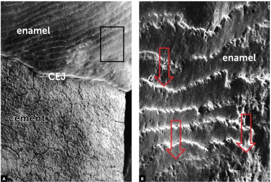
com-monly thought to be an inactive and amorphous tis-sue. Once the enamel is produced by the ameloblasts, it is formed by the rods, truly twisted and laterally connected beams. In cross section, the enamel rods are geometrically compared to a keyhole. Among the rods, there is an interrod sheath. Each rod is made up of millions of hexagonal crystals that may be com-pared to bricks or stones (Fig 1) among which the “adamantine fluid” flows.
In addition to taking part in the hydration process of each crystal, the adamantine luid brings and takes ions from the enamel surface as well as from other parts of it. Due to the low of adamantine luid, the ions are incorporated into the enamel surface and may achieve its deepest parts, as it happens with the topic use of luoride. This ion exchange allows enamel rem-ineralization in the white spots caused by caries ater external dental bleaching. At tooth eruption, the pro-portion of minerals between the deepest and surface enamel layers is similar, however, once it is exposed to the oral environment, the enamel surface becomes
The wavy enamel surface, or perikymata surface, exhibits development pores (Fig 1), millions and true depressions that represent the marks left by the am-eloblast Tomes’ process when the last layer of amelo-blasts is deposited. Fluids and ions enter through the surface pores, renewing the crystals. However, these pores also allow that other substances, including un-desirable ones, enter the inner parts of the enamel.
When bacterial as well as other acids enter the enamel via pores and adamantine fluid, they pro-mote demineralization of crystals, dissolving them as an ice cube melts inside a glass, reducing its struc-ture and altering its form. The intercrystal space will increase in size, therefore, increasing enamel poros-ity, allowing even more undesirable agents, such as bacterial acids and pigments, to enter. Many of these products or agents may reach the dentin or even the dental pulp. At this stage, in enamel decay still in the form of white spots, there are subtle and subclinical localized pulp alterations.1
The enamel is alive! When leveling, cutting, removing, smoothing, bleaching and chemically changing the enamel, one aspect must be borne in mind: we are working with a living tissue. Similarly to the care given to the dentin, and even more, to the pulp, one must be careful when working with the enamel.1
When external dental bleaching is finished, the enamel surface pores are more open; resin bonding is weaker; orthodontic appliances are more prone to detaching; and the splits more open. Should acids be applied to the enamel for technical purposes, it must be done only if necessary. Demineralized enamel ar-eas exposed to the oral environment are more prone to incorporating pigments and allowing toxic prod-ucts and acids to enter, thus, promoting alterations in color and increased sensibility in the affected tooth.
Similarly, one must be selective and demanding when indicating or recommending cosmetic prod-ucts, dentifrices, antiseptics, eating habits and special care related to the teeth, since these aspects influence enamel porosity, composition and structure as well as the dentin and the pulp. The flow of adamantine fluid among the enamel crystals connects the dental surface with the dentinal fluid. What we do to the enamel reflects on the dentin and, in smaller propor-tions, on the dental pulp. This is due to the fact that
not only tooth color depends on the dentin, but also because there is a specific flow within these tissues.1
Women, who are careful about their jewels, wash them with seawater, handling, caring and keeping their pearls at special places because they strongly believe that the pearls are alive! They do not expose their pearls to perfume; neither allow them to rub against other surfaces. They want to prevent their pearls from dying and, as a consequence, losing their brightness, vigor and fascination.1
Hydrogen peroxide is highly penetrative in enamel; perhaps, it is the most penetrative chemical substance in dental structures. Should it be applied on enamel nearby, to which a bracket is adhered, hydrogen peroxide may reach the enamel subsurface and, thus, bleach the area of enamel that is covered by the bracket (Figs 1 and 2).
However, we must be careful, since we do not know how the hydrogen peroxide flows through the adamantine fluid: Does it flow uniformly? The bleached surface would become irregular, would it not? What is the speed of the bleaching agent dif-fusibility? Would hydrogen peroxide extend to lat-eral areas when applied on a surface? Wouldn’t the bleaching procedures performed on enamel below the bracket weaken it and cause splinters to be bro-ken off the enamel when brackets are removed?
Why are these questions in this article? This “In-sight” sections aim at suggesting, highlighting and instigating new researches that prove or disprove theories, confirming hypothesis and producing sci-entific knowledge on the basis of concrete data and evidence! The “how”, “why” and “if” of whether or not it is convenient to perform dental bleaching before orthodontic treatment are still a matter of clinical suggestion, as it is a procedure that is under analysis, empirical knowledge waiting for scientific proof or disproof!
FINAL CONSIDERATIONS
The best moment to recommend dental bleach-ing associated with orthodontic treatment coincides with treatment finishing because that is when all dental surfaces are uniformly exposed. What had to be extruded, intruded or leveled, had already been.
precau-Figure 1 - Morphological aspects of human enamel surface in scanning electron microscopy: with periky-mata, or wavy, and very porous. The cementoenamel junction (CEJ) is also observed. (SEM: 100x in A and 500x in B).
Figure 2 - A) Cross section of a human tooth. Morphological aspects of enamel surface and its relationship with the dentin and the cementum. (CEJ = cemen-toenamel junction). B) Transmission electron microscopy reveals the enamel crystals and the spaces where the adamantine fluid flows (arrows), which may carry the dental bleaching agent.
bleaching
agent
enamel
dentin
CEJ
b
enamel
cement
CEJ
enamel
A
A
tions and exceptions must be clarified when dental bleaching is exceptionally associated with orthodon-tic treatment before the latter is finished.
Frequent bracket detachment, white spots, loss of enamel color uniformity after orthodontic appli-ance is removed, retention of bacterial plaque and potential detachment of surface parts during brack-ets removal are among the consequences of dental bleaching associated with orthodontic treatment, due to the fact that bleaching agents promote enamel demineralization. Associating dental bleaching with orthodontic treatment is an exception that requires a lot of care and scientific knowledge. Additionally, it is of paramount importance that the patient knows about its potential risks.
It is worth noting that dental bleaching should al-ways be performed by a qualiied professional properly
trained to act directly on the patient, always protect-ing the tooth cervical region as well as the oral mucosa with mechanical barriers — among which the resin barrier is the most widely used — in order to prevent the hydrogen peroxide from reaching the cementoe-namel junction and its action on the oral mucosa.
The “how”, “why” and “if” of whether or not it is convenient to perform dental bleaching before orthodontic treatment are still a matter of clinical suggestion, as it is a procedure that is under analy-sis, empirical knowledge waiting for scientific proof or disproof. Adamantine fluid flow in the dental enamel, with its own specific metabolism, may be the scientific basis to explain and justify the applica-tion of dental bleaching before and during orthodon-tic treatment. However, we must be very careful and wait for tests, results and evidences!
1. Camargo WR. Análise do potencial carcinogênico de dentifrício com
peróxido de hidrogênio e de agente clareador dentário. Avaliações clínico-macroscópica e microscópica em hamsters em modelo de carcinogênese bucal DMBA-induzida [tese]. Bauru (SP): Universidade de São Paulo; 1999.
2. Consolaro A. Reabsorções dentárias nas especialidades clínicas. 3ª ed.
Maringá: Dental Press; 2012.
3. Consolaro A. Há vida no esmalte: metabolismo e circulação própria. Rev
Dental Press Estét. 2004;1(1):123-7.
4. Francischone LA, Consolaro A. Clareação dentária externa: importância
e tipos de proteção da junção amelocementária. Rev Clín Ortod Dental Press. 2005;4(5):88-98.
REFERENCES
5. Francischone LA, Consolaro A. Morphology of the cement-enamel
junction of primary teeth. J Dent Child. 2008;75(3):252-9.
6. Esberard R, Esberard RR, Esberard RM, Consolaro A, Pameijer CH.
Efect of bleaching on the cemento-enamel junction. Am J Dent. 2007;20(4):245-9.
7. Neuvald L, Consolaro A. Cemento-enamel junction: microscopic analysis
and external cervical resorption. J Endod. 2000;26(9):503-8.
8. Pieroli DA. Avaliação do potencial carcinogênico dos agentes clareadores
dentais [dissertação]. Bauru (SP): Universidade de São Paulo; 1997.
9. Consolaro A, Consolaro RB, Francischone L. Clareação dentária e o