D Y N A M I C M A N U S C R I P T S
Transvesical peritoneoscopy with rigid scope: feasibility study
in human male cadaver
Frederico Branco•Giovannalberto Pini•Luı´s Oso´rio•
Victor Cavadas•Rui Versos •Ma´rio Gomes•
Riccardo Autorino•J. Correia-Pinto •Estevao Lima
Received: 27 May 2010 / Accepted: 22 October 2010 / Published online: 22 December 2010 ÓSpringer Science+Business Media, LLC 2010
Abstract
Background Transvesical port refers to the method of accessing the abdominal cavity through a natural orifice (i.e., urethra) under endoscopic visualization. Since its introduction in 2006, various reports have been published describing different surgical interventions using a rigid ureteroscope in a porcine model. The aim of this study was to test the access and feasibility of peritoneoscopy by using a rigid ureteroscope in a human male cadaver.
Methods Two adult male cadavers were used to perform the procedures. A rigid ureteroscope was used for the creation of transvesical access into the peritoneal cavity. Peritoneoscopy, liver biopsy, and identification and manipulation of the ileocecal appendix were performed. Results Transvesical access into the peritoneal cavity was quickly established. The rigid ureteroscope easily allowed visualization of the abdominal cavity with good image quality. Liver biopsy and manipulation of ileocecal appendix were carried out without difficulties.
Conclusions Peritoneoscopy, liver biopsy, and ileocecal appendix manipulation using a rigid ureteroscope through a transvesical port is feasible in a cadaver model. The development of a specific rigid scope for the transvesical port might herald a promising future for this NOTES access. Keywords CadaverEndoscopySurgeryTransvesical NOTES
The craving for the discovery of new, minimally invasive surgical procedures allowed a new surgery concept to emerge: natural orifice transluminal endoscopic surgery (NOTES). The main challenge of this new concept is the execution of numerous surgical procedures through natural orifices, with the consequent advantages that may result, such as cosmetic benefits due to the absence of a surgical incision. The absence of a surgical incision also means less risk of wound infection and potentially less pain. In 2004, Kalloo et al. [1] described access to the peritoneal cavity through the transgastric port in a porcine model. Since then, several studies have been performed using transga-stric access [2–6]. However, many limitations were described, particularly when singly performed by transga-stric port.
Electronic supplementary material The online version of this
article (doi:10.1007/s00464-010-1496-x) contains supplementary material, which is available to authorized users.
F. BrancoL. Oso´rioV. CavadasM. Gomes
J. Correia-PintoE. Lima (&)
Life and Health Research Institute, School of Health Sciences, University of Minho, Braga, Portugal
e-mail: estevaolima@ecsaude.uminho.pt
G. Pini
Department of Urology, University of Modena and Reggio Emilia, Modena, Italy
R. Versos
Department of Urology, Centro Hospitalar do Alto Ave, Guimara˜es, Portugal
J. Correia-Pinto
Department of Pediatric Surgery, Hospital de Sa˜o Joa˜o, Porto, Portugal
R. Autorino
Glickman Urological and Kidney Institute, Cleveland Clinic, Cleveland, OH, USA
E. Lima
guidance in choosing the site of entry into the peritoneal cavity via the transgastric port [9, 10]. The transvesical port, although at the lower end of the abdomen, also allowed the execution of thoracic procedures in the porcine model [11].
Some critics question the feasibility and reproducibility of these procedures in the human being, particularly regarding the use of rigid instruments. The distance from the bladder to other organs in the abdominal cavity is larger than in the animal model, which could limit the imaging and manipulation of the organs of the upper abdominal cavity. Another questioned aspect is the possibility of obtaining images of the upper abdomen using rigid instruments without angulation, which might preclude the use of the scopes currently on the market in the transvesical approach in humans. Therefore, transvesical access to the peritoneal cavity might be a reality not only in the animal model, but also in the human model in the near future, especially if it is possible to use rigid instruments in this procedure.
The aim of this study was to describe and test the fea-sibility of NOTES procedures performed in a human male cadaver, with access to the abdominal cavity made through the transvesical port and by using rigid instruments.
Material and methods
The procedures were performed at the Institute of Forensic Medicine, North Delegation, Porto, Portugal. The experi-mental protocol was approved by the Institutional Review Committee. Two adult male cadavers were used.
Access
The cadaver was placed in the lithotomy position. An HD platform (Image 1 HD, Storz) was used. The procedure was started with the transurethral introduction of a 9.5 Fr rigid ureteroscope (Storz 27002L) connected to a saline
gation was replaced by gas (CO2) insufflation and creation of the pneumoperitoneum was obtained with a maximum pressure of 12 mmHg.
Procedure
Peritoneoscopy was performed with visualization of the entire peritoneal cavity, including bowel loops, omentum, stomach, liver, and gallbladder. At the liver edge, a biopsy was performed by introducing a 5 Fr Pe´res Castro forceps (Storz 274525R). To identify the ileocecal appendix, bowel loops and omentum were lifted and the blind loops were explored, with the ileocecal appendix identified and iso-lated/manipulated at the end. After the peritoneoscopy, the ureteroscope was removed from the peritoneal cavity through the previously created bladder orifice, without it being dilated or lacerated. The same procedure was repe-ated on the other cadaver (see video).
Results
The mean duration of the entire procedure, including ure-throscopy, cystoscopy, creation of bladder access, peri-toneoscopy with liver biopsy, and ileocecal appendix identification with isolation, was 11 min. During the entire procedure the clarity of the images obtained was remark-able, with the use of either saline or gas. The pneumo-peritoneum was obtained quickly resulting in an excellent view of intra-abdominal organs by keeping a maximum CO2 pressure of 12 mm Hg. The progression of the ure-teroscope within the peritoneal cavity was found to be safe, and no difficulty was encountered in identifying the structures of the upper abdomen (Fig.1).
mobilized by using forceps, without the need to change the position of the cadaver. It was also possible to identify the various segments of the intestine and the ileocecal appen-dix with its subsequent manipulation (Fig.3).
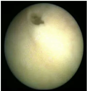
At the end of the procedure the bladder orifice main-tained the same diameter it had at the beginning of the procedure, i.e., a diameter of about 10 Fr (Fig.4).
Discussion
The current study shows that peritoneoscopy with liver biopsy and ileocecal appendix manipulation is feasible in a human cadaver model by using rigid instruments exclu-sively through a transvesical approach.
Transvesical access to the abdominal cavity either alone or in combination with transgastric access has been used for a broad range of procedures such as peritoneoscopy, liver biopsy, lung biopsy, cholecystectomy, nephrectomy, and partial cystectomy [7–12]. More recently, Gettman et al. [13] reported the clinical feasibility of transvesical peritoneoscopy by using a flexible ureteroscope (DUR-8) during a case of robotic radical prostatectomy. They used the same technical mode of transvesical approach in a porcine model with a few modifications. However, in this study it was recognized that the flexibility of the scope was a disadvantage and a limitation mainly when applying force to tissue because it was very difficult to both push and pull at the same time. In fact, it was recognized that
Fig. 1 Endoscopic view of upper abdominal organs provided by a
ureteroscope
Fig. 2 Representative endoscopic views of liver biopsy
Fig. 3 Manipulation of the ileocecal appendix with the forceps
introduced by the ureteroscope working channel
Fig. 4 Endoscopic view of the bladder orifice at the end of the
bladder perforations after five nephroureterectomies with an endoloop via a 15-mm umbilical trocar with the assis-tance of a 2-mm transurethrally placed endoscopic clamp. Thus, the transvesical approach has been shown to be an excellent way to access the peritoneal cavity by providing several advantages: (1) it is naturally sterile, (2) its location is advantageous, i.e., it in the most anterior portion of the pelvic cavity allowing peritoneal access above the bowel loops, (3) it is possible to introduce rigid instruments via the working channels of the scopes, thus enhancing the possibility of retracting and grasping structures, (4) pneu-moperitoneum is achieved quickly and easily, (5) the procedure can be performed on both genders. The unique disadvantage is that the diameter of the urethra limits the size of the devices used and the size of the specimen retrieved at the end of the procedure. Despite these potential advantages, several issues remain to be answered: (a) Would it be possible to use rigid instruments through the transvesical port in a human model? (b) Would it be possible to obtain images with the same clearness of the abdomen in the human model? (c) Would it be possible to easily handle and even perform procedures in the human model using rigid instruments through the transvesical port?
Our study revealed that it was easy to perform vesical access to the peritoneal cavity with the current rigid ure-teroscope. Perforation of the bladder wall was rapid, easily manageable, and safe. It should be emphasized that the image provided by the ureteroscope allowed us to have a good view of the perivesical anatomy and to achieve the parietal peritoneum easily and then the peritoneal cavity. After entrance into the peritoneal cavity, saline irrigation was replaced by CO2 insufflation and the ureteroscope working channel allowed the creation of a pressure-con-trolled pneumoperitoneum with a maximum pressure of 12 mmHg, as in swine experiments.
Regarding peritoneoscopy, we were able to identify easily the whole abdomen with good image quality. The length of ureteroscope used (43 cm) reached the edge of
used with them (the endoscope shaft should contain a lar-ger channel so other instruments can be introduced with better efficiency, and (4) the current rigid ureteroscope is fine but the axis between the pelvis and abdomen suggests that there should be a slight curve in the endoscope to allow better visualization of other organs that are not in the axis where the scope is introduced.
In conclusion, peritoneoscopy, liver biopsy, and ileo-cecal appendix manipulation using a rigid ureteroscope through a transvesical port is feasible in a human male cadaver. The development of a specific rigid scope for a transvesical port might herald a promising future for this NOTES access.
Disclosures Frederico Branco, Giovannalberto Pini, Luı´s Oso´rio,
Victor Cavadas, Rui Versos, Ma´rio Gomes, Riccardo Autorino, J. Correia-Pinto, and Estevao Lima have no conflicts of interest or financial ties to disclose.
References
1. Kalloo AN, Singh VK, Jagannath SB, Niiyama H, Hill SL, Vaughn CA, Magee CA, Kantsevoy SV (2004) Flexible trans-gastric peritoneoscopy: a novel approach to diagnostic and ther-apeutic interventions in the peritoneal cavity. Gastrointest Endosc 60:114–117
2. Park PO, Bergstro¨m M, Ikeda K, Fritscher-Ravens A, Swain P (2005) Experimental studies of transgastric gallbladder surgery: cholecystectomy and cholecystogastric anastomosis (videos). Gastrointest Endosc 61:601–606
3. Kantsevoy SV, Jagannath SB, Niiyama H, Chung SS, Cotton PB, Gostout CJ, Hawes RH, Pasricha PJ, Magee CA, Vaughn CA, Barlow D, Shimonaka H, Kalloo AN (2005) Endoscopic gastro-jejunostomy with survival in a porcine model. Gastrointest En-dosc 62:287–292
4. Bergstro¨m M, Ikeda K, Swain P, Park PO (2006) Transgastric anastomosis by using flexible endoscopy in a porcine model (with video). Gastrointest Endosc 63:307–312
6. Merrifield BF, Wagh MS, Thompson CC (2006) Peroral trans-gastric organ resection: a feasibility study in pigs. Gastrointest Endosc 63:693–697
7. Lima E, Rolanda C, Peˆgo JM, Henrique-Coelho T, Silva D, Carvalho JL, Correia-Pinto J (2006) Transvesical endoscopic peritoneoscopy: a novel 5 mm port for intra-abdominal scarless surgery. J Urol 176:802–805
8. Lima E, Rolanda C, Peˆgo JM, Henriques-Coelho T, Silva D, Oso´rio L, Moreira I, Carvalho JL, Correia-Pinto J (2007) Third-generation nephrectomy by natural orifice transluminal endo-scopic surgery. J Urol 178:2648–2654
9. Rolanda C, Lima E, Correia-Pinto J (2007) Searching the best approach for third-generation cholecystectomy. Gastrointest En-dosc 65:354–355
10. Rolanda C, Lima E, Peˆgo JM, Henriques-Coelho T, Silva D, Moreira I, Macedo G, Carvalho JL, Correia-Pinto J (2007) Third-generation cholecystectomy by natural orifices: transgastric and transvesical combined approach. Gastrointest Endosc 65:111–117 11. Lima E, Henriques-Coelho T, Rolanda C, Pego JM, Silva D, Carvalho JL, Correia-Pinto J (2007) Transvesical thoracoscopy: a
natural orifice translumenal endoscopic approach for thoracic surgery. Surg Endosc 21:854–858
12. Sawyer MD, Cherullo EE, Elmunzer BJ, Schomisch S, Ponsky LE (2009) Pure natural orifice translumenal endoscopic surgery partial cystectomy: intravesical transurethral and extravesi-cal transgastric techniques in a porcine model. Urology 74: 1049–1053
13. Gettman MT, Blute ML (2007) Transvesical peritoneoscopy: initial clinical evaluation of the bladder as a portal for natural orifice translumenal endoscopic surgery. Mayo Clin Proc 82: 843–845
14. Lima E, Rolanda C, Oso´rio L, Peˆgo JM, Silva D, Henriques-Coelho T, Carvalho JL, Bergstro¨m M, Park PO, Mosse CA, Swain P, Correia-Pinto J (2009) Endoscopic closure of transmural bladder wall perforations. Eur Urol 56:151–157
