* Corresponding Author: Ali Ghavidel, Email: [email protected] © 2014 The Authors; Tabriz University of Medical Sciences
This is an Open Access article distributed under the terms of the Creative Commons Attribution License (http://creativecommons.org/licenses/by/3.0), which permits unrestricted use, distribution, and reproduction in any medium, provided the original work is properly cited.
Case Series
Aortoenteric fistula as a rare manifestation of rupture of an abdominal
aortic aneurysm: Case report a diagnostic dilemma
Ali Ghavidel*1
1 Assistant Professor, Liver and Gastrointestinal Diseases Research Centre, Imam Reza Hospital, Tabriz University of Medical Sciences, Tabriz, Iran
Citation: Ghavidel A. Aortoenteric fistula as a rare manifestation of rupture of an abdominal aortic aneurysm: Case report a diagnostic dilemma. J Anal Res Clin Med 2014; 2(4): 217-20.
Introduction
Sudden hematemesis is a life-threatening emergency that directs a physician’s attention toward various causes of gastrointestinal bleeding. Aortoenteric fistula is an uncommon but life-threatening complication of aortic reconstructive surgery.1 Communications
between the aorta and the intestine resulting from disease at either site are referred to as aortoenteric fistulas. Fistula formation between the aorta and the intestinal tract was first described in 1839 in reference to a man with a “pulsating tumor and a discharge of bloody stool,” who died suddenly. At autopsy, it was noted that “the jejunum had adhered to the aneurismal bag and that sac had ulcerated into the intestine.”2
Fistulas occurring after aortic reconstructive surgery, also called aortic graft-enteric fistulas, are considered secondary aortoenteric fistulas
(SAF). Before 1960, the most common cause of abdominal aortoenteric fistulas was aortic aneurysm, followed by infectious aortitis due to syphilis or tuberculosis.3 However, over the
past three decades or so, erosion of the intestine by prosthetic vascular grafts has become a much more common cause, with an incidence of up to 4%.4,5 The complication often occurs
months to years after the original surgery. Bastounis et al. that the mean interval from the initial operation to the onset of upper gastrointestinal bleeding was 32 months.5 The
20 years’ experience with SAF at the Johns Hopkins Medical institution showed the average to be 2.8 years.6 The first reported SAF
was reported by Brock in a case involving an aortic homograft and the duodenum.7
Claytor et al. presented the first aortoenteric fistula caused by a prosthetic graft of the aorta.8
In Mac Kenzie et al.9 demonstrated the first Ghavidel A, J Anal Res Clin Med, 2014, 2(4), 217-20.
doi: 10.5681/jarcm.2014.036, http://journals.tbzmed.ac.ir/JARCM
Abstract
Introduction: Secondary aortoenteric fistula (SAF) is an uncommon, but very important
complication of abdominal aortic reconstruction. The complication often occurs months to years after aortic surgery. The clinical manifestation of the aortoenteric fistula is always upper gastrointestinal bleeding. Treatment of the disease is early surgical intervention. If operative treatment is not performed promptly, the mortality is high.
Case Report: A case of secondary aortoduodenal fistula found 6 years after aortic
reconstructive surgery, with the clinical presentation of upper gastrointestinal bleeding because of the increasing number of elective aortic aneurysm repairs in the aging population, it is likely that more patients with SAF will present to the clinical physicians in the future. Hence, a high index of suspicion is necessary for prompt diagnosis and treatment of this life-threatening event. The patient treated medically and finally expiration of the patient and review of the literature currently available in Medline.
Conclusion: The aim of this case report is to emphasize early diagnosis and management of
all gastrointestinal bleeding in patients who have a history of aortic reconstructive surgery. Article info
Article History: Received: 15 June. 2014 Accepted: 14 Sep. 2014 ePublished: 30 Nov. 2014
Keywords:
Abdominal Aortic, Aneurysm, Allograft,
Aorto-enteric fistula, aortic aneurysm
218 JARCM/ Autumn 2014; Vol. 2, No. 4
successful repair of an SAF between a synthetic graft and the intestine. Owing to the anatomic proximity, the majority of cases involve the duodenum, with the proximal suture line of an aortic prosthesis. Prompt diagnosis with surgical intervention is the only possible treatment that preserves the patient’s life. Due to the nonspecific nature of the clinical history and physical findings, diagnosis of aortoenteric fistula is difficult to make preoperatively. There is no single diagnostic investigation that has a very high specificity and sensitivity, including upper computed tomography (CT), angiography, or gallium-67 CT.
Gastrointestinal endoscopy is the most helpful method for diagnosis. If findings are negative, this test is meaningless unless another source of bleeding is found. Nevertheless, exploratory laparotomy is the only method that can definitely confirm the diagnosis.
Case Report
We present a case of secondary aortoduodenal fistula found 6 years after aortic reconstructive surgery, with the clinical presentation of upper gastrointestinal bleeding. The patient was a 70-year-old man who complained of hematemesis and melena. He gave a history of aorto-femoral reconstructive surgery in Tehran, Iran, 6 years ago. There was no history of peptic ulcer disease or any other gastrointestinal pathology.
On physical examination, the patient appeared pale with cold, clammy skin in a pre-shock condition. His vital signs were recorded as pulse rate 112 beats per minute regular, respiratory rate 22 breaths/min, and
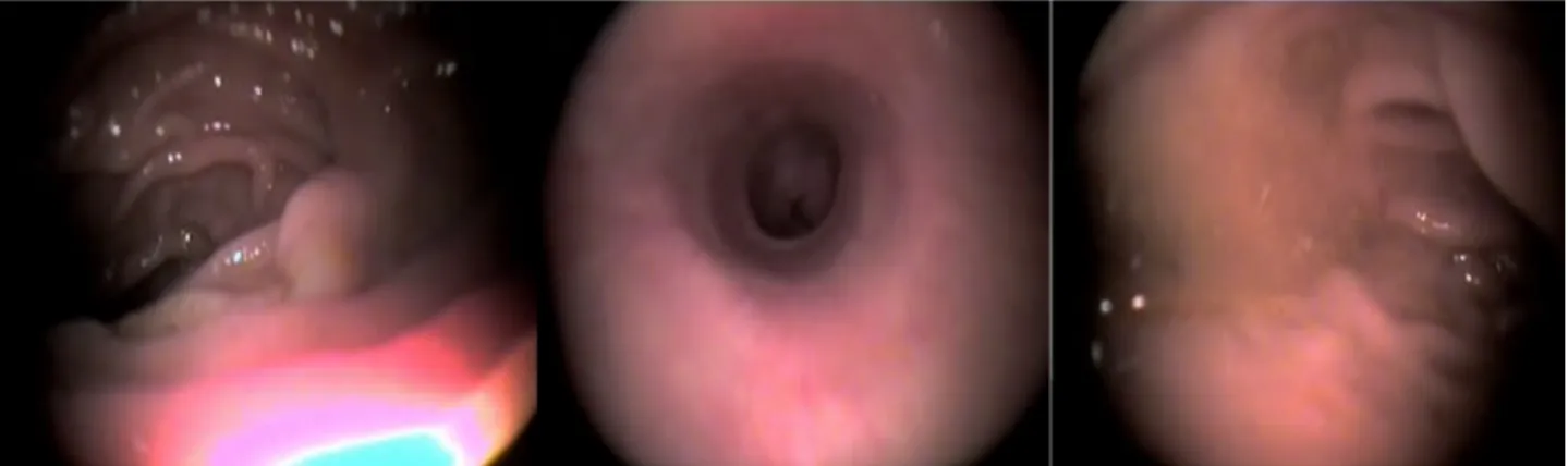
blood pressure 90/60 mm Hg. Chest wall, heart, and lungs were normal on physical examination. There was a median linear scar on his abdomen showing previous abdominal surgery. Epigastrium was tender on palpation. None of the abdominal viscera was palpable. His past surgical records revealed aorto-bifemoral graft 6 years ago. CT showed an aneurysmal mass around the graft (Figure 1). He was admitted with massive hematemesis and hypovolemic shock. The clinical examination, blood tests, electrocardiogram, and chest roentgenogram were unremarkable only with abdominal tenderness emergency gastroscopy could not identify a bleeding source due to massive amounts of clot and fresh blood in the stomach. However, bright red blood was seen in the stomach and injection therapy with repeated upper endoscopy was not possible due to massive amounts of clot and fresh blood in the stomach (Figure 2).
Figure 1. There is a defect in the anterior wall of the (repaired) aortic aneurysm
Ghavidel A
JARCM/ Autumn 2014; Vol. 2, No. 4 219
Surgical consultation was requested, but the surgeon refused operation due to inappropriate condition for surgery. The patient rebleeded, and another upper endoscopy was performed, but with no successful results and the patient was expired. This case illustrates intensive care treatment of a life-threatening hemorrhage from an aortoentric fistula.
There is a defect in the anterior wall of the (repaired) aortic aneurysm contiguous with the third part of the duodenum. Increased attenuation material is present in the third part of the duodenum (the patient was not given oral contrast). Small bubbles of gas in the aneurysm sac and periaortic inflammatory tissue are also present. Note also the presence of peritoneal fluid with some dilated small bowel loops may be due to massive bleeding. The esophagogastroduodenoscopy showed no pathologies except for a clot in the stomach with mucosal tearing at the esophagogastric junction (Mallory-Weiss syndrome). This patient did not survive probably due to massive blood loss, very old age, and infection.
Discussion
The diagnosis and the treatment of aortoenteric fistula are difficult and represent a big problem for a vascular surgeon.10 Nevertheless in a
patient with hematemesis and melena who underwent an aortobifemoral bypass or aortic interposition grafting without esophagogastroduodenal pathologies, a diagnosis of aortoenteric fistula should not be overlooked.11 In the present clinical case, the
available clinical, instrumental, and radiological supports made the hypothesis of such a diagnosis very much presumable.
The esophagogastroduodenoscopy showed no pathologies except for a clot in the stomach with mucosal tearing at the esophagogastric junction (Mallory-Weiss syndrome). These signs, associated with high gastroesophageal bleeding and the history of aortobifemoral bypass grafting 6 years previously lead to the diagnosis of aortoenteric fistula.
The longest postoperative interval for an aortoenteric fistula was 23 years after
aortofemoral bypass surgery; the shortest postoperative interval was 2 days, recorded in 1974, in which a para-prosthetic enteric fistula developed after resection of a ruptured abdominal aortic aneurysm with graft interposition.12 In our case, the
complication presented 6 years after aortic aneurysm reconstruction.
Both in situ and extra-anatomic bypass grafting have been described in the literature.13,14 The treatment of choice is aortic
ligature and axillofemoral bypass. It was reported that once the fistula identified, the surgical procedures most commonly used are graft excision, over sewing of the aortic stump, repair of the gut defect, and placing a new graft in situ or use of extra-anatomic bypass. The mortality rate during surgery and in the postoperative period is relatively high, averaging approximately 50-60%.13,15
Chang et al. from Taiwan reported a similar case. An SAF developed in an 80-year-old patient as an immediate postoperative complication after aortic reconstruction surgery; the patient died on the 20th day after
primary surgery. This patient did not survive probably due to massive blood loss, very old age, and infection.15 Our patient is similar and
presented after 6 years with melena and hematemesis, which was diagnosed and managed with delay and the patient died on the 7th day.
In general 2 types of SAF have been described. Type 1, termed a true aortoenteric fistula or graft enteric fistula, with or without a pseudoaneurysm, develops between the proximal aortic suture line and the bowel. This type of fistula is the most common and often initiates massive gastrointestinal hemorrhage. The main clinical manifestation of this type is always upper gastrointestinal bleeding (76%), which might be either hematemesis or melena with equal frequency. Sepsis and abdominal pain are relatively rare with this type of fistula. The present case is appearing 6 years after aortic surgery was of this type.
Aorto-enteric fistula, aortic aneurysm
220 JARCM/ Autumn 2014; Vol. 2, No. 4
SAF. In this type of fistula, bleeding occurs from the edges of the eroded bowel by mechanical pulsations of the aortic graft. Sepsis is more frequently associated with this type of fistula (75%). In addition to sepsis, gastrointestinal hemorrhage (30%), abdominal pain (20%), septic emboli in the lower extremities, septic arthritis, multicentric osteomyelitis, and hypertrophic osteoarthropathy (HOA) have been described.13,15
The aim of this case report is to emphasize early diagnosis and management of all gastrointestinal bleeding in patients who have a history of aortic reconstructive surgery. Possibility of aortoenteric fistula should be considered in such cases. In selected cases, aortic reconstruction with
patch graft, duodenorrhaphy, and omentoplasty can represent a valid alternation and easy choice for aortoenteric fistula without any complication.
Conflict of Interests
Authors have no conflict of interest.
Acknowledgments
I am using this opportunity to express my gratitude to everyone who supported me throughout the course of this article. I am thankful for their aspiring guidance, invaluably constructive criticism and friendly advice during the article work. I am sincerely grateful to them for sharing their truthful and illuminating views on a number of issues related to the article.
References
1. Mitchel MB, Rutherford RB, Krupski WE.
Infrarenal aortic aneurysms. In: Rutherford RB, Editor. Vascular surgery. Philadelphia, PA: WB Saunders; 1995. p. 1032-59.
2. Diethrich EB, Campbell DA, Brandt RL.
Gastrointestinal hemorrhage. Presenting symptom of aortoduodenal fistulization. Am J Surg 1966; 112(6): 903-7.
3. Grande JP, Ackermann DM, Edwards WD.
Aortoenteric fistulas. A study of 28 autopsied cases spanning 25 years. Arch Pathol Lab Med 1989; 113(11): 1271-5.
4. Reckless JP, McColl I, Taylor GW. Aorto-enteric
fistulae: an uncommon complication of abdominal aortic aneurysms. Br J Surg 1972; 59(6): 458-60.
5. Bastounis E, Papalambros E, Mermingas V,
Maltezos C, Diamantis T, Balas P. Secondary aortoduodenal fistulae. J Cardiovasc Surg (Torino) 1997; 38(5): 457-64.
6. O'Mara CS, Williams GM, Ernst CB. Secondary
aortoenteric fistula. A 20 year experience. Am J Surg 1981; 142(2): 203-9.
7. Brock RC. Aortic homografting; a report of six
successful cases. Guys Hosp Rep 1953; 102(3): 204-28.
8. Claytor H, Birch L, Cardwell E, Zimmerman SL.
Suture-Line rupture of a nylon aortic bifurcation graft
into the small bowel. Arch Surg 1956; 73(6): 947-50.
9. Mac Kenzie RJ, Buell A, Pearson SC. Aneurysm of
aortic homograft with rupture into the duodenum. Arch Surg 1958; 77(6): 965-9.
10.Seeger JM, Back MR, Albright JL, Carlton LM,
Harward TR, Kubulis PS, et al. Influence of patient characteristics and treatment options on outcome of patients with prosthetic aortic graft infection. Ann Vasc Surg 1999; 13(4): 413-20.
11.Reilly LM, Ehrenfeld WK, Goldstone J, Stoney RJ.
Gastrointestinal tract involvement by prosthetic graft infection. The significance of gastrointestinal hemorrhage. Ann Surg 1985; 202(3): 342-8.
12.Shindo S, Tada Y, Sato O, Idezuki Y, Nobori M,
Tanaka N. A case of an aortocolic fistula occurring 27 years after aorto-femoral bypass surgery, treated successfully by surgical management. Surg Today 1993; 23(11): 993-7.
13.Dachs RJ, Berman J. Aortoenteric fistula. Am Fam
Physician 1992; 45(6): 2610-6.
14.Jacobs MJ, Reul GJ, Gregoric I, Cooley DA. In-situ
replacement and extra-anatomic bypass for the treatment of infected abdominal aortic grafts. Eur J Vasc Surg 1991; 5(1): 83-6.
15.Chang MW, Chan Y, Hsieh H, Chang S. Secondary