Inhibitory action of antioxidants
(ascorbic acid or
α
-tocopherol)
on seizures and brain damage
induced by pilocarpine in rats
Adriana da Rocha Tomé1,Paulo Michel Pinheiro Ferreira2, Rivelilson Mendes de Freitas2
ABSTRACT
Temporal lobe epilepsy is the most common form of epilepsy in humans. Oxidative stress is a mechanism of cell death induced by seizures. Antioxidant compounds have neuroprotective effects due to their ability to inhibit free radical production. The objectives of this work were to comparatively study the inhibitory action of antioxidants (ascorbic acid or α-tocopherol) on behavioral changes and brain damage induced by high doses of pilocarpine, aiming to further clarify the mechanism of action of these antioxidant compounds. In order to determinate neuroprotective effects, we studied the effects of ascorbic acid (250 or 500 mg/ kg, i.p.) and α-tocopherol (200 or 400 mg/kg, i.p.) on the behavior and brain lesions observed after seizures induced by pilocarpine (400 mg/kg, i.p., P400 model) in rats. Ascorbic acid or α-tocopherol injections prior to pilocarpine suppressed behavioral seizure episodes. These findings suggested that free radicals can be produced during brain damage induced by seizures. In the P400 model, ascorbic acid and α-tocopherol significantly decreased cerebral damage percentage. Antioxidant compounds can exert neuroprotective effects associated with inhibition of free radical production. These results highlighted the promising therapeutic potential of ascorbic acid and α-tocopherol in treatments for neurodegenerative diseases.
Key words: seizures, status epilepticus, pilocarpine, ascorbic acid, α-tocopherol.
Ação inibitória de antioxidantes (ácido ascórbico e α-tocoferol) nas convulsões e dano cerebral em ratos induzidos pela pilocarpina
RESUMO
A epilepsia de lobo temporal é a mais comum forma de epilepsia em humanos. O estresse oxidativo é um dos mecanismos de morte celular induzida pelas crises convulsivas. Os compostos antioxidantes apresentam efeitos neuroprotetores devido à sua capacidade de inibir a produção de radicais livres. Os objetivos do presente trabalho foram estudar de forma comparativa a ação inibitória de antioxidantes (ácido ascórbico e α-tocoferol) sobre as alterações comportamentais e histopatológicas no hipocampo de ratos após convulsões induzidas pela pilocarpina. A fim de determinar os efeitos neuroprotetores destas drogas, o presente trabalho estudou os efeitos do ácido ascórbico (250 ou 500 mg/kg, i.p.) e do α-tocoferol (200 ou 400 mg/kg, i.p.) sobre o comportamento e as lesões cerebrais observados após convulsões induzidas pela pilocarpina (400 mg/kg, i.p., P400), em ratos. As injeções de ácido ascórbico ou α-tocoferol antes da administração de pilocarpina reduzem o número de animais que convulsionam. Estes achados sugerem que os radicais livres podem induzir o desenvolvimento de lesão cerebral durante as crises epilépticas. No modelo P400, o ácido ascórbico e o α-tocoferol, diminuem significativamente os danos cerebrais. Os compostos antioxidantes podem exercer efeitos neuroprotetores, e esses resultados podem estar associados à inibição da produção de radicais livres. Estes resultados sugerem um promissor potencial terapêutico tanto para o ácido ascórbico quanto para o α-tocoferol no tratamento de doenças neurodegenerativas.
Palavras-chave: crises epilépticas, estado de mal epiléptico, pilocarpina, ácido ascórbico,
α-tocoferol.
Correspondence
Rivelilson Mendes de Freitas Rua Cícero Eduardo s/n 64600-000 Picos PI - Brasil E-mail: [email protected]
Received 29 June 2009
Received in final form 18 September 2009 Accepted 29 September 2009
1State University of Ceará, Fortaleza CE, Brazil; 2Laboratory of Experimental Research in Biological Sciences of the Federal
Status epilepticus (SE) is a severe form of continuous seizure attacks and a medical emergency associated with brain damage and signiicant mortality1. he common se-quels of SE include continuing recurrent seizures, per-manent neurological deicit and brain injury. he SE can be induced by administration of pilocarpine or lithium-pilocarpine2,3. Systemic injection of pilocarpine induces SE in rodents associated to histopathological alterations, which are most prominent in the limbic structures4.
Pilocarpine administration induces seizures with three distinct phases: [A] an acute period, which lasts 1-2 days which is associated to repetitive seizures and SE; [B] a sei-zufree (silent period) characterized by a progressive re-turn to normal electroencephalography (EEG) and behav-ior that lasts 4 to 44 days; [C] a chronic period character-ized by spontaneous recurrent seizures (SRS) that starts 5 to 45 days after pilocarpine administration and persists until the animal dies. Histopathological examinations during the acute phase of seizures induced by pilocarpine show extensive hippocampal brain damage, pyriform, en-torhinal, frontal, temporal and parietal cortices and in the striatum and amygdaloid nucleus5. Cerebral lesions dur-ing the acute period are characterized by neuronal loss, gliosis and vacuolation, although there are contradictory data with respect to the severity and relative distribution of brain damage6,7. Brain necrosis is associated with the occurrence of seizures, although studies have demonstrat-ed that this association is not obligatory, especially in the pilocarpine model8. he seizures induced by pilocarpine can be blocked by atropine, pointing towards involvement of the cholinergic system. On the other hand, atropine did not act after seizure onset, suggesting that others neu-rotransmitters and oxidative stress may participate in the maintenance and/or propagation of seizures and brain damage well2. Oxidative stress mediated by free radical produces lipid peroxidation9, increases the nitrite content in the hippocampus, striatum and frontal cortex2 and may play a major role in the neuronal injury development after seizures induced by pilocarpine10.he biological efects of free radicals are controlled in vivo by a wide range of anti-oxidants, such as α-tocopherol, ascorbic acid, vitamin A, and reduced glutathione11,12. Acid ascorbic (ascorbic acid, AA) and α-tocopherol (α-tocopherol) have many functions in the brain and in the neuronal microenvironment. hey work as neuromodulators as well as antioxidant/free radi-cal scavengers13-16. It has been suggested that ascorbic acid and α-tocopherol have neuroprotective properties in some experimental models of excitotoxic neurological disor-ders, including seizure activity induced by pilocarpine15-18.
he objectives of the present study were to compara-tively study the inhibitory action of antioxidants (ascor-bic acid or α-tocopherol) on behavioral changes and brain damage induced by high doses of pilocarpine, in order to
further clarify the mechanism of action of these antioxi-dant compounds.
METHOD
88 adult male Wistar rats (250-280 g, 2 months old) were used. he animals were housed in cages with free access to food and tap water and were kept with stan-dard light-dark cycling (12 h with alternate day and night cycles). he experiments were performed in accordance with the guide for the care and use of laboratory animals of the US Department of Health and Human Services, Washington, DC.
behav-ioral changes and convulsive state. After 24 h of obser-vation, the animals were sacriiced by decapitation 24 h after the treatment, and their brains were dissected out and ixed in 10% formalin20,21.After an initial coronal sec-tion at the level of the optic nerve, secsec-tions of 3-5 μm in thickness were prepared and stained with hematoxylin & eosin (H&E) for optical microscopy analysis (100x). he degree of brain damage was deined on a scale ranging from 0 (none) to 100 (total) using optical microscopy and previously deined to be reliable for morphological anal-ysis22. Brain damage presence was conirmed if one or more structures showed at least 50% involvement.
Cholinergic reactions, seizures and mortality rate were presented as percentages (% seizures and % death, respec-tively) and compared with a nonparametric test (c2). For statistical analyses of histopathological abnormalities, the results were compared using ANOVA and the Student-Newman-Keuls test as a post hoc test. In order to deter-mine diferences between groups, the results were com-pared by one-way analysis of variance (ANOVA) followed by Newman-Keuls (p<0.05) using the GraphPad program (Intuitive Software for Science, San Diego, CA).
RESULTS
Pilocarpine induced the first seizure at 35.00±0.70 min. All animals showed generalized tonic-clonic con-vulsions with SE leading to a survival rate of 40%. Animals treated with saline or atropine 30 min before pilocarpine injection as well as those that received saline, atropine, ascorbic acid or α-tocopherol manifested no behavior al-terations (Table 1). On the other hand, in the AA250 plus P400 group, changes in behavior were observed, such as peripheral cholinergic signs (100%), staring spells, facial automatisms, wet dog shakes, rearing and motor seizures (50%), which developed progressively within 1-2 h into a long-lasting SE (50%), revealing a survival rate from the seizures of 50%. In the AA500 plus P400 group, periph-eral cholinergic signs (100%), staring spells, facial autom-atisms, wet dog shakes, rearing and motor seizures (25%) were observed, which progressively developed (1-2 h) into long-lasting SE (25%), revealing a survival rate of 75% (Ta-ble 1). In the VITE200 plus P400 group, we found changes in behavior, such as peripheral cholinergic signs (100%), staring spells, facial automatisms, wet dog shakes, rear-ing and motor seizures (32%), which progressively
devel-Table 1. Behavioral changes in rats treated intraperitoneally with pilocarpine, atropine, ascorbic acid, or their combinations.
Groups n Cholinergic reactions (%) Seizures (%) Status epilepticus (%) Death (%)
P400 16 100 75 75 60
A 12 0a 0a 0a 0a
A plus P400 12 0a,b 0a,b 0a,b 0a,b
AA250 plus P400 12 100 50a 50a 50
AA250 12 0a,b 0a,b 0a,b 0a,b
AA500 plus P400 12 100 25a 25a 25a
AA500 12 0a,b 0a,b 0a,b 0a,b
Pilocarpine was administered in a single dose (400 mg/kg, P400), AA groups with ascorbic acid (250 or 500 mg/kg,) and A group with atropine (50 mg/kg). The A plus P400 group was treated with atropine (50 mg/kg) and 30 min before P400. The AA plus P400 groups were treated with ascorbic acid (250 or 500 mg/kg) and 30 min before P400. Results are expressed as percentages of the number of animals. The animals treated with saline, ascorbic acid, or atropine 30 min before P400 did not show any behavioral changes. ap<0.05 compared with P400 group (c2 test). bp<0.05 compared with A plus P400, AA250 plus P400
groups or AA500 plus P400 groups (c2 test).
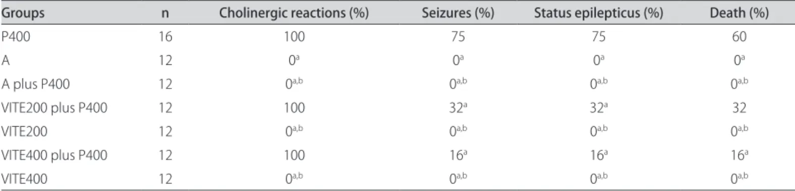
Table 2. Behavioral changes in rats treated intraperitoneally with pilocarpine, atropine, α-tocopherol, or their combinations.
Groups n Cholinergic reactions (%) Seizures (%) Status epilepticus (%) Death (%)
P400 16 100 75 75 60
A 12 0a 0a 0a 0a
A plus P400 12 0a,b 0a,b 0a,b 0a,b
VITE200 plus P400 12 100 32a 32a 32
VITE200 12 0a,b 0a,b 0a,b 0a,b
VITE400 plus P400 12 100 16a 16a 16a
VITE400 12 0a,b 0a,b 0a,b 0a,b
Pilocarpine was administered in a single dose (400 mg/kg, P400), VITE groups with α-tocopherol (200 or 400 mg/kg,) and A group with atropine (50 mg/kg). The A plus P400 group was treated with atropine (50 mg/kg) and 30 min before P400. The VITE plus P400 groups were treated with α-tocopherol (200 or 400 mg/kg) and 30 min before P400. Results are expressed as percentages of the number of animals. The animals treated with saline, α-tocopherol, or atropine 30 min before P400 did not show any behavioral changes. ap<0.05 compared with P400 group (c2 test). bp<0.05 compared with A plus P400, VITE200 plus
oped (1-2 h) into a long-lasting SE (32%), revealing a sur-vival rate of 68%. he VITE400 plus P400 group manifest-ed alterations in behavior, such as peripheral cholinergic signs (100%), staring spells, facial automatisms, wet dog shakes, rearing and motor seizures (16%), which progres-sively developed (1-2 h) into long-lasting SE (16%), reveal-ing a survival rate of 84% from the seizures (Table 2).
Brain tissue examinations of the control (saline 0.9%) (Figs 1A and 2A), atropine (50 mg/kg) (Figs 1C and 2C), atropine plus pilocarpine (A plus P400) (Figs 1D and 2D), AA (250 or 500 mg/kg) and α-tocopherol groups (VITE 200 or 400 mg/kg) did not reveal hippocampal histo-pathological changes (Tables 3 and 4). On the other hand,
pilocarpine-treated animals (400 mg/kg) presented neu-ronal loss, gliosis, and typical vacuolar degeneration in hippocampus region (Figs 1B and 2B). Histopathologi-cal damage was observed in three (50%), two (33%), two (33%) and in one animal (17%) in the AA250 plus P400, AA500 plus P400, VITE200 plus P400 and VITE400 plus P400 groups, respectively (Tables 3 and 4).
Brain tissue examination of AA 250 (Fig 1E), AA 500 (Fig 1G), VITE200 (Fig 2E), and VITE400 (Fig 2G) did not show any hippocampus histological alterations. Howev-er, the AA250 plus P400 (Fig 1F), AA500 plus P400 (Fig 1H), VITE200 plus P400 (Fig 2F), and VITE400 plus P400 groups (Fig 2H) showed typical histopathological chang-Table 3. Histopathological abnormalities in the hippocampus treated intraperitoneally with pilocarpine, atropine,
ascorbic acid, or their combinations.
Groups
Histopathological abnormalities in the hippocampus (%) Rats with
brain lesion (%) Severity of lesion Number of animals with brain damage Total number of animals
P400 85 59.92±0.23 5 6
A 0a 0a 0 6
A plus P400 0a,b 0a,b 0 6
AA250 plus P400 50a 20.00±0.32a 3 6
AA250 0a,b 0a,b 0 6
AA500 plus P400 33a,b 17.66±0.33a 2 6
AA500 0a,b 0a,b 0 6
Pilocarpine was administered in a single dose (400 mg/kg, P400), AA groups with ascorbic acid (250 or 500 mg/kg,) and A group with atropine (50 mg/kg). The A plus P400 group was treated with atropine (50 mg/kg) and 30 min before P400. The AA plus P400 groups were treated with ascorbic acid (250 or 500 mg/kg) and 30 min before P400. Severity of lesion was expressed as mean ± S.E.M. of scores of damage based on a scale from zero (none) to 100 (total) percent of structural involvement. Brain damage was deined as present if there was at least 50% hippocampal involvement. Results for % rats with brain lesion and % severity of lesion are expressed as percentages of the number of animals. ap<0.05 compared with P400 group (c2 test). bp<0.05 compared with A
plus P400, AA250 plus P400 groups or AA500 plus P400 groups (c2 test).
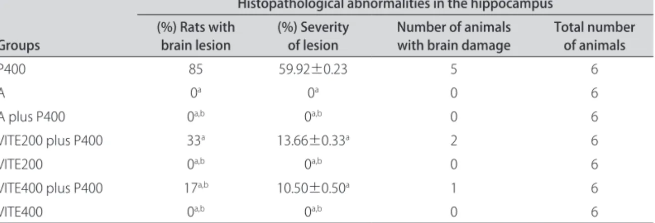
Table 4. Histopathological abnormalities in the hippocampus treated intraperitoneally with pilocarpine, atropine,
α-tocopherol or their combinations.
Groups
Histopathological abnormalities in the hippocampus (%) Rats with
brain lesion (%) Severity of lesion Number of animals with brain damage Total number of animals
P400 85 59.92±0.23 5 6
A 0a 0a 0 6
A plus P400 0a,b 0a,b 0 6
VITE200 plus P400 33a 13.66±0.33a 2 6
VITE200 0a,b 0a,b 0 6
VITE400 plus P400 17a,b 10.50±0.50a 1 6
VITE400 0a,b 0a,b 0 6
Pilocarpine was administered in a single dose (400 mg/kg, P400), VITE groups with α-tocopherol (200 or 400 mg/kg,) and A group with atropine (50 mg/kg). The A plus P400 group was treated with atropine (50 mg/kg) and 30 min before P400. The VITE plus P400 groups were treated with α-tocopherol (200 or 400 mg/kg) and 30 min before P400. Severity of lesion was expressed as mean ± S.E.M. of scores of damage based in a scale from zero (none) to 100 (total) percent of structural involvement. Brain damage was deined as present if there was at least 50% hippocampal involvement. Results for % rats with brain lesion and % severity of lesion are expressed as percentages of the number of animals. ap<0.05 compared with P400 group (ANOVA and Student-Newman-Keuls
es characterized by neuronal loss, gliosis and vacuola-tion afecting 50, 33, 33 and 17% of the rats, respective-ly (Tables 3 and 4).
DISCUSSION
Cholinergic mechanisms play an important role in ac-tivation of limbic seizures and dopaminergic, serotoner-gic, GABAergic and glutamatergic systems are responsible for the propagation and/or maintenance of seizures and SE induced by pilocarpine2. Previous studies described a model of limbic seizures followed by brain damage pro-duced by systemic injection of a high dose of pilocarpine
(400 mg/kg) in rats5,23. The evidence included tempo-ral correlation among free radical generation, develop-ment of seizures and neuroprotective efects of antioxi-dant drugs against neuronal damage caused by seizures24. Others studies showed that pilocarpine-induced seizures and brain damage in various cerebral regions21,25,26.
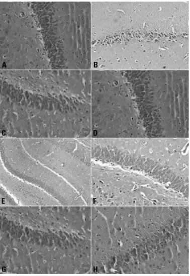
In our work, a signiicant hippocampal injury was ob-served in the P400 group. he anticonvulsant efect in the absence of anticholinergic drugs subsequent to the sei-zure onset suggests that muscarinic receptor activation is involved directly on the onset of seizures by pilocarpine. However, the oxidative stress might also play an essential Fig 1. Histopathological alterations in rat hippocampus treated
with pilocarpine, atropine, ascorbic acid or their combinations. [A] Control group; [B] P400 group; [C] Atropine group; [D] A plus P400 group was treated with atropine (50 mg/kg) and 30 min be-fore P400; [E] AA plus 250 group; [F] AA250 plus P400 groups was treated with ascorbic acid (250 mg/kg) and 30 min before P400; [G] AA500 group; [H] AA500 plus P400 group was treated with ascorbic acid (500 mg/kg) and 30 min before P400. Severity of le-sion was expressed as mean ± S.E.M. of scores of damage based in a scale from zero (none) to 100 (total) percent of structural in-volvement. Brain damage was considered positive if there was at least 50% hippocampal involvement. Hematoxylin & eosin stain-ing (H&E). Magniication, 100 X. One representative experiment with n=6 is shown.
Fig 2. Histopathological alterations in rat hippocampus treated with pilocarpine, atropine, α-tocopherol or their combinations. [A] Control group; [B] P400 group; [C] Atropine group; [D] A plus P400 group was treated with atropine (50 mg/kg) and 30 min before P400; [E] VITE200 group; [F] VITE200 plus P400 group was
treat-ed with α-tocopherol (200 mg/kg) and 30 min before P400; [G]
VITE400 group; [H] VITE400 plus P400 group was treated with α
role in the production of neuronal damage, which can be justiied by neuroprotective actions of antioxidant com-pounds according to previous studies2,14,27,28. Previous re-search indicates that anticonvulsant efects of noradrener-gic antagonist drugs have a fundamental role in the mech-anisms responsible for seizure beginning, severity and du-ration. In fact, the reduction of severity and duration of sei-zures are protective against neurotoxicity caused by seisei-zures induced by hemoconvulsants (e.g. pilocarpine, kainic acid and others). hese data, although conirming a pivotal role of anticonvulsant drugs in modulating seizure threshold and neuronal death, ofer a novel target, which may be used to develop anticonvulsant and neuroprotective agents29.
here are several indications that free radical plays a role in epileptogenesis2,10,26. During seizures, the reac-tive oxygen species (ROS) concentration and brain lipid peroxidation increase2. It is currently hypothesized that any pathological process such as SE, which releases dop-amine and glutamate, activates D2 and NMDA receptors.
his may lead to neuronal necrosis by elevating intracel-lular calcium and activating potentially destructive calci-um-dependent enzymes31, augmenting the production of free radicals during seizures induced by pilocarpine10,31. hus, it could be expected that antioxidant drugs such as ascorbic acid and α-tocopherol, can be used as scav-engers of free radicals, reducing brain injury induced by pilocarpine. In the present work, we showed that the tioxidants ascorbic acid and α-tocopherol protected an-imals against seizures, SE and brain damage induced by pilocarpine, thus decreasing the percentage of seizures, SE and death in relation to both doses tested.
A variety of epilepsy models relect the efects of acid ascorbic and α-tocopherol and specify their action13,15. Previously, it had been demonstrated that these com-pounds reduced the frequency of penicillin-induced epileptiform activity12,27. In recent years, many roles of α-tocopherol have been discovered, including not only an antioxidant function, but also pro-oxidant, cell signaling, and gene regulatory functions. Some studies have report-ed that α-tocopherol is considerreport-ed to be the main antiox-idant substance in the human body, interfering with the production of hydroxyl radical and also with the oxygen in cell membranes, thereby reducing lipid peroxidation9. Our results demonstrated that seizure pattern and brain damage observed in pilocarpine-treated animals differ from those treated with α-tocopherol plus pilo-carpine (VITE400/P400). They reproduced the syn-drome with lower intensity of histopathological changes and mortality rate, in comparison with the VITE200 plus P400 and P400 groups, thus corroborating the outcomes obtained by Ribeiro et al.32 and Ayyildiz et al.12. he per-centage of SE (75%) that was found further corroborated prior investigations5,33,34.
Ascorbic acid is probably the most important water-soluble antioxidant in the brain extracellular luid, and it is essential in regenerating reduced α-tocopherol in mem-branes35. Although ascorbic acid has an antioxidant role to counter oxidative stress, ascorbic acid also form re-active oxidants, especially in the presence of transition metals. he evidence suggest that ascorbic acid partici-pates in pro-oxidant reactions under certain conditions36. In the present work, the outcomes conirm that ascor-bic acid (250 and 500 mg/kg) decreased the frequency of pilocarpine-induced seizures, SE and brain lesions in rats. In addition, ascorbic acid decreases the severity of hippocampal lesions and mortality rate caused by pilo-carpine. Yamamoto et al.37 demonstrated that injection of ascorbate, 60 min before FeCl3 administration,
prevent-ed the occurrence of epileptic discharges. Since there are wide variations of α-tocopherol and ascorbic acid dos-es used in diferent models of seizure, more detailed in-vestigations are necessary before an ultimate conclusion on the efects of ascorbic acid and α-tocopherol on pilo-carpine-induced seizures can be achieved.
In conclusion, we suggest that there is an accumula-tion of free radicals after SE induced by pilocarpine, and oxidative changes in other parameters during the acute phase. This finding suggests that the seizures, SE and deaths induced by pilocarpine have a large participation of brain oxidative stress, which is closely related to the mechanism of propagation and/or maintenance of the ep-ileptic focus by pilocarpine. he results from the present work suggest that free radicals as well as the muscarinic receptor activation seem to be involved in the genesis of seizures and brain damage obtained with pilocarpine. On the other hand, the muscarinic activation seems to play a major role in the neuronal damage produced by pilo-carpine. Antioxidant compounds can exert neuroprotec-tive function during acute phase of seizures, thereby de-creasing the severity of hippocampal lesions. All these outcomes indicate the promising therapeutic potential of ascorbic acid and α-tocopherol in treatments for neuro-degenerative diseases.
aCkNowleDGmeNTS– his work was supported in part by grants from the Brazilian National Research Council (CNPq), Brazil. R.M.F is fellow from CNPq. We would like to thank Stenio Gardel Maia for her technical assistance.
REFERENCES
1. Aminof MJ, Simon RP. Status epilepticus. Causes, clinical features and con-sequences in 98 patients. Am J Med 1980;12:657-666.
2. Freitas RM, Sousa FCF, Vasconcelos SMM, Viana GSB, Fonteles MMF. Pilo-carpine-induced seizures in adult rats: lipid peroxidation level, nitrite forma-tion, GABAergic and glutamatergic receptor alterations in the hippocampus, striatum and frontal cortex. Pharmacol Biochem Behav 2004;78:327-332. 3. Hirsh E, Baran TZ, Snead OC. Ontogenic study of lithium-pilocarpine induced
4. Cavalheiro EA, Leite JP, Bortolotto ZA, Turski WA, Ikonomidou C, Turski L. Long-term effects of pilocarpine in rats: structural damage of the brain triggers kindling and spontaneous recurrent seizures. Epilepsia 1991;32: 778-782.
5. Marinho MMF, Sousa FCF, Bruin VMS, Aguiar LMV, Pinho RSN, Viana GSB. In-hibitory action of a calcium channel blocker (nimodipine) on seizures and brain damage induced by pilocarpine and lithium-pilocarpine in rats. Neu-rosci Lett 1997;235:13-16.
6. Mello LEAM, Cavalheiro EA, Tan AM, et al. Circuit mechanisms of seizures in the pilocarpine model of chronic epilepsy: cell loss and mossy iber sprout-ing. Epilepsia 1993;34:985-995.
7. Mello LEAM, Del Bel EA, Gomes ELT, Oliveira JAC, Wakamatsu H, Cairasco NG. Neuroethological and morphological (Neo-Timm staining) correlates of lim-bic recruitment during the development of audiogenic kindling in seizures susceptible Wistar rats. Epilepsy Res 1996;26:177-192.
8. Peredery O, Blomme MA, Parker G. Absence of maternal behaviour in rats with lithium/pilocarpine seizure induced brain damage: support of macleans triune brain theory. Physiol Behav 1992;52:665-671.
9. Ferreira PMP, Farias DF, Oliveira JTA, Carvalho AFU. Moringa oleifera: bioac-tive compounds and potential nutritional. Rev Nutr 2008;21:431-437. 10. Freitas RM, Nascimento VS, Sousa FCF, Vasconcelos SMM, Viana GSB,
Fon-teles MMF. Catalase activity in cerebellum, hippocampus, frontal cortex and striatum after status epilepticus induced by pilocarpine in Wistar rats. Neu-rosci Lett 2004;365:102-105.
11. Halliwell B, Gutteridge JMC. The antioxidants of human extracellular luids. Arch Biochem Biophys 1990;280:1-8.
12. Ayyildiz M, Yildirim M, Agar E. The efects of vitamin E on penicilin-induced epileptiform activity in rats. Exp Brain Res 2006;174:109-113.
13. Koza R, Ayyildiz M, Coskun S, Yildirim M, Agar E. The inluence of ethanol in-take and its withdrawal on the anticonvulsant efect of α-tocopherol in the penicillin-induced epileptiform activity in rats. Neuro Toxicol 2007;28:463-470. 14. Xavier SML, Barbosa CO, Barros DO, Silva RF, Oliveira AA, Freitas RM. Vitamin
C antioxidant in hippocampus of adult Wistar rats after seizures and status epilepticus induced by pilocarpine. Neurosci Lett 2007;420:76-79. 15. Gaby AR, Natural approaches to epilepsy. Altern Med Rev 2007;12:9-24. 16. Devi PU, Manocha A, Vohora D. Seizures, antiepileptics, antioxidants and
ox-idative stress: an insight for researchers. Expert Opin Pharmacother 2008;9: 3169-3177.
17. Ranganathan LN, Ramaratnam S. Vitamins for epilepsy. Ophthalmology 1994; 101:1347-1352.
18. Barros DO, Xavier SML, Freitas RLM, et al. Efeccts of the vitamin E in catalase activities in hippocampus after status epilepticus induced by pilocarpine in Wisar rats. Neurosci Lett 2007;416:227-230.
19. Turski WA, Cavalheiro EA, Schwarz M, Czuczwar SJ, Kleinronk Z, Turski, L. Lim-bic seizures produced by pilocarpine in rats: behavioural, eletroencephalo-graphic and neuropathological study. Behav Brain Res 1983;9:315-336. 20. Marinho MMF, Sousa FCF, Bruin VMS, Vale MR, Viana GSB. Efects of lithium,
alone or associated with pilocarpine, on muscarinic and dopaminergic
re-ceptors and on phosphoinositide metabolism in rat hippocampus and stri-atum. Neurochem Int 1998;33:299-306.
21. Szyndler J, Bobtowicz TW, Skórzewska A, et al. Behavioral, biochemical and histological studies in a model of pilocarpine-induced spontaneous recur-rent seizures. Pharmacol Biochem Behav 2005;1:15-23.
22. Paxinos G, Watson C. The rat brain in stereotaxie coordenates. Second Edi-tion. New York, Academic Press, 1986.
23. Turski L, Ikonomidou C, Turski WA, Bortolotto ZA, Cavalheiro EA. Cholinergic mechanisms and epileptogenesis. The seizures induced by pilocarpine: a novel experimental model of intractable epilepsy. Synapse 1989;3:154-171. 24. Kabuto H, Yokoi I, Ogawa N. Melatonin inhibits iron-induced epileptic
dis-charges in rats by suppressing peroxidation. Epilepsia 1998;30:237-243. 25. Turski L, Cavalheiro EA, Leite JP, Bortolotto ZA, Turski WA, Ikonomidou C.
Long-term efects of pilocarpine in rats: structural damage of the brain trig-gers kindling and spontaneous recurrent seizures. Epilepsia 1991;32:778-782. 26. Curia G, Longo D, Biagini G, Jones RSG, Avoli M, The pilocarpine model of
temporal lobe epilepsy. J Neurosc Methods 2008;172:143-157.
27. Ayyildiz M, Coskun S, Yildirim M, Agar E. The involvement of nitric oxide in the anticonvulsant efects of α-tocopherol on penicillin-induced epilepti-form activity in rats. Epilepsy Res 2007;73:166-172.
28. Ayyildiz M, Coskun S, Yildirim M, Agar E. The efects of ascorbic acid on pen-icillin-induced epileptiform activity in rats. Epilepsia 2007;48:1388-1395. 29. Pizzanelli C, Lazzeri G, Fulceri F, et al. Lack of alpha 1b-adrenergic receptor
protects against epileptic seizures. Epilepsia 2009;50:59-64.
30. Vanin A, Vitskova G, Narkevich V, Bashkatova V. The inluence of anticonvul-sant and antioxidant drugs on nitric oxide level and lipid peroxidation in the rat brain during penthylenetetrazole-induce epileptiform model seizures. Prog Neuro-Psychopaharm Biol Psychiatry 2003;27:487-492.
31. Michotte Y, Ebinger G, Manil J, Khan GM, Smolders I. NMDA receptor-medi-ated pilocarpine-induced seizures: characterization in freely moving rats by microdialysis. Br J Pharmacol 1997;121;1171-1179.
32. Ribeiro MCP, Avila DS, Scheneider CYM, et al. α-tocopherol protects against pentylenetetrazol- and methylmalonate-induced convulsions. Epilepsy Res 2005;66:185-194.
33. Cliford DB, Podolsky A, Zorumski CP. Acute efects of lithium on hippocam-pal kindled seizures. Epilepsia 1985;26:689-692.
34. Cliford DB, Olney JW, Maniotis A, Collins RC, Zorumski CF. The functional anatomy and pathology of lithium-pilocarpine and high-dose pilocarpine seizures. Neuroscience 1987;23:953-968.
35. Niki E. Action of ascorbic acid as a scavenger of active and stable oxygen rad-icals. Am J Clin Nutr 1991;54(Suppl):S1119-S1124.