51
Introduction
An emerging paradigm in the pathophysiology of sickle cell disease is the strong link between hemolysis-related nitric oxide system dysfunction and risks for pulmonary hypertension, leg ulcers, priapism, and death.1,2 In sickle cell disease hemolysis is the consequence of hemoglobin S (Hb S) polymerization, which causes red cell rigidity and sickling. Mechanical injury to the membrane of these rigid, Hb S polymer-containing red cells shortens their intravascular survival.
The strongest factor determining Hb S polymerization both in solution and within red cells is Hb S concentration.3 Because iron deficiency lowers the Hb concentration (MCHC) within erythrocytes, Lincoln et al. hypothesized in 1973 that inducing iron depletion could be beneficial in sickle cell disease.4 More recently, and with the same rationale, Koduri5 suggested that iron restriction could be explored as a
Iron deficiency decreases hemolysis in sickle cell anemia
Anemia ferropriva diminui hemólise em anemia falciforme
Oswaldo Castro1
Adriana Medina2
Peter Gaskin3
Gregory J. Kato4
Victor R. Gordeuk5
A woman with homozygous sickle cell disease developed severe iron deficiency due to long-standing uterine bleeding. At this point, the serum lactic dehydrogenase level was normal and the reticulocyte count was only minimally elevated. This suggested that the low red cell hemoglobin concentration that resulted from iron deficiency also decreased Hb S polymerization and lowered the hemolytic rate. Iron replacement led first to a substantially improved hemoglobin concentration with only a minimal increase in the hemolytic rate and secondarily to a modest further improvement in the hemoglobin concentration and a marked increase in the hemolytic rate. The hematologic changes observed in this patient, and those in other iron deficient sickle cell patients reported in the literature, suggest that it may be appropriate to consider the induction of an intermediate iron deficient stage as experimental treatment in adult sickle cell patients. Rev. Bras. Hematol. Hemoter. 2009;31(1):51-53.
Key words: Sickle cell anemia; hemolysis; iron deficiency.
Relato de Caso / Case Report
REVISTA BRASILEIRA D E H E M ATO L O G I A E H E M O T E R A P I A
1M.D. Professor Emeritus of Medicine - Consultant, Howard University Center for Sickle Cell Disease, Washington, D.C. USA 2M.D. Department of Medicine, Howard University College of Medicine, Washington, D.C. USA
3M.D. Department of Pediatrics, Howard University College of Medicine, Washington, D.C., USA
4M.D. Pulmonary and Vascular Medicine Branch - National Heart, Lung and Blood Institute National Institutes of Health, Bethesda, MD, USA 5M.D. Director - Center for Sickle Cell Disease Howard University College of Medicine, Washington, D.C. USA
Howard University Center for Sickle Cell Disease, Howard University College of Medicine, Washington, DC, USA
Correspondência: Oswaldo Castro, M.D.,
HURB-1 1840 7th. St. NW
Washington DC, 20001, USA Email: [email protected]
52
Rev. Bras. Hematol. Hemoter. 2009;31(1):51-53 Castro O et al
restriction in sickle cell disease have shown that low MCHC and lower reticulocyte counts can occur without further decreases in the hemoglobin level. This suggests that such an intermediate iron deficiency stage may in fact develop in these patients. We report here a patient with sickle cell disease and severe iron deficiency ane-mia whose laboratory findings also indicated a marked reduction of hemolysis. In this patient, serial laboratory parameters during iron therapy demonstrated that correction of the iron deficiency component of her ane-mia could take place without significant worsening of the hemolytic rate.
Case summary
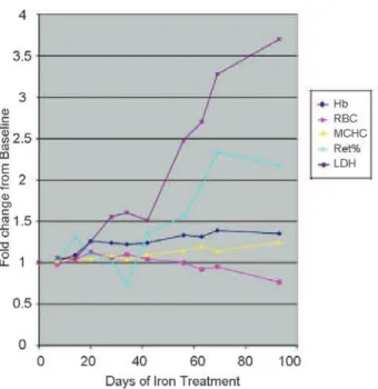
A 47 year-old woman with homozygous sickle-cell disease was seen in our clinic with severe and long-standing iron deficiency from of heavy menstrual flow associated with uterine fibroids. Her stools for occult blood and endoscopic examination of the gastrointestinal tract were negative. Two years earlier iron deficiency had to be corrected with intravenous iron and red cell transfusion because severe nausea and vomiting precluded the use of standard oral iron preparations even at low doses. She declined gynecologic surgery. This time, to avoid parenteral iron and transfusions, she was prescribed prenatal vitamin tablets (Cal-NateTM, Ethex Corporation, St. Louis, MO, USA), which contain 27 mg of carbonyl iron, one twice daily. With this gradual, low dose iron supplementation there were no problems with gastrointestinal intolerance or treatment adherence. Table 1 shows the patient's laboratory values at (a) baseline, before iron treatment, (b) an early stage of iron treatment during which anemia improved without an excessive increase in the hemolytic rate (LDH and reticulocytes were used as surrogates for hemolysis), and (c) iron repletion, in which hemolysis increased markedly but with only a modest additional improvement of the anemia.
Figure 1 displays some of the laboratory results observed during iron-treatment normalized to their baseline values. During the early stages of iron repletion, there was a 24% improvement of the anemia (over baseline) with only a modest increase in hemolytic parameters: 51% increase in LDH and 36% increase in reticulocytes. By treatment day 93 there was a 370% increment in the LDH and a 270% percent increment in reticulocytes, but still only a 25% improvement in the anemia and MCHC. Hemolysis apparently limited the more robust additional improvement of the hemoglobin level that would have been expected with increased erythropoiesis in response to iron treatment. In fact, the highest red cell counts (3.43-3.69 x10⁶/µl) in this patient were seen during partial iron repletion. At this intermediate stage, the patient's hemolytic rate seems to have been low enough to allow longer survival of the red cells, produced at a higher rate following partial iron repletion.
Discussion
Our experience with this single case report suggests that a state of partial iron deficiency can develop in sickle cell disease and that it could be beneficial by reducing the hemolytic rate without making the anemia substantially worse. We believe that this was possible because modest reductions in the MCHC (30.1-31.7%, see Table 1) during partial iron deficiency reduced intracellular polymerization and hemolysis to a greater extent than its inhibition of erythropoiesis. Direct red cell survival measurements were not carried out, but the following parameters were used as surrogates to define the degree of hemolysis: relative and absolute reticulocyte counts, and serum levels of LDH, AST and bilirubin. The potential benefit of limited iron deficiency suggested by our case is also supported by published reports of spontaneous and induced8,9 iron restriction in sickle cell anemia.
This sickle cell patient's serial hematologic and hemolytic parameters, measured during slow iron repletion, also allowed characterization of the partial iron deficiency stage as a potential therapeutic "window". An MCHC lower than 32% but higher than 30% (Table 1 and Figure 1) appeared to keep hemolysis at a relatively low rate: the patient's hemoglobin levels ranged from 6.6 to 6.8 g/dl (and the RBC counts from 3.4 to 3.7 x10⁶/
µl) despite the moderate iron
deficiency state. The changes in RBC counts were particularly interesting, since, with continued iron supplementation, they actually decreased by 16% (2.99 x10⁶/
µl) below those seen at
the baseline, severe iron deficiency state (3.28 x10⁶/
µl).
53
Finantial support: This work was supported in part by NIH research grant no 5R25-HL003679-10 from the National Heart, Lung and Blood Institute and the Office of Research on Minority Health, NIH grant no 5 RO1 HL079912-03, and Howard University General Clinical Research Center Grant no MO1-RR10284
Avaliação: Editor e dois revisores externos Conflito de interesse: não declarado
Recebido: 01/08/2008 Aceito: 13/08/2008 achievable by phlebotomy.8,10-12 An RBC count rise above
40% (over non-iron deficiency values), as seen in our patient, may turn out be a useful predictor of the partial iron deficiency state, in which hemolysis decreases without substantial marrow suppression.
However, iron deficiency, while generally a more benign disorder than sickle cell disease at least in adults, can be associated with rare but serious problems such as cerebral sinovenous thrombosis.13,14 On the other hand, iron deficiency may have a direct, vaso-protective effect in sickle cell patients, apart from its inhibitory effect on hemolysis: iron deficiency anemia up-regulates vascular nitric oxide synthase in animals,15 and in humans it increases NO production even in the absence of anemia.16
Resumo
Uma mulher com anemia falciforme homozigose para a Hb S evo-luiu com anemia ferropriva grave devido a sangramento uterino prolongado. A dosagem de dehidrogenase lática era normal e a contagem de reticulócitos estava levemente aumentada. Isto sugere que concentrações baixas de hemoglobina, que resulta de anemia ferropriva, também diminuem a polimeração de Hb S e reduz a taxa de hemólise. O complemento de ferro levou, primeiramente, a uma concentração substancialmente maior de hemoglobina com apenas um aumento mínimo na taxa hemolítica e subsequentemente a um aumento leve adicional na concentração da hemoglobina e um au-mento notável na taxa hemolítica. As mudanças hematológicas ob-servadas nesta paciente e aquelas em outras pacientes com anemia falciforme e também deficientes de ferro relatadas na literatura suge-rem que pode ser interessante considerar a indução de deficiência de
ferro como tratamento experimental em pacientes adultos com ane-mia falciforme. Rev. Bras. Hematol. Hemoter. 2009;31 (1):51-53.
Palavras-chave: Anemia falciforme; hemólise; anemia ferropriva.
References
1. Kato GJ, McGowan V, Machado RF, Little JA, Taylor J 6th, Morris CR, et al. Lactate dehydrogenase as a biomarker of hemolysis-associated nitric oxide resistance, priapism, leg ulceration, pulmonary hypertension, and death in patients with sickle cell disease. Blood. 2006;107(6):2279-85.
2. Jeffers A, Gladwin MT, Kim-Shapiro DB. Computation of plasma hemoglobin nitric oxide scavenging in hemolytic anemias. Free Radic Biol Med. 2006;41(10):1557-65.
3. Eaton WA, Hofrichter J. Hemoglobin S gelation and sickle cell disease. Blood. 1987;70(5):1245-66.
4. Lincoln TL, Aroesty J, Morrison P. Iron-deficiency anemia and sickle-cell disease: a hypothesis. Lancet. 1973;2(7823):260-1. 5. Koduri PR. Iron in sickle cell disease: a review why less is better.
Am J Hematol. 2003;73(1):59-63.
6. Castro O, Haddy TB. Improved survival of iron-deficient patients with sickle erythrocytes. N Engl J Med. 1983;308(9):527. 7. Ballas SK, Marcolina MJ. Determinants of red cell survival and
erythropoietic activity in patients with sickle cell anemia in the steady state. Hemoglobin. 2000;24(4):277-86.
8. Castro O, Poillon WN, Finke H, Massac E. Improvement of sickle cell anemia by iron-limited erythropoiesis. Am J Hematol. 1994; 47(2):74-81.
9. King L, Reid M, Forrester TE. Iron deficiency anaemia in Jamaican children, aged 1-5 years, with sickle cell disease. West Indian Med J. 2005;54(5):292-6.
10. Bouchair N, Manigne P, Kanfer A, Raphalen P, de Montalembert M, Hagage I, et al. Prèvention des crises douloureuses drèpanocytaires par saignees itèratives. Arch Pèdiatr 2000;7:249-55
11. Rombos Y, Tzanetea R, Kalotychou V, Konstantopoulos K, Simitzis S, Tassiopoulos T, et al. Amelioration of painful crises in sickle cell disease by venesections. Blood Cells Mol Dis. 2002;28(2):283-7. 12. Markham MJ, Lottenberg R, Zumberg M. Role of phlebotomy in
the management of hemoglobin SC disease: case report and review of the literature. Am J Hematol. 2003;73(2):121-5.
13. Benedict SL, Bonkowsky JL, Thompson JA, Van Orman CB, Boyer RS, Bale JF Jr, et al. Cerebral sinovenous thrombosis in children: another reason to treat iron deficiency anemia. J Child Neurol. 2004;19(7):526-31.
14. Balci K, Utku U, Asil T, Büyükkoyuncu N. Deep cerebral vein thrombosis associated with iron deficiency anaemia in adults. J Clin Neurosci. 2007;14(2):181-4.
15. Ni Z, Morcos S, Vaziri ND. Up-regulation of renal and vascular nitric oxide synthase in iron-deficiency anemia. Kidney Int. 1997; 52(1):195-201.
16. Choi JW, Pai SH, Kim SK, Ito M, Park CS, Cha YN. Iron deficiency anemia increases nitric oxide production in healthy adolescents. Ann Hematol. 2002;81(1):1-6.
Figure 1. Changes in laboratory parameters during iron treatment in iron-deficient sickle cell anemia patient. All data points are relative to the baseline (day 0) values. Hb: blood Hb level, RBC: red blood cell count, MCHC: mean corpuscular hemoglobin concentration, Ret%: percent reticulocytes, LDH: serum lactic dehydrogenase