ABSTRACT
Acromegaly is a systemic disease with various etiologies. It can occur as a sporadic or, more rarely, as a familial disease. Numerous complica-tions such as endocrine, cardiovascular, respiratory, metabolic, osteoarticular and neoplastic disturbances occur and must be taken into account when establishing a therapeutic strategy. For this reason, the decision as to a treatment modality of acromegaly must be followed by a thorough evaluation of the patient and once the diagnosis of com-plications is settled, adequate treatment should be instituted. Follow up of the patients requires periodical re-assessment of complications’ sta-tus.(Arq Bras Endocrinol Metab 2005;49/5:626-640)
Keywords:Acromegaly; Etiology; Treatment; Management; Complica-tions
RESUMO
Aspectos Etiológicos e Manejo da Acromegalia.
A acromegalia é uma doença sistêmica com diversas etiologias. A grande maioria dos casos se manifesta de forma esporádica, e uma minoria tem transmissão familiar. Além disso, a acromegalia pode ser acompanhada de várias complicações como alterações endócrinas, cardiovasculares, respiratórias, metabólicas, osteoarticulares e neo-plásicas que devem ser consideradas quando a estratégia de trata-mento for estabelecida. O acompanhatrata-mento dos pacientes requer reavaliações periódicas do status das complicações. Neste artigo serão abordados os aspectos etiológicos e o manejo da acromegalia. (Arq Bras Endocrinol Metab 2005;49/5:626-640)
D e s c r i t o r e s : Acromegalia; Etiologia; Manejo; Tratamento; Compli-cações
A
CROMEGALY IS A WELL-CHARACTERIZED syndrome resulting from ele-vated levels of growth hormone (GH) and insulin-like growth factor-I (factor-IGF-factor-I). However, this proves to be a heterogeneous disease with vari-ous etiologies, although some of them are quite rare, as it will be discussed below. Manifestations include acral enlargement, increased perspiration, arthralgia and paresthesias. The disease also presents systemic complica-tions such as hypertension, cardiomyopathy, respiratory and metabolic dis-turbances. For this reason, a thorough evaluation must be made once the diagnosis is settled and adequate follow up requires periodical re-assess-ment of complications’ status. The managere-assess-ment of the acromegalic patient will be discussed next.Giselle F. Taboada
Flávia R. van Haute
Lívia L. Corrêa
Alessandra F. Casini
Mônica R. Gadelha
Endocrine Unit – Hospital Universitário Clementino Fraga Filho (GFT, FRvH, LLC, AFC & MRG), Universidade Federal do Rio de Janeiro (UFRJ); and Endocrine Unit – Instituto Estadual de Diabetes e Endocrinologia Luiz
Capriglione do Rio de Janeiro (IEDE – RJ) (LLC & MRG), Rio de Janeiro, RJ.
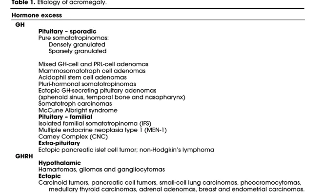
ETIOLOGY
GH Hypersecretion
GH hypersecretion of pituitary origin – spo -radic
Acromegaly, in the majority of the cases, occurs as a sporadic disease usually caused by a GH-secreting pituitary adenoma (somatotropinoma) or rarely as part of the McCune-Albright syndrome (table 1).
Different types of GH-secreting pituitary ade-nomas, characterized in accordance with their hor-mone expression and ultrastructural features, may be responsible for distinct clinical presentations of acromegaly (table1). Somatotropinomas are mono-clonal in origin (1) and the most common genetic alteration involved in their pathogenesis is the activat-inggspmutation. This somatic mutation is found in up to 40% of the patients (2). Other genes that may be involved arepRb,p27/KIP1,PTTGand a tumor sup-pressor gene located at chromosome region 11q13 distinct fromMEN1(3).
Pure somatotropinomas are the most frequently found (60% of the GH-secreting pituitary adenomas) and can harbor densely or sparsely distributed cytoplas-mic granules that stain positive for GH. Densely-granu-lated somatotropinomas are acidophilic, occur in older individuals, grow slowly and present in an insidious manner. On the other hand, sparsely-granulated soma-totropinomas are chromophobic, occur in younger indi-viduals and grow faster. Mixed GH-cell and prolactin (PRL)-cell adenomas are formed by two distinct cell types and may appear acidophilic, partly acidophilic or chromophobic, depending on the granularity of the two components. They correspond to 25% of the GH-secreting pituitary adenomas and cause acromegaly with moderately increased serum PRL levels. Mammosoma-totroph cell adenomas are the most common tumor type in children and adolescents with gigantism and constitute 10% of the GH-secreting pituitary adenomas. They are acidophilic, the cells are well differentiated and contain both GH and PRL granules. Serum PRL levels are normal or moderately increased. Acidophil stem cell adenomas are very infrequent (< 5%), rapidly growing and invasive tumors. They originate in the acidophil stem cells, the common precursors of somatotrophs and lactotrophs, and express both GH and PRL. T h e s e tumors have relatively low hormonal activity and the clinical presentation may be similar to that of a non-functioning pituitary adenoma or marked by hyperpro-lactinemia, since PRL is the major product of tumor cell
secretion. Pluri-hormonal somatotropinomas, which are either monomorphous or plurimorphous, are rare (< 5%) and may express GH with any combination of adrenocorticotrophic hormone (ACTH), glycoproteic hormones and/or a-subunit (4) (table 1).
Ectopic GH-secreting adenomas may originate from pituitary remnant tissues in the sphenoid sinus (5), temporal bone and nasopharynx (6). The presence of this ectopic tissue is explained by pituitary develop-ment from Rathke’s pouch, which originates in the nasopharynx and migrates to its normal location in the sella turcica (table 1).
Somatotroph carcinomas are extremely rare and their diagnosis is based on the identification of distant metastases (7). Tumors exhibiting mitotic activity, hy-percellularity and nuclear pleomorphism without metas-tases should not be misdiagnosed as malignant, even if they are rapidly growing and invasive (7,8) (table 1).
The McCune-Albright syndrome is caused by an early somatic activating mutation in the gene
GNAS1that encodes thesubunit of the GTP-bind-ing protein (Gs). The severity of the disease depends on the percentage of mutant cells in different embry-onic tissues (9). It is characterized by the triad of polyostotic fibrous dysplasia, café-au-lait spots and sexual precocity. Other endocrine manifestations are hyperthyroidism, hypercortisolism, acromegaly/ gi-gantism, hyperprolactinemia, hyperparathyroidism and hypophosphatemic rickets/osteomalacia. Acromegaly may be due to an adenoma or to mammosoma-totrophic hyperplasia (10,11) (table 1).
GH hypersecretion of pituitary origin – famil -ial
syndrome is due to loss of function of the tumor sup-pressor geneMEN-1. Carney complex also exhibits an autosomal dominant inheritance pattern and arises from inactivation of the tumor suppressor gene
PRKAR1A(protein kinase A type 1 alpha regulatory subunit) or by a genetic alteration in an oncogene, not yet identified, located at chromosome 2p16 (16-18). It is manifested by heart, skin and breast myxomas, spotty mucocutaneous pigmentation, schwannomas, primary pigmented nodular adrenocortical disease (PPNAD), testicular and thyroid tumors, as well as somatotropinomas (12,15,16). In patients with CNC, histopathological examination has revealed adenomas, tipically multicentric, with staining for GH (predomi-nantly), PRL and occasionally for other hormones. Adenohypophyseal hyperplasia has also been seen (19). The diagnois of CNC is based on the recogni-tion of at least two of the components of the complex (15,16). Rarely, MEN-1 and CNC may occur as spo-radic diseases (12).
GH hypersecretion of extra-pituitary origin
Acromegaly has been described in a patient harboring a GH-secreting intramesenterioc islet cell pancreatic tumor (20) and in another with a non-Hodgkin’s lym-phoma (21) (table1). In these extremely rare cases, normal sized or reduced pituitary is seen on magnetic resonance imaging (MRI) and GH is not responsive to TRH administration (20). Finally, other tumors may
stain positively for GH, like lung, breast and ovarian adenocarcinomas (22,23). However, acromegaly has not been described under these circumstances to date.
GHRH HYPERSECRETION
GHRH hypersecretion of hypothalamic origin
Hypothalamic tumors like hamartomas, gliomas and gangliocytomas may secrete GHRH and cause soma-totrophic hyperplasia or even a somatotropinoma (24,25) (table 1).
GHRH hypersecretion of ectopic origin
Carcinoid tumors, pancreatic cell tumors, small-cell lung carcinomas, pheocromocytomas, medullary thy-roid carcinomas, adrenal adenomas, breast and endometrial carcinomas may also secrete GHRH. In these cases, acromegaly is caused by somatotrophic hyperplasia. Carcinoid tumors, mainly bronchial in ori-gin, represent most of the tumors associated with ectopic GHRH secretion (26-28) (table 1). Acromegaly in MEN-1 patients is rarely caused by GHRH production from a pancreatic islet cell tumor rather than by a somatotropinoma (29).
Management of acromegaly
As stated before, acromegaly is a systemic disease and causes endocrine, cardiovascular, respiratory, metabo-lic, osteoarticular and neoplastic morbidities besides increasing mortality (1.26 - 3 times the general
popu-Table 1.Etiology of acromegaly.
Hormone excess GH
Pituitary – sporadic
Pure somatotropinomas: Densely granulated Sparsely granulated
Mixed GH-cell and PRL-cell adenomas Mammosomatotroph cell adenomas Acidophil stem cell adenomas Pluri-hormonal somatotropinomas Ectopic GH-secreting pituitary adenomas
(sphenoid sinus, temporal bone and nasopharynx) Somatotroph carcinomas
McCune Albright syndrome
Pituitary – familial
Isolated familial somatotropinoma (IFS) Multiple endocrine neoplasia type 1 (MEN-1) Carney Complex (CNC)
Extra-pituitary
Ectopic pancreatic islet cell tumor; non-Hodgkin’s lymphoma
GHRH
Hypothalamic
Hamartomas, gliomas and gangliocytomas
Ectopic
lation mortality) (30-32). On the other hand, diagno-sis is made 7 to 10 years after symptoms begin. There-fore, as soon as the diagnosis is settled and treatment of acromegaly is defined, a careful evaluation of the anterior pituitary function and of acromegaly related complications are important aspects of the manage-ment of this disease.
Defining the treatment of acromegaly
Treating acromegaly is a challenging task and should be carried on by a multi-professional team, including an endocrinologist, a neurosurgeon and a radiotherapist.
Treatment options include: surgical resection of the adenoma by transsphenoidal approach, medical management and conventional or stereotaxic radio-therapy. Craniotomy for the adenomectomy is rarely necessary (33). Further details on each of these treat-ment modalities can be obtained at Donangelo et al. (34). The objectives of the treatment are to restore GH secretion and/or action to normal, reduce IGF-I levels to age- and gender-matched controls, relieve signs and symptoms of the disease, minimize compli-cations, control tumor growth, preserve anterior pitu-itary function and prevent tumor recurrence. The ulti-mate goal is to reduce mortality to general population rates which can be accomplished by obtaining GH lev-els less then 2.5ng/mL and normalization of IGF-I for age and sex (30,31,35). Recent epidemiological stud-ies have suggested that lower GH levels should be pur-sued (1ng/mL) (32,36).
The best treatment option for each patient should be chosen based on clinical, biochemical and
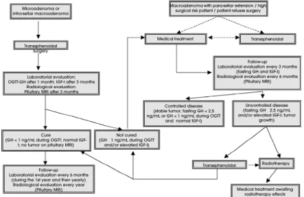
radiological characteristics of the disease, as well as the patient’s preference. An algorithm for the treatment of acromegaly is showed in figure 1. The first question to be answered is whether surgical approach has curative potential. If this is the case, transsphenoidal adenomec-tomy should be performed and the disease status re-assessed in the following 3 months. If the patient is cured, then he/she should be regularly followed and evaluated for co-morbidities. If the cure is not achieved, then adjuvant medical therapy should be started. Hor-mone hypersecretion is controlled (GH< 2.5ng/mL) with surgical management in 86-91% of the patients bearing microadenomas and in 46-52% of the patients with non-invasive macroadenomas (35,37,38). On the other hand, if surgical cure is improbable, medical ther-apy should be the first option. Two questions that still awaits for controlled-randomized clinical trials to be designed for this purpose are: 1) If pre-treatment with somatostatin analogs (SA) can improve surgical out-come, and 2) whether surgical debulking of the tumor should be done, even if surgical cure is unlikely. In a re-trospective study which involved 52 patients, Colao et al. (39) showed that patients with uncontrolled disease by somatostatin analogs as a primary therapy (mean ± SEM GH: 22.7 ± 4.5ng/ml; mean ± SEM IGF-I: 2.2 ± 0.1 Upper Limit Reference Values - ULRV) presented better results in terms of biochemical control after sur-gical debulking of the tumor (7.7 ± 1.6ng/ml and 1.3 ± 0.1 ULRV, respectively).
Three classes of drugs are available for the treat-ment of acromegaly: dopamine agonists, SA and GH receptor antagonists (40) (table 2). During medical
Microadenoma or intra-sellar macroadenoma
Transsphenoidal surgery
Laboratorial evaluation: OGTT-GH after 1 month; IGF-I after 3 months
Radiological evaluation: Pituitary MRI after 3 months
Cure
(GH < 1 ng/mL during OGTT; normal IGF-I; no tumor on pituitary MRI)
Follow-up Laboratorial evaluation every 6 months
(during the 1st year and then yearly) Radiological evaluation every year
(Pituitary MRI)
Not cured (GH 1 ng/mL during OGTT
and/or elevated IGF-I)
Controlled disease (stable tumor; fasting GH < 2,5 ng/mL or GH < 1 ng/mL during OGTT
and normal IGF-I)
Follow-up Laboratorial evaluation every 3 months
(fasting GH and IGF-I) Radiological evaluation every 6 months
(Pituitary MRI)
Uncontrolled disease (fasting GH 2,5 ng/mL and/or elevated IGF-I; tumor
growth) Macroadenoma with para-sellar extension / high
surgical risk patient / patient refuses surgery
Medical treatment Transsphenoidal
Transsphenoidal Radiotherapy
Medical treatment awaiting radiotherapy effects
treatment, hormonal levels should be assessed every 3 months and imaging studies (preferentially MRI) should be performed every 6 months. If the disease is controlled, this routine should be maintained and MRI can be done yearly. However, if the disease is not controlled, drug doses should be increased, the drug class should be changed and/or combination therapy (2 drug classes) should be started.
Somatostatin analogs are the most commonly used drugs for the medical treatment of acromegaly (table 2). Octreotide LAR adequately controls GH and IGF-I levels in 60-75% of the patients (41,42) and tumor shrinkage has been seen in up to 61% of the patients when used as an adjunctive therapy and up to 100% of the patients when used as a primary therapy (43). In some instances, cabergoline, a dopamine ago-nist, may be successfully used, such as in patients bear-ing mixed GH/PRL secretbear-ing tumors and in patients with slight elevations of GH and IGF-I levels (GH< 20ng/mL and IGF-I< 750ng/mL), in which GH and IGF-I control rates are around 50 – 57% (44,45). The control rates with the use of bromocriptine are unac-ceptably low, making it an unsuitable therapeutic option (46). Cabergoline can also be used in combina-tion with depot SA in patients partially-responsive to the latter (47). Pegvisomant represents the newest class of drugs available for treatment of acromegaly and has proven to be the most effective in terms of IGF-I nor-malization (97%) (48). However it exerts no direct action over the tumor mass in such a way that questions have been raised as whether pegvisomant treatment could lead to tumor growth. This seems not to be the case, but more experience with the use of this drug should be accumulated to enlighten this subject (48).
Selective estrogen receptor modulators (SERMs) like tamoxifen and raloxifene have been shown to reduce IGF-I in small series of acromegalic patients. Tamoxifen was able to normalize IGF-I in
31% (4/13) of the patients (males and females) and raloxifene in 54% (7/13) of the female and 25% (2/8) of the male patients (49-51). Also, estroprogestinic pill has been shown to normalize IGF-I levels in 50% (4/8) of the female patients (52).
Radiotherapy is, in most cases, a third-line treatment for acromegaly, being indicated to patients not cured by surgery and resistant or intolerant to medical treatment. Even if normalization of IGF-I lev-els occurs in up to 89%, this takes approximately 10 years and may be accompanied by hypopituitarism, radionecrosis, cognitive deficits, optic nerve damage and other central nervous system malignancies (40,53,54). Radiosurgery seems to be a better option when radiotherapy is required because the results are seen earlier than with conventional radiotherapy (33,55) and probable side-effects are minimized by lesser exposure of normal brain tissue to radiation.
High cost is an inconvenience of the treatment with SA. Therefore, the ability to predict which patients will achieve “safe” GH levels and IGF-I nor-malization would be advantageous. The usefulness of the acute test with subcutaneous octreotide has been extensively investigated (56-63) and even intravenous octreotide has been used for this purpose (64). A pos-itive response is considered when GH levels fall at least 50%. Some authors use 50mg and others use 100mg of octreotide as a test dose and distinct results are found. Therefore, it is still under debate whether the test is capable to distinguish the patients that will achieve “safe” GH and normal IGF-I levels during long term SA therapy. As an alternative, once octreotide LAR binds with high affinity to somato-statin receptor subtypes 2 and 5 (SSTR2 and 5), the analysis of the SSTR gene expression profile in the tumor may help select the patients with better chance to respond to these drugs (65).
New drugs have been developed and are under
Table 2.Drug options for the medical treatment of acromegaly.
Drug class Drugs Suitable patients Dopamine agonists
Cabergoline Mixed GH/PRL secreting tumors; patients with slight eleva-tions of GH and IGF-I levels (GH < 20ng/mL and IGF-I < 750ng/mL); combination therapy in SST analogs partial responders.
SST analogs
Octreotide LAR
As a primary or adjunctive treatment (see figure 1). Lanreotide SR
GH-R antagonist
Pegvisomant Patients resistant to SST analogs (isolated or combined use).
investigation for clinical use, such as SOM-230, a “universal ligand” of somatostatin receptors (SSTR), with high affinity with SSTR1, SSTR2, SSTR3 and SSTR5 and somatostatin analogs with selective speci-ficity for SSTR1 (BIM-23296 and CH 275) and SSTR5 (BIM-23206 and BIM-23268) (66,67). A chimeric molecule with ligand properties to both SSTR2 and D2 dopamine receptor (BIM23A387) has also been developed and proved to be highly effective in reducing GH secretion in vitro (68). More recent-ly, tri-selective chimeric molecules with activity at SSTR2, SSTR5 and D2 dopamine receptor (BIM-23A758, BIM-23A760 and BIM-23A761) have shown enhanced efficacy in suppressing GH and PRL from SA-resistant somatotropinomas (69).
Assessment of GH/IGF-I axis
As stated before, during medical treatment, GH/IGF-I axis should be evaluated every 3 months. Although GH supression to less than 1ng/mL during an oral glucose tolerance test (OGTT) is one of the biochem-ical criteria of disease control (70), performing this test every 3 months is very uncomfortable for the patients. It is important to mention that with the highly sensi-tive GH assays that are being used nowadays, the cut-off nadir of 1ng/mL during an OGTT is too high and it has been demonstrated that a more appropriate cut-off nadir is approximately 0.25ng/mL (71). Since GH is released in a pulsatile manner, whether a single ran-dom blood sample is representative of the 24-hr GH secretion by the somatotropinoma is discussed. Sam-pling during a GH surge or during a valley could result in falsely discordant GH and IGF-I results. To cir-cumvent this problem a number of “GH profile pro-tocols” has been used in the literature, but no stan-dardization has been proposed (72-74). In our outpa-tient clinic, GH mean is calculated from 5 samples col-lected over a 2-hr period (every 30 min) and IGF-I is assessed in the first sample. However, we observed that the first GH from the profile correlates quite well with the mean (r2= 0.953; p= 0.000) (data not
pub-lished) and concluded that this test should only be applied to patients with GH levels around 2.5ng/mL and discordant GH and IGF-I levels in order to be useful and cost-effective.
Real discordance between GH and IGF-I levels has been seen in 8-23% of the patients during SA treatment (42,75). A previous study from our group revealed discordant results in 19.2% (10/50) of the patients (76). In all of them, GH levels were < 2.5ng/mL and IGF-I was increased. This can be explained by tonic GH stimulation over IGF-I
secre-tion, delayed normalization of IGF-I values following treatment or by the presence of non-immunoreactive GH molecules with biological activity. On the other hand, discordance with elevated GH and normal IGF-I levels seems to be the result of bioinnactive GH molecules being secreted by the tumor and detected by the GH assays. When discordant GH and IGF-I are detected, a careful evaluation of symptoms and signs of disease activity should guide the therapeutic d e c i s i o n .
Evaluation of the anterior pituitary function
Anterior pituitary function should be evaluated at diagnosis because compression of the normal pituitary tissue, stalk and/or hypothalamus by the macroadeno-ma macroadeno-may cause hypopituitarism, which macroadeno-may add signifi-cant morbidity to the patients. Hypogonadism, mani-fested by amenorrhea or reduced libido, is found in over 50% of the patients (77,78) and may result in reduced bone mass (79). Thyrotroph and corticotroph failure are present in nearly 15% and 5% of the patients, respectively (80).
Surgical management and radiation therapy may also cause hypopituitarism, although sometimes surgery or even somatostatin analogs therapy may cor-rect hypopituitarism by reducing compression.
Anterior pituitary function should be re-assessed every 6 months during follow-up. For thy-rotrophic and corticotrophic evaluation, measure-ment of serum free T4, thyroid-stimulating hormone (TSH) and 8AM plasma cortisol, respectively, should be carried out. A cortisol value greater than 18µg/dL invariably indicates an intact corticotrophic axis and a value lower than 5µg/dL indicates hypocortisolism. Values between 5 and 10µg/dL indicate the possi-bility of hypocortisolism and a stimulation test should be done, optimally with insulin induced hypoglycemia and alternatively with a low dose syn-thetic ACTH. Cortisol values between 10 and 18µg/dL should be further investigated according to clinical signs and symptoms. Gonadotrophic evalua-tion, with measurement of estradiol or testosterone, besides FSH and LH, is indicated only in patients with irregular menses or sexual dysfunction. In menopaused women, low LH and FSH levels already confirm hypogonadism. Measurement of prolactin levels is also recommended. Hyperprolactinemia is observed in 30-40% of the patients as a result of GH-PRL-secreting adenomas or stalk compression and galactorrhea may or may not ensue.
acromegaly should aim at maintaining normal anterior pituitary function and, if necessary, adequate hormone replacement therapy should be given.
Evaluation of acromegaly related complications
Cardiovascular system
Mortality in acromegaly is increased mostly because of cardiovascular complications, responsible for 50-60% of the deaths (30,32). The cardiovascular system com-plications include: hypertension, acromegalic car-diomyopathy, arrhythmias, coronary artery disease (CAD) and endothelial dysfunction.
Hypertension is considered one of the major negative predictors of survival in acromegaly (36,81). Its prevalence has been reported to range from 18 to 60% in different clinical series (81-83), with a mean prevalence of about 35% (84). In a study from our group its prevalence was 47.5% (85). Hypertension in acromegaly is generally mild, uncomplicated, and eas-ily controlled with standard antihypertensive medica-tions (84).
Chronic GH and IGF-I excess affects cardiac morphology and performance inducing a specific car-diomyopathy (86-88). Its prevalence is variable, between 25% and 100% in different series, which is related to the different populations studied (86,89-92). Its most common feature is left ventricular hypertrophy (LVH) (93,94), which was present in 57.5% of our patients (85). In that study, hypertension and IGF-I were independent determinant factors of LHV (85).
Systolic and diastolic dysfunction are functional consequences of acromegalic cardiomyopathy. At diagnosis, alteration of the diastolic filling at rest is common. Impairment of the ejection fraction after exercise can be recorded in 73% of patients (87). In the advanced stage, systolic disorders become more relevant. Ventricular dilatation is a less common com-plication, with poor prognosis, that occurs in later stages of the disease (95).
Cardiac valve disease, specially mitral and aortic are other cardiac abnormalities reported (92). In a recent study, Colao et al. (96) demonstrated a high prevalence of mitral and aortic valve dysfunction in acromegalic patients, compared with controls (86% vs. 24%).
Ectopic rhythm, paroxysmal atrial fibrillation, paroxysmal supraventricular tachycardia, sick sinus syndrome and ventricular tachycardia are more fre-quently recorded in acromegalic patients than in con-trol subjects (97). In particular, complex ventricular arrhythmias were found in 48% of acromegalic patients as compared with 12% of controls (98). Herrmann et
al. (99) found a prevalence of late potentials signifi-cantly higher in patients with active acromegaly than in the control group.
The prevalence of CAD varies between 3 and 37% in different series (100). Studies of post-mortem heart-catheterization showed involvement of small vessels with thickening of the intramural vessels in up to 22% of cases (101). Holdaway et al. (36) recently showed that myocardial infarct was the main cause of death in acromegalic patients.
Significant increase of the carotid intima-media thickness without an increased prevalence of athero-sclerotic plaques have been reported in acromegalics (102). Increased concentration of IGF-I might be involved in the lack of susceptibility to atherosclerosis in some acromegalic patients (103).
The management of acromegaly should include a careful evaluation of cardiovascular function and morphology at the diagnosis and during the follow-up. Our patients have their blood pressure measured in every appointment and ECG, Holter ECG, Doppler echocardiogram, and Doppler ultrasound of the carotids are done at diagnosis (table 3). Coronary artery disease is investigated if signs and/or symptoms of ischemic cardiopathy are present. Other diagnostic methods are: equilibrium radionuclide angiography, coronary angiography and cardiac magnetic resonance. The Doppler echocardiogram and Doppler ultrasound of carotid should be done every 12 months during the follow-up of an uncontrolled acromegalic patient.
Treatment of the cardiovascular complications of acromegaly is based on controlling the activity of acromegaly, once studies suggested that its progression can be arrested by achieving biochemical control of the disease (104-107). In addition, hypertension, arrhyth-mias and systolic dysfunction requires specific treat-ment. Their follow-up and therapeutic management are similar to that of the general population (108).
Respiratory system
to the presence of ischemic cardiopathy, hypertension, stroke, pulmonary hypertension, and cardiac arrhyth-mias (112-114). With the more extensive use of polysomnography, an increasing prevalence of SA has been recorded in acromegaly over the years: from 20-30% to 60-70% or more nowadays (115).
There are two types of SA: a central type man-ifested by apneic events without an inspiratory effort, indicating reduced central respiratory drive; and an obstructive type characterized by repetitive obstruction of the upper airway, leading to apneic events despite ongoing inspiratory efforts. Both types of SA result in oxygen desaturation and arousals from sleep. The mixed type of SA, previ-ously described, is not regarded anymore (116). The polysomnographic evaluation of 25 of our de novo
acromegalic patients revealed a SA prevalence of 88% (22 patients), higher than described in the literature. In addition, out of the 22 patients with SA on polysomnography, 21 had predominantly obstruc-tive apnea and only one had predominantly central apnea (data not published).
Therefore, in our acromegalic patients, we rec-ommend to perform overnight polysomnography and cavum MRI at diagnosis in order to identify the pres-ence of SA and possible related anatomic abnormalities (table 3). In patients with confirmed SA, we also screen for SAS with the Epworth Scale Questionaire
(117). A score greater than 10 is considered a risk fac-tor for the performance of regular activities, due to excessive daytime sleepiness.
The relationship between SAS and the bio-chemical activity of acromegaly is not clear. Some authors have reported the persistence of SAS in acromegalic patients, besides reduction or normaliza-tion of GH levels after treatment (118-122). Howev-er, some studies showed a marked improvement in SAS after treatment with both short and long-acting somatostatin analogs, even in patients not properly controlled (119,123), suggesting that octreotide may exert direct effects on the respiratory control (124) or on the upper airway (reducing soft tissues edema) (120,125). This action seems to be unrelated to the GH-lowering effects of octreotide (124).
Thus, it is advisable to repeat polysomnography and cavum MRI in acromegalic patients 6 months after disease control with surgery or medical therapy with octreotide LAR. The use of continuous positive airway pressure (CPAP) maintains the upper airway permeability, preventing its collapse, mainly in the inspiratory phase. Its use should be recommended in acromegalic patients with persitent SAS despite treat-ment of acromegaly. A new polysomnography should be performed after 1 year of continuous therapy with CPAP.
Table 3.Screening of acromegaly complications.
Complications Evaluation/Diagnostic Tests Cardiovascular System BP measurement
Eletrocardiogram (ECG) Doppler Echocardiogram Doppler ultrasound of the carotids Holter ECG
Respiratory System Epworth score Polysomnography Cavum MRI
Glucose metabolism
Non-diabetic OGTT
Fasting insulin (HOMA-IR)
Diabetic Fasting glucose
HbA1c C peptide
Osteoarticular Clinical evaluation X-ray*
Ultrasonography*
Cancer Colonoscopy
BP= blood pressure; MRI= magnetic resonance imaging; OGTT= oral glucose tolerance test; HbA1c= hemoglobin A1c.
* If necessary.
Screening for prostate, breast and female genital tract cancer should
Glucose metabolism
The anti-insulin effects of GH cause carbohydrate intol-erance and secondary diabetes mellitus (DM) in, respec-tively, 50% and 10-30% of the acromegalic patients (126-129). Data from 72 newly diagnosed patients fol-lowed in our outpatient clinic revealed impaired glucose tolerance (IGT) in 18.1% and DM in 27.8%.
Active acromegaly is frequently characterized by the presence of insulin resistance (IR). Although the precise mechanisms remain poorly understood, the IR in acromegaly is characterized by defects in the ability of insulin to suppress hepatic glucose production and by impairment of glucose uptake and oxidation into peripheral tissues (130). A study using homeostasis model assessment (HOMA) to evaluate IR demon-strated that HOMA-IR was higher in acromegalic patients than in healthy controls (131). In a study of our group in which 20 newly diagnosed acromegalic patients without diabetes were evaluated, mean HOMA-IR was 4.0 ± 3.2 (RV:2.1 ± 0.7) (data not published). Hyperinsulinemia and insulin resistance may play an important role on the cardiovascular risk of acromegalic patients (132).
Recently, Kasayama et al. (133) described that insulin sensitivity is reduced to a similar extent in acromegalic patients with normal glucose tolerance and those with impaired glucose tolerance or diabetes. However, compensatory beta cell hyperfunction appears to counterbalance the reduced insulin sensitiv-ity only in those with normal glucose tolerance.
At diagnosis of acromegaly, carbohydrate me-tabolism should be assessed by fasting and two-hour glucose during an OGTT, and fasting insulin in order to obtain HOMA-IR (table 3). Oral glucose tolerance test should not be undertaken if the patient already has the diagnosis of DM. Laboratorial evaluation should be done every 6 months in patients with uncontrolled disease. In diabetic patients, we currently perform the measurement of fasting glucose, glycosylated hemo-globin (HbA1c) and C peptide at diagnosis and every 3 months (table 3). At physical examination, the abdominal circumference, height and weight should be recorded.
Carbohydrate intolerance and DM improve rapidly with lowering of GH after surgery. Somato-statin analogues therapy are able to inhibit in a similar manner pancreatic insulin and glucagon secretion, as well as GH secretion in acromegalic patients. These metabolic effects are responsible for complex results on overall glycemic control, mainly depending on the balance between the improvement of GH-dependent insulin resistance and the suppression of insulin and
glucagon secretion (131). Therefore, acromegalic patients in regular use of somatostatin analogues must be evaluated more carefully (131,134).
Treatament of impaired glucose tolerance and DM in acromegaly should include dietary approach (135), physical activity (136) and pharmacologic treat-ment in the same way that for the regular type 2 dia-betic patient (137).
Osteoarticular system
Articular manifestations are frequent clinical complica-tions and a leading cause of morbidity and functional disability in acromegaly (97). At presentation, 60-70% of the patients have involvement of large peripheral joints (shoulder, knee, hip) and about 50% have axial arthropathy affecting mainly the lumbar area (138-140). The most common sign of acromegalic arthro-pathy on examination is crepitus (97).
Excess circulating GH and IGF-I, as well as locally produced IGF-I, stimulate the articular chon-drocytes leading to replication and matrix synthesis. As a result, thickening of the cartilage occurs, accompa-nied by widening of the articular space and alteration of joint geometry. The latter is worsened by synovial hypertrophy that results from GH and IGF-I action over the periarticular structures (141). At this point, arthropathy can be reversed by adequate treatment of acromegaly and control of disease activity (97). As dis-ease progresses, the cartilage surface develops progres-sively enlarging fissures, the fibrocartilage proliferates and calcifies, resulting in osteophyte formation. Final-ly, an irreversible thinning of the articular cartilage occurs, resulting in narrowed joint space (141). Radi-ological evaluation reveals joint space widening early in the disease, whereas long-standing acromegaly is characterized by the narrowing of joint spaces, osteo-phytosis, and other features of osteoarthritis (141).
Periodic clinical evaluation of arthropathy is recommended and imaging studies such as X-rays and/or ultrasonography should be done if necessary (table 3). Treatment with somatostatin analogs have already been shown to improve signs and symptoms of arthropathy. Colao et al. (142) showed that treatment with octreotide decreased cartilage thickness and, in a subsequent study with lanreotide, clinical improve-ment was observed in all patients with normalization of plasma IGF-I (143).
such as physical therapy, local steroid injections or, in advanced cases, surgical intervention (including joint replacement).
Cancer
The association between acromegaly and cancer has been extensively investigated and is still under debate. Evidence from a large series (31) suggests acromegaly modifies the progression of existing malignancies but their role in tumorigenesis is still unproven (145). The most well established associa-tion between acromegaly and neoplasia is with colon cancer. It appears that acromegalics are at risk for benign and malignant colonic tumors that occur at a younger age than in the general population (146-150). In addition, patients who are found to harbor a colonic neoplasia are at risk for recurrence (149). In a study by our group, the evaluation of 30 patients (22 women) with 39.6 years (± 14.3), revealed hyperplastic polyps in 16.7% (5 patients) and adeno-matous polyps in 13.3% (4 patients). No adenocarci-nomas were found (151).
Hyperplastic polyps have little malignant potential. Risk factors for progression to colonic cancer include: multiplicity (> 20), size (> 10mm), high grade dysplasia and concomitant adenomas (152). Adenomatous polyps are considered pre-malignant lesions and the pathogenesis of colon can-cer seems to involve the progression from adenomas to carcinomas, a process that takes approximately 10 years (153).
In our outpatient clinic, every acromegalic patient undergoes colonoscopic examination at diag-nosis (table 3). If colonoscopy is normal or only a few hyperplastic polyps are found, the patient has no fam-ily history of colon cancer and GH/IGF-I levels are under control, colonoscopy is repeated every 5 years. However, if adenomatous polyps are found or the patient has a positive family history or hormonal levels are not adequately controlled, repeat examination is done every 3 years (154). The colonoscopic examina-tion is always performed by the same experienced colonoscopist. This is a very important issue because large bowel lenght and loop complexity makes colono-scopic examination in acromegalics a technically diffi-cult task which can increase the risk of serious compli-cations (155).
Screening for breast and female genital tract cancers as well as for prostate cancer should follow the recommendations for the general population.
In conclusion, acromegaly is a heterogeneous disease and numerous aspects should be considered before deciding the treatment modality. In addition,
careful evaluation of its associated complications should be done and appropriate therapeutic interven-tions undertaken.
ACKNOWLEDMENT
This work was supported by a grant from CNPq to MRG.
REFERENCES
1. Levy A, Stafford L. Molecular defects in the pathogene-sis of pituitary tumours.Frontiers in Neuroendocrinolo-gy 2003;24:94-127.
2. Yang I, Park S, Ryu M, Woo J, Kim S, Kim J, et al. Char-acteristics of g s p-positive growth hormone-secreting pituitary tumors in Korean acromegalic patients.Eur J Endocrinol 1996;134:720-6.
3. Donangelo I, Gadelha M. Bases moleculares dos ade-nomas hipofisários com ênfase nos somatotropiade-nomas.
Arq Bras Endocrinol Metab 2004;48:464-79.
4. Kovacs K, Horvath E. Adenomas with growth hormone production. Hartmann WH and Sobin LH editors. Tumors of the pituitary gland. 2ndedition. Washington: Armed
Forces Institute of Pathology,1986. p.70-133.
5. Madonna D, Kendler A, Soliman AM. Ectopic growth-hormone secreting pituitary adenoma in the sphenoid sinus.Ann Otol Rhinol Laryngol 2001;110:99-101. 6. Lloyd RV, Chandler WF, Kovacs K, Ryan N. Ectopic
pitu-itary adenomas with normal anterior pitupitu-itary glands.
Am J Surg Pathol 1986;10:546-52.
7. Asa SL, Kovacs K. Pituitary pathology in acromegaly.
Endocrinol Metab Clin North Am 1992;21:553-74. 8. Melmed S, Kleiberg D. Anterior pituitary. In: Larsen PR,
Kronenberg HM, Melmed S, Polonsky KS, editors.
Williams Textbook of Endocrinology. 10thed.
Philadel-phia: Saunders,2003. p.177-279.
9. Happle R. The McCune-Albright syndrome: a lethal gene surviving by mosaicism.Clin Genet 1986 ;29:321-4.
10. Grumbach MM, Styne DM. Puberty: Ontogeny, neu-roendocrinology, physiology and disorders. In: Larsen PR, Kronenberg HM, Melmed S, Polonsky KS, editors.
Williams Textbook of Endocrinology. 10thed.
Philadel-phia: Saunders,2003. p.1115-286.
11. Gadelha MR, Beserra I, Oliveira HP. Síndrome de McCune-Albright: atualização bibliográfica e relato de um caso.Arq Bras Pediat 1995;2:11-4.
12. Gadelha MR, Kineman RD, Frohman LA. Familial soma-totropinomas: clinical and genetic aspects. T h e Endocrinologist 1999;9:277-85.
13. Gadelha MR, Prezant TR, Une KN, Glick RP, Moskal SF 2nd, Vaisman M, et al. Loss of heterozygosity on chro-mosome 11q13 in two families with acromegaly/gigan-tism is independent of mutations of the multiple endocrine neoplasia type 1 gene.J Clin Endocrinol Metab 1999;84:249-56.
RD, Frohman LA. Isolated familial somatotropinomas: establishment of linkage to chromosome 11q13.1-11q13.3 and evidence for a potential second locus at chromosome 2p16-12. J Clin Endocrinol Metab 2000;85:707-14.
15. Gagel RF, Marx SJ. Multiple Endocrine Neoplasia. In: Larsen PR, Kronenberg HM, Melmed S, Polonsky KS, edi-tors.Williams Textbook of endocrinology. 10th ed. Philadelphia: Saunders,2003. p.1717-62.
16. Stratakis CA, Kirschner LS, Carney JA. Clinical and mol-ecular features of the Carney complex: Diagnostic cri-teria and recommendations for patient evaluation.J Clin Endocrinol Metab 2001;86:4041-6.
17. Stratakis CA, Carney JA, Lin JP, Papanicolaou DA, Karl M, Kastner DL, et al. Carney complex: a familial multiple neoplasia and lentiginosis syndrome: Analysis of 11 kin-dreds and linkage to the short arm of chromosome 2.J Clin Invest 1996;97:699-705.
18. Kirschner LS, Carney JA, Pack SD, Taymans SE, Giatzakis C, Cho YS, et al. Mutations of the gene encoding the protein kinase A type I-alpha regulatory subunit in patients with the Carney complex. Nat Genet 2000;26:89-92.
19. Pack SD, Kirschner LS, Pak E, Zhuang Z, Carney JA, Stratakis CA. Genetic and histologic studies of somatomammotropic pituitary tumors in patients with the “complex of spotty skin pigmentation, myxomas, endocrine overactivity and schwannomas” (Carney Complex).J Clin Endocrinol Metab 2000;85:3860-5. 20. Melmed S, Ezrin C, Kovacs K, Goodman RS, Frohman LA.
Acromegaly due to secretion of growth hormone by an ectopic pancreatic islet-cell tumor. N Engl J Med 1985;312:9-17.
21. Beuschlein F, Strasburger CJ, Siegerstetter V, Moradpour D, Lichter P, Bidlingmaier M, et al. Acromegaly caused by secretion of growth hormone by a non-Hodgkin’s lymphoma.N Engl J Med 2000;342:1871-6.
22. Kaganowicz A, Farkouh NH, Frantz AG, Blaustein AU. Ectopic human growth hormone in ovaries and breast cancer.J Clin Endocrinol Metab 1979;48:5-8.
23. Greenberg PB, Martin TJ, Beck C, Burger HG. Synthesis and release of human growth hormone from lung car-cinoma in cell culture.Lancet 1972;1:350-2.
2 4 . Sano T, Asa SL, Kovacs K. Growth hormone-releasing hor-mone-producing tumors: clinical, biochemical and morphological manifestations.Endocr Rev 1988; 9 : 3 5 7 -7 3 .
25. Sabel MC, Hans VHJ, Reifenberg G. Mixed gangliocy-toma/pituitary adenoma.Arch Neurol 2000;57:587-8. 26. Faglia G, Arosio M, Bazzoni N. Ectopic acromegaly.
Endocrinol Metab Clin North Am 1992;21:575-95. 2 7 . Frohman LA, Szabo M, Berelowitz M, Stachura ME. Partial
purification and characterization of a peptide with growth hormone-releasing activity from extrapituitary tumors in patients with acromegaly.J Clin Invest 1980; 6 5 : 4 3 - 5 4 . 28. Reuters VS, Dias EM, Pupo MR, Gadelha MR.
Acromegaly secondary to ectopic growth hormone-releasing hormone-secreting bronchial carcinoid cured after pneumectomy. The Endocrinologist 2003;13:376-9.
29. Liu SW, van de Veldecj, Heslinga JM, Kievit J, Roelfsema F. Acromegaly caused by growth hormone releasing hormone in a patient with multiple endocrine neoplasia type 1.Jpn J Clin Oncol 1996;26:49-52.
30. Bates AS, Vant’Hoff W, Jones JM, Clayton R. An audit of outcome of treatment in acromegaly. Q J Med 1993;86:293-9.
31. Orme SM, McNally RJQ, Cartwright RA, Belchetz PE, and the United Kingdom Acromegaly Study Group. Mortali-ty and cancer incidence in acromegaly: a retrospec-tive cohort study. J Clin Endocrinol Metab 1998;83:2730-4.
32. Ayuk J, Clayton RN, Holtder G, Sheppard MC, Stewart PM, Bates AS. Growth hormone and pituitary radiother-apy, but not serum insulin-like growth factor-I concen-trations, predict excess mortality in patients with acromegaly.J Clin Endocrinol Metab 2004;89:1613-7. 33. Gadelha M, Donangelo I, Taboada G. Acromegalia. In: Antunes-Rodrigues J, Moreira AC, Elias LLK, Castro M, editors.Neuroendocrinologia básica e aplicada. 1a
ed. Rio de Janeiro: Guanabara Koogan,2005. p.411-33.
34. Donangelo I, Une K, Gadelha M. Diagnóstico e trata-mento da acromegalia no Brasil.Arq Bras Endocrinol Metab 2003;47:331-46.
35. Swearingen B, Barker FG, Katznelson L, Biller BMK, Grin-spoon S, Klibanski A, et al. Long-term mortality after transsphenoidal surgery and adjunctive therapy for acromegaly.J Clin Endocrinol Metab 1998 ;83:3419-26.
36. Holdaway IM, Rajasoorya RC, Gamble GD. Factors influencing mortality in acromegaly.J Clin Endocrinol Metab 2004;89:667-74.
37. Gittoes NJL, Sheppard MC, Johnson AP, Stewart PM. Out-come of surgery for acromegaly – the experience of a dedicated pituitary surgeon.Q J Med 1999;92:741-5. 38. Ahmed S, Elsheikh M, Page RCL, Adams CBT, Wass JAH.
Outcome of transsphenoidal surgery for acromegaly and its relationship to surgical experience. C l i n Endocrinol 1999;50:561-7.
39. Colao A, Cozzi R, Pivonello R, Attanasio R, Cappabian-ca P, Gaccione M, et al. Does surgiCappabian-cal debulking improve GH/IGF-I suppression induced by somatostatin analogue (SSA) therapy in acromegalic patients resis-tant to SSA? The Endocrine Society’s 87th Annual
Meeting 2005;P1-527.
40. Freda PU. How effective are current therapies for acromegaly?Growth Horm IGF Res 2003;13:144-51. 41. Freda PU. Somatostatin analogs in acromegaly.J Clin
Endocrinol Metab 2002;87:3013-8.
42. Cozzi R, Attanasio R, Montini M, Pagani G, Lasio G, Lodri-ni S, et al. Four-year treatment with octreotide-long-act-ing repeatable in 110 acromegalic patients: predictive value of short-term results?J Clin Endocrinol Metab 2003;88:3090-8.
43. Bevan JS. The antitumoral effects of somatostatin ana-log therapy in acromegaly.J Clin Endocrinol Metab 2005;90:1856-63.
Coolens JL, et al. Cabergoline in the treatment of acromegaly: a study in 64 patients.J Clin Endocrinol Metab 1998;83:374-8.
45. Freda PU, Reyes CM, Nuruzzaman AT, Sundeen RE, Khandji AG, Post KD. Cabergoline therapy of growth hormone & growth hormone/prolactin secreting pitu-itary tumors.Pituitary 2004;7:21-30.
46. Jaffe CA, Barkan AL. Treatment of acromegaly with dopamine agonists.Endocrinol Metab Clin North Am 1992;21:713-35.
47. Cozzi R, Attanasio R, Lodrini S, Lasto G. Cabergoline addition to depot somatostatin analogues in resistant acromegalic patients: efficacy and lack of predictive value of prolactin status.Clin Endocrinol 2004 ;61:209-15.
48. Trainer PJ. Lessons from 6 years of GH receptor antago-nist therapy for acromegaly. J Endocrinol Invest 2003;26:S44-52.
49. Attanasio R, Barausse M, Cozzi R. Raloxifene lowers IGF-I levels in acromegalic women. Eur J Endocrinol 2003;148:443-8.
50. Dimaraki EV, Symons KV, Barkan AL. Raloxifene decreases serum IGF-I in male patients with active acromegaly.Eur J Endocrinol 2004;150:481-7.
51. Cozzi R, Attanasio R, Oppizzi G, Orlandi P, Giustina A, Lodrini S, et al. Effects of tamoxifen on GH and IGF-I lev-els in acromegaly.Endocrinol Invest 1997;20:445-51. 52. Cozzi R, Barausse M, Lodrini S, Lasio G, Attanasio R.
Estro-progestinic pill normalizes IGF-I levels in acromegalic women.Endocrinol Invest 2003;26:347-52.
53. Barkan AL. Radiotherapy in acromegaly: the argument against.Clin Endocrinol 2003;58:132-5.
54. Wass JAH. Radiotherapy in acromegaly: a protagonist viewpoint.Clin Endocrinol 2003;58:128-31.
55. Jackson IMD, Norén G. Role of gamma knife manage-ment of pituitary tumors.Endocrinol Metab Clin North Am 1999;28:133-42.
56. Lamberts SWJ, Uitterlinden P, Schuijff PC, Klijn JG. Thera-py of acromegaly with sandostatin: the predictive value of an acute test, the value of serum somatomedin-C measurements in dose adjustment and the definition of a biochemical cure.Clin Endocrinol 1988;29:411-20. 57. Schmidt K, Althoff PH, Harris A, Hofmeister-Wagner W,
Shifferdecker E, Schoffling K. Long-term treatment of acromegaly with the somatostatin analog octreotide (Sandostatin). On the predictive significance of acute tests.Med Klin 1990;85:700-6.
58. Halah FPB, Elias LLK, Martinelli Jr CE, Castro M, Moreira AC. A utilização da octreotida subcutânea ou LAR como teste preditivo e no tratamento da acromegalia.
Arq Bras Endocrinol Metab 2004;48:245-52.
59. Colao A, Ferone D, Lastoria S, Marzullo P, Cerbone G, Di Sarno A, et al. Prediction of efficacy of octreotide thera-py in patients with acromegaly. J Clin Endocrinol Metab 1996;81:2356-62.
60. Lindsay JR, McConnell EM, Hunter SJ, McCance DR, Sheridan B, Atkinson AB. Poor responses to a test dose of subcutaneous octreotide predict the need for adjuvant therapy to achieve “safe” growth hormone levels.
Pitu-itary 2005;Apr 28 [Epub ahead of print].
61. Karavitaki N, Botusan I, Radian S, Coculescu M, Turner HE, Wass JA. The value of an acute octreotide suppres-sion test in predicting long-term responses to depot somatostatin analogues in patients with active acromegaly.Clin Endocrinol 2005;62:282-8.
62. de Herder WW, Taal HR, Uitterlinden P, Feelders RA, Janssen JA, van der Lely AJ. Limited predictive value of an acute test with subcutaneous octreotide for long-term IGF-I normalization with Sandostatin LAR in acromegaly.Eur J Endocrinol 2005;153:67-71.
63. Taboada GF, Donangelo I, Guimarães RFC, Silva MO, Fontes R, Gadelha MR. Teste agudo com octreotide subcutâneo como preditor de resposta ao tratamento com octreotide LAR. Arq Bras Endocrinol Metab 2005;in press.
64. Biermasz NR, Pereira AM, Smit JW, Romijn JA, Roelfsema F. Intravenous octreotide test predicts the long term out-come of treatment with octreotide-long-acting repeat-able in active acromegaly. Growth Horm IGF Res 2005;15:200-6.
65. Taboada GF. Perfil de expressão dos genes dos recep-tores da somatostatina em somatotropinomas humanos e correlação com a resposta ao tratamento com octreotide LAR. Tese de Mestrado. Rio de Janeiro, UFRJ, Faculdade de Medicina,2005.
66. Boerlin V, van der Hoek J, Beglinger Ch, Poon KW, Hart-mann S, Dutreix C, et al. New insights on SOM230, a uni-versal somatostatin receptor ligand. J Endocrinol Invest 2003;26:14-6.
67. Hannon JP, Nunn C, Stolz B, Bruns C, Weckbecker G, Lewis I, et al. Drug design at peptide receptors: somato-statin receptor ligands.J Mol Neurosci 2002;18:15-27. 68. Ren S-G, Kim S, Taylor J, Dong J, Moreau J-P, Culler MD,
et al. Supression of rat and human growth hormone and prolactin secretion by a novel somatostatin/dopaminergic chimeric ligand. J Clin Endocrinol Metab 2003;88:5414-21.
69. Jaquet P, Gunz G, Saveanu A, Dufour H, Taylor J, Shen Y, et al. Tri-selective chimeric molecules with activity at somatostatin receptors 2 and 5, and the dopamine D2 receptor, display enhanced efficacy and potency in suppressing GH and PRL secretion from somatostatin analog-resistant, acromegaly-producing pituitary ade-nomas.The Endocrine Society’s 87thAnnual Meeting
2005;OR20-1.
70. Giustina A, Barkan A, Casanueva F, Cavagnini F, Frohman L, Ho K, et al. Criteria for cure of acromegaly: a consensus statement. J Clin Endocrinol Metab 2000;85:526-9.
71. Dimaraki EV, Jaffe CA, DeMott-triberg R, Chandler WF, Barkan AL. Acromegaly with apparently normal GH secretion: implications for diagnosis and follow-up.J Clin Endocrinol Metab 2002;87:3537-42.
72. Lamberts SWJ, Uitterlinden P, del Pozo E. SMS 201-995 induces a continuous decline in circulating growth hor-mone and somatomedin-C levels during therapy of acromegalic patients for over two years. J Clin Endocrinol Metab 1987;65:703-10.
Harris PE, et al. Primary medical therapy for acromegaly: an open, prospective, multicenter study of the effects of subcutaneous and intramuscular slow-release octreotide on growth hormone, insulin-like growth factor-I, and tumor size. J Clin Endocrinol Metab 2002;87:4554-63.
74. Ayuk J, Stewart SE, Stewart PM, Sheppard MC. Efficacy of S a n d o s t a t i n®L A R® (long-acting somatostatin ana-logue) is similar in patients with untreated acromegaly and in those previously treated with surgery and/or radiotherapy.Clin Endocrinol 2004;60:375-81.
75. Wass JAH. Growth hormone, insulin-like growth factor-I and its binding proteins in the follow-up of acromegaly.
J Endocrinol 1997;155 (Suppl):s17-s19.
76. Taboada GF, Casini AF, van Haute F, Correa L, Balarini G, Vieira Neto L, et al. Níveis de GH e IGF-I discordantes em acromegálicos. 2 6o Congresso Brasileiro de
Endocrinologia e Metabologia 2004;TLP 354.
77. Katznelson L, Kleinberg D, Vance ML. Hypogonadism in patients with acromegaly: data from the multi-centre acromegaly registry pilot study. Clin Endocrinol 2001;54:183-8.
78. Kaltsas GA, Mukherjee JJ, Jenkins PJ, Satta MA, Islam N, Monson JP, et al. Menstrual irregularity in women with acromegaly.J Clin Endocrinol Metab 1999;84:2731-5. 79. Lesse GP, Fraser WD, Farquharson R. Gonadal status is an important determinant of bone density in acromegaly.Clin Endocrinol 1998;48:59-65.
80. Aron DC, Findling JW, Tyrrell JB. Hypothalamus and pitu-itary gland. In: Greenspan FS, Gardner DG, editors.
Basic and clinical endocrinology. 7thedition. McGraw Hill,2004. p.106-75.
81. Rajassorya C, Holdaway IM, Wrightson P, Scott DJ, Ibbertson HK. Determinants of clinical outcome and sur-vival in acromegaly.Clin Endocrinol 1994;41:95-102. 82. Otsuki M, Kasayama S, Yamamoto H, Saito H, Sumitani
S, Kouhara H, et al. Characterization of premature ath-erosclerosis of carotid arteries in acromegalic patients.
Clin Endocrinol 2001;54:791-6.
83. Gordon DA, Hill FM, Erzin C. Acromegaly: A review of 100 cases.Can Med Assoc J 1962;87:1106-9.
84. Bondanelli M, Ambrosio MR, Uberti EC. Pathogenesis and prevalence of hypertension in acromegaly. Pitu-itary 2001;4:239-49.
85. Casini AF. Alterações morfofuncionais cardíacas e análise dos fatores determinantes de hipertrofia ventri-cular esquerda em 40 pacientes com acromegalia.
Tese de Mestrado. Rio de Janeiro, UFRJ, Faculdade de Medicina,2005.
86. López-Velasco R, Escobar-Morreale HF, Veja B, Villa E, Sancho JM, Moya-Mur JL, et al. Cardiac involvement in acromegaly: specific myocardiopathy or conse-quence of systemic hypertension?J Clin Endocrinol Metab 1997;82:1047-53.
87. Fazio S, Cittadini A, Cuocolo A, Merola B, Sabatini D, Colao A, et al. Impaired cardiac performance is a dis-tinct feature of uncomplicated acromegaly. J Clin Endocrinol Metab 1994;79:441-5.
88. Colao A, Marzullo P, Di Somma C, Lombardi G. Growth
hormone and heart.Clin Endocrinol 2001;54:137-54. 89. Lie JT, Grossman SJ. Pathology of the heart in
acro-megaly: anatomic findings in 27 autopsied patients.
Am Heart J 1980;100:41-52.
90. Colao A, Baldelli R, Marzullo P, Ferretti E, Ferono D, Gargiulo P, et al. Systemic hypertension and impaired glucose tolerance are independently correlated to the severity of the acromegalic cardiomyopathy.J Clin Endocrinol Metab 2000;85:193-9.
91. Rodrigues EA, Caruana MP, Lahiri A, Nabaro JD, Jacobs HS, Raftery EB. Subclinical cardiac dysfunction in acromegaly evidence for a specific disease of heart muscle.Br Heart J 1989;62:185-94.
92. Pereira AM, Van Thiel SW, Linder JR, Roelfsema F, Van Der Wall EE, Morreau H, et al. Increased prevalence of regurgitant valvular heart disease in acromegaly.J Clin Endocrinol Metab 2004;89:71-5.
93. Clayton RN. Cardiovascular function in acromegaly.
Endocr Rev 2003;24:272-7.
94. Vitale G, Galderisi M, Pivonello R, Spinelli L, Ciccarelli A, De Divittis A, et al. Prevalence and determinants of left ventricular hypertrophy in acromegaly: impact of dif-ferent methods of indexing left ventricular mass.Clin Endocrinol 2004;60:343-9.
95. Bihan H, Espinosa C, Valdes-Socian H, Salenare S, Young J, Levasseur S, et al. Long-term outcome of patients with acromegaly and congestive heart failure.
J Clin Endocrinol Metab 2004;89:5308-13.
96. Colao A, Spinelli L, Marzullo P, Pivonello R, Petretta M, Di Somma C, et al. High prevalence of cardiac valve dis-ease in acromegaly: an observational analytical case-control study.J Clin Endocrinol Metab 2003;88:3196-201.
97. Colao A, Ferone D, Marzullo P, Lombardi G. Systemic complications of acromegaly: epidemiology, patho-genesis, and management.Endocr Rev 2004 ;25:102-52.
98. Kahaly G, Olshausen KV, Mohr-Kahaly S, Erbel R, Boor S, Beyer J, et al. Arrhythmia profile in acromegaly.Eur Heart J 1992;3:51-6.
99. Herrmann BL, Bruch C, Saller B, Ferdin S, Dagrest N, Ose C, et al. Occurence of ventricular late potentials in patients with active acromegaly. Clin Endocrinol 2001;55:201-7.
100.Vitale G, Pivonello R, Galderisi M, D’Errico A, Spinelli L, Lupoli G, et al. Cardiovascular complications in acro-megaly: methods of assessment.Pituitary 2001; 4 : 2 5 1 - 7 . 101.Lie JT, Grossman SJ. Pathology of the heart in
acromegaly: anatomic findings in 27 autopsied patients.Am Heart J 1980;100:41-52.
102.Colao A, Spiezia S, Cerbone G, Pivonello R, Marzullo P, Ferone D, et al. Increased arterial intima-media thick-ness by B-M mode echodoppler ultrasonography in acromegaly.Clin Endocrinol 2001;54:515-24.
103.Otsuki M, Kasayama S, Yamamoto H, Saito H, Sumitani S, Kouhara H, et al. Characterization of premature ath-erosclerosis of carotid arteries in acromegalic patients.
Clin Endocrinol 2001;54:791-6.
Bonaduce D, et al. Cardiovascular effects of depot long-acting somatostatin analogue Sandostatin LAR in acromegaly.J Clin Endocrinol Metab 2000 ;85:3132-40.
105.Vianna CB, Vieira MC, Mady C, Liberman B, Durazzo AS, Knoepfelmacker M, et al. Treatment of acromegaly improves myocardial abnormalities. Am Heart J 2002;143:873-6.
106.Chanson P, Timsit J, Masquet C, Warnet A, Guillausseau PJ, Birman P, et al. Cardiovascular effects of the somato-statin analog octreotide in acromegaly. Ann Intern Med 1990;113:921-5.
107.Colao A, Cuocolo A, Marzullo P, Nicolai E, Ferone D, Florimante L, et al. Effects of 1-year treatment with octreotide on cardiac performance in patients with acromegaly.J Clin Endocrinol Metab 1999;84:17-23. 108.IV Diretriz Brasileira de Hipertensão Arterial.
Diag-nóstico e classificação. Campos de Jordão, Rio de Janeiro,2002;1-7.
109.Etxabe J, Gaztambide S, Latorre P, Vasquez JA. Acromegaly: an epidemiological study.J Endocrinol Invest 1993;16:181-7.
110.Silverberg DS, Iaina A, Oksenberg A. Treating obstruc-tive sleep apnea improves essential hypertension and quality of life.Am Fam Phys 2002;65:229-36.
111.Blankfield RP, Tapolyai AA, Zyzanski SJ. Left ventricular dysfunction, pulmonary hypertension, obesity, and sleep apnea.Sleep Breath 2001;5:57-62.
112.Tilkian G, Guilleminault C, Schroeder JS. Sleep-induced apnea syndrome: Prevalence of cardiac arrythmias and their reversal after tracheostomy. Am J Med 1977;63:348-55.
113.Shepard JW Jr, Garrison M, Grither DA, Dolan GF. Rela-tionship of ventricular ectopy to oxyhemoglobin desat-uration in patients with obstructive sleep apnea.Chest 1985;88:335-40.
114.He J, Kryger M, Zorick F, Conway W, Roth T. Mortality and apnea index in obstructive sleep apnea: experi-ence in 385 male patients.Chest 1988;94:9-14. 115.Fatti LM, Scacchi M, Pincelli AI, Loavezzi E, Cavagnini F.
Prevalence and pathogenesis of sleep apnea and lung disease in acromegaly.Pituitary 2001;4:259-62. 116.American Academy of Sleep Medicine Task Force.
Sleep related breathing disorders in adults: recommen-dations for syndrome definition and measurement tech-niques in clinical research.Sleep 1999;22:667-89. 117.Perez BJJ, Blanco-Ramos MA, Zamarron SC, Souto FA,
Mato MA, Lamela LJ. Acromegaly and sleep apnea.
Arch Bronconeumol 2004;40:355-9.
118.Grunstein RR, Ho KY, Sullivan CE. Sleep apnea in acromegaly.Ann Intern Med 1991;115:527-32. 119.Leibowitz G, Shapiro MS, Slameh M, Glaser B.
Improve-ment of sleep apnea due to acromegaly during short term treatment with octreotide.J Int Med 1994; 2 3 6 : 2 3 1 -5 .
120.Chanson P, Timsit J, Benoit O, Augendre B, Moulonguet M, Guillausseau PG, et al. Rapid improvement in sleep apnoea of acromegaly after short-term treatment with somatostatin analogue SMS 201-995. L a n c e t
1986;1:1270-1.
121.Ziemer DC, Dunlap DB. Relief of sleep apnea in acromegaly by bromocriptine. Am J Med Sci 1988;295:49-51.
122.Pelttari L, Polo O, Rauhala E. Nocturnal breathing abnormalities in acromegaly after adenomectomy.
Clin Endocrinol 1995;43:175-82.
123.Grunstein RR, Ho KY, Sullivan CE. Effect of octreotide, a somatostatin analog, on sleep apnea in patients with acromegaly.Ann Intern Med 1994;121:478-83. 124.Buyse B, Michiels E, Bouillon R, Bobbaers H, Demedts M.
Relief of sleep apnea after treatment of acromegaly: report of three cases and review of the literature.Eur Respir J 1997;10:1401-4.
125.Rosenow F, Renter S, Szelies B, Hildebrandt G, Schneider D, Winkelmam W, et al. Sleep apnea in acromegaly – prevalence, pathogenesis and therapy.Presse Med 1994;23:1203-8.
126.Melmed S. Acromegaly. N Engl J Med 1990;322:966-77. 127.Davidson MB. Effect of growth hormone on carbohy-drate and lipid metabolism.Endocr Rev 1987;8:115-31. 128.Sonksen PH, Russel-Jones D, Jones RH. Growth hormone and diabetes mellitus. A review of sixty-three years of medical research and a glimpse into the future ?Horm Res 1993;40:68-79.
129.Ronchi C, Epaminonda P. Effects of two different somatostatin analogs on glucose tolerance in acromegaly.J Endocr Invest 2002;25:502-7.
130.Hansen I, Tsalikian E, Beaufrere B, Gerich J, Haymond M, Rizza R. Insulin resistance in acromegaly: defects in both hepatic and extrahepatic insulin action. Am J Physiol 1986;250:269-73.
131.Ronchi C, Orsi E. Evaluation of insulin resistance in acromegalic patients before and after treatment with somatostatin analogues.J Endocrinol 2003;26:533-8. 132.Baldelli R, Battista C, Leonetti F. Glucose homeostasis in
acromegaly. Effects of long-acting somatostatin ana-logues treatment.Clin Endocrinol 2003;59:492-9. 133.Kasayama S, Otsuki M. Impaired beta cell function in
the presence of reduced insulin sensitivity determines glucose tolerance status in acromegalic patients.Clin Endocrinol 2000;52:549-55.
134.Koop BL, Harris AG, Ezzat S. Effect of octreotide on glu-cose tolerance in acromegaly. Eur J Endocrinol 1994;130:581-6.
135.American Diabetes Association Position Statement. Dia-betes Nutrition Recommendations for Health Care Insti-tutions.Diabetes Care 2004;27 (Suppl. 1):S55-S57. 136.American Diabetes Association Position Statement.
Physical Activity/Exercise and Diabetes.Diabetes Care 2004;27 (Suppl. 1):S58-S62.
137.American Diabetes Association Position Statement. The pharmacological treatment of hyperglycemia in NIDDM.Diabetes Care 1995;18:1510-8.
139.Podgorski M, Robinson R, Weissberger A, Stiel J, Wang S, Brooks PM. Articular manifestations of acromegaly.
Aust N Z Med 1988;18:28-35.
140.Layton MW, Fudman EJ, Barkan A, Braunstein EM, Fox IH. Acromegalic arthropathy: characteristics and response to therapy.Arthritis Rheum 1988;31:1022-7. 141.Lieberman SA, Bjorkengren AG, Hoffman AR.
Rheuma-tologic and skeletal changes in acromegaly.
Endocrinol Metab North Am 1992;21:615-31.
142.Colao A, Marzullo P, Valone G, Marino V, Annecchino M, Ferone D, et al. Reversibility of joint thickening in acromegalic patients: an ultrasonography study. J Clin Endocrinol Metab 1998;83:2121-5.
143.Colao A, Marzullo P, Vallone G, Giaccio A, Ferone D, Rossi E, et al. Ultrasonographic evidence of joint thick-ening reversibility in acromegalic patients treated with lanreotide for 12 months.Clin Endocrinol 1999 ;51:611-8.
144.Colao A, Cannavo S, Marzullo P, Pivonello R, Squadrito S, Vallone G, et al. Twelve months of treatment with octreotide LAR reduces joint thickness in acromegaly.
Eur J Endocrinol 2003;148:31-8.
145.Melmed S. Acromegaly and cancer: not a problem?J Clin Endocrinol Metab 2001;86:2929-34.
146.Ezzat S, Strom C, Melmed S. Colon polyps in
acromegaly.Ann Intern Med 1991;114:754-5.
147.Vasen HF, van Erpecum KJ, Roelfsema F, Raue F, Koppeschaar H, Griffioen G, et al. Increased preva-lence of colonic adenomas in patients with acromegaly.Eur J Endocrinol 1994;131:235-7.
148.Jenkins PJ, Fairclough PD, Richards T, Lowe DG, Monson J, Grossman A, et al. Acromegaly, colonic polyps and carcinoma.Clin Endocrinol 1997;47:17-22.
149.Terzolo M, Reimondo G, Gasperi M, Cozzi R, Pivonello R, Vitale G, et al. Colonoscopic screening and follow-up in patients with acromegaly: a multicenter study in Italy.J Clin Endocrinol Metab 2005;90:84-90.
150.Matano Y, Okada T, Suzuki A, Yoneda T, Takeda Y, Mabuchi H. Risk of colorectal neoplasm in patients with acromegaly and its relationship with serum growth hor-mone levels.Am J Gastroenterol 2005;100:1154-60. 151.Alegria AR. Acromegalia e lesões de cólon.Tese de
Mestrado. Rio de Janeiro, UFRJ, Faculdade de Medici-na,2002.
152.Jass JR. Hiperplastic polyps of the colorectum – inno-cent or guilty?Dis Colon Rectum 2001;44:163-6. 153.Kronborg O. Colon polyps and cancer. Endoscopy
2000;32:124-30.
154.Jenkins PJ, Fairclough PD. Screening guidelines for col-orectal cancer and polyps in patients with acromegaly.
Gut 2002;51(Suppl V):v13-v14.
155.Renehan AG, Painter JE, Bell GD, Rowland RS, O’Dwyer ST, Shalet SM. Determination of large bowel length and loop complexity in patients with acromegaly undergo-ing screening colonoscopy. Clin Endocrinol 2 0 0 5; 6 2 : 3 2 3 - 3 0 .
Endereço para correspondência:
Mônica R. Gadelha