CLINICAL SCIENCE
Supine sleep and positional sleep apnea after acute
ischemic stroke and intracerebral hemorrhage
Millene R. Camilo,IRegina M. F. Fernandes,IHeidi H. Sander,IFernando Nobre,II Taiza Santos-Pontelli,I Antonio C. dos Santos,III Draulio B. de Araujo,IVJoa˜o P. Leite,IOctavio M. Pontes-NetoI
IUniversity of Sa˜o Paulo, Ribeira˜o Preto School of Medicine, Neurology Division, Department of Neuroscience and Behavioral Sciences, Ribeira˜o Preto/SP, Brazil.IIUniversity of Sa˜o Paulo, Ribeira˜o Preto School of Medicine, Cardiology, Ribeira˜o Preto/SP, Brazil.IIIUniversity of Sa˜o Paulo, Ribeira˜o Preto School of Medicine, Cardiology, Radiology Division, Department of Internal Medicine, Ribeira˜o Preto/SP, Brazil.IVFederal University of Rio Grande do Norte, Brain Institute/Onofre Lopes University Hospital, Natal/RN, Brazil.
OBJECTIVE:Obstructive sleep apnea is frequent during the acute phase of stroke, and it is associated with poorer outcomes. A well-established relationship between supine sleep and obstructive sleep apnea severity exists in non-stroke patients. This study investigated the frequency of supine sleep and positional obstructive sleep apnea in patients with ischemic or hemorrhagic stroke.
METHODS:Patients who suffered their first acute stroke, either ischemic or hemorrhagic, were subjected to a full polysomnography, including the continuous monitoring of sleep positions, during the first night after symptom onset. Obstructive sleep apnea severity was measured using the apnea-hypopnea index, and the NIHSS measured stroke severity.
RESULTS:We prospectively studied 66 stroke patients. The mean age was 57.6¡11.5 years, and the mean body mass index was 26.5¡4.9. Obstructive sleep apnea (apnea-hypopnea index$5) was present in 78.8% of patients, and the mean apnea-hypopnea index was 29.7¡26.6. The majority of subjects (66.7%) spent the entire sleep time in a supine position, and positional obstructive sleep apnea was clearly present in the other 23.1% of cases. A positive correlation was observed between the NIHSS and sleep time in the supine position (rs= 0.5;p,0.001).
CONCLUSIONS:Prolonged supine positioning during sleep was highly frequent after stroke, and it was related to stroke severity. Positional sleep apnea was observed in one quarter of stroke patients, which was likely underestimated during the acute phase of stroke. The adequate positioning of patients during sleep during the acute phase of stroke may decrease obstructive respiratory events, regardless of the stroke subtype.
KEYWORDS: Stroke; Sleep Apnea; Supine; Positional; Intracerebral Hemorrhage.
Camilo MR, Fernandes RM, Sander HH, Nobre F, Santos-Pontelli T, dos Santos AC, et al. Supine sleep and positional sleep apnea after acute ischemic stroke and intracerebral hemorrhage. Clinics. 2012;67(12):1357-1360.
Received for publication onJuly 6, 2012;Accepted for publication onAugust 6, 2012 E-mail: [email protected]
Tel.: 55 16 3602-2556
INTRODUCTION
Obstructive sleep apnea (OSA) is frequent during the acute phase of stroke; it occurs in 62% of patients with ischemic stroke and 59.4% of patients with primary intracerebral hemorrhage (ICH) (1,2). The impact of OSA is clinically significant after ischemic stroke because it produces early neurological deterioration, poor functional outcome, and increased long-term mortality (3,4). The apnea-hypopnea index (AHI), which measures apnea severity, is an independent predictor of mortality in these patients (5). We recently reported a relationship between
OSA and perihematoma edema in patients with hyperten-sive ICH (2).
The severity of breathing abnormalities in the general OSA population is frequently related to the body position during sleep. The supine position influences the occurrence of sleep-disordered breathing by increasing the number and severity of apneic events (6). The collapsibility of the upper airways is higher in the supine position than in the lateral decubitus position during all sleep stages (7). Positional OSA occurs when patients exhibit an AHI while sleeping in the supine position that is at least two times higher than their lateral AHI (8). Positional OSA and supine position during sleep are frequent in ischemic stroke patients (9,10). However, the frequency of supine sleep and positional OSA within the first 24 hours after stroke and in ICH patients are controversial. Therefore, we investigated the frequency of supine sleep and positional OSA during the first night after an acute ischemic stroke or primary ICH.
Copyrightß2012CLINICS– This is an Open Access article distributed under
the terms of the Creative Commons Attribution Non-Commercial License (http:// creativecommons.org/licenses/by-nc/3.0/) which permits unrestricted non-commercial use, distribution, and reproduction in any medium, provided the original work is properly cited.
No potential conflict of interest was reported.
CLINICS 2012;67(12):1357-1360 DOI:10.6061/clinics/2012(12)02
SUBJECTS AND METHODS
Patients were prospectively recruited from the Emergency Unit of our University Hospital. Subjects were eligible for the study if they were over 18 years of age and presented with their first ischemic stroke or primary ICH. The following exclusion criteria were used: orotracheal intubation; severe chronic obstructive pulmonary disease; decompensated heart failure; recent myocardial infarction;
.24 hours between the onset of stroke symptoms and hospital admission; and secondary causes of ICH. Demographic data and vascular risk factors of all patients were recorded. The Ethics Committee at our institution approved this study. Written informed consent was obtained from all of the patients or their relatives.
Subjects underwent a full polysomnography (PSG) during the first night after stroke symptoms onset from 11:00 PM to 7:00 AM using a digital system (BioLogic Sleepscan IITM; Mundelein, Il., USA). The PSG did not interfere with the conventional care of the patient. System variables included six EEG channels (F3-A2, F4-A1, C3-A2, C4-A1, O1-A2 and O2-A1 of the international electrode placement system), two electro-oculographic leads, chin and bilateral anterior tibialis surface electromyograms, electrocardiogram, nasal and oral airflow, thoracic and abdominal movements and finger pulse oximetry. Sleep stages and respiratory events were scored using standard criteria (11).
Apnea was defined as the absence of airflow for at least 10 seconds. Respiratory effort was maintained in obstructive apnea, but breathing movements were absent in central apnea. Mixed apnea was defined as a combination of central and obstructive apnea. Hypopnea was defined as a thoracoabdominal amplitude decrease of$50% of baseline for at least 10 seconds with an arousal or oxygen desaturation $3%. Cheyne-Stokes respiration was defined as a periodic crescendo and decrescendo breathing pattern with central apnea or hypopnea. The AHI was calculated as the average number of apnea and hypopnea episodes per hour of sleep. Sleep apnea was further classified as obstructive or central in each patient according to the type of predominating event. OSA was defined as an AHI$5 with a predominance of obstructive events (1,3).
The sleep positions (e.g., prone, supine, left or right side) were recorded continuously using an electronic position sensor and were confirmed by the sleep technologist during the PSG. The subjects were not provided with instructions for their sleep position.
Positional OSA was defined when AHI$5; the AHI was at least 50% lower in lateral positions than the AHI in the supine position. Possible positional OSA was defined as an AHI$5 in the supine position with no recorded sleep in lateral positions. Nonpositional OSA was defined as an AHI$5 with less than a 50% reduction in AHI in the lateral position (left or right) compared to supine, which required sleep in a nonsupine position (8,9).
Certified study personnel measured stroke severity using the NIH Stroke Scale (NIHSS) at the time of study enrollment (12).
Statistical analyses
We collected demographic, clinical, and polysomno-graphic data from all study subjects. The means¡standard deviations (SD) or medians with interquartile ranges (IR) were calculated for numeric variables. The Chi-square test
was used for categorical data, and the t test or the Mann-Whitney U test compared the means or the distribution of AHI values between supine and lateral positions, respec-tively. The percentage of the total sleep time spent in the supine position was tested for rank correlation with stroke severity (NIHSS) using the Spearman’s rank correlation coefficient (rs). All statistical analyses were performed using
the SPSS software package, version 17.0 (Chicago, IL, USA).
RESULTS
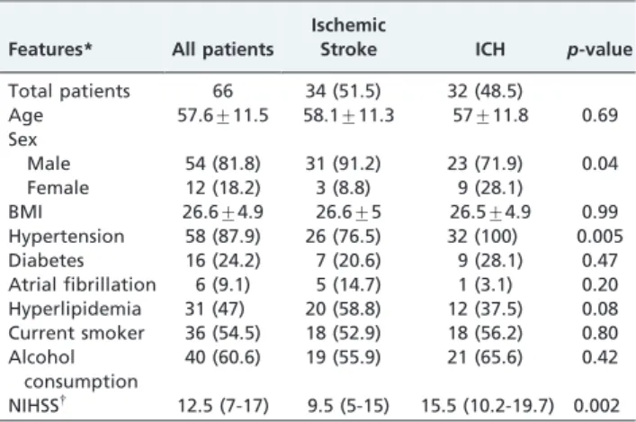
Thirty-four of the 66 subjects (51.5%) presented with ischemic stroke, and 32 subjects (48.5%) suffered ICH. Demographics and major risk factors between the stroke subtypes are presented in Table 1. The median baseline NIHSS was 12.5 (IR: 7-17), which was significant between ICH and ischemic stroke patients (p= 0.002).
The mean recorded total sleep time per stroke case was 206.9¡93.8 minutes, and the mean total recording time was
338.1¡69.6 minutes. The majority of sleep time was spent in
the supine position in all subjects; the median percentage of total sleep time in the supine position was 100% (IR: 85.9-100). The majority (66.7%) of subjects spent the entire sleep time in a supine posture. Fifty-seven patients (86.4%) spent no time sleeping in the prone position, 52 patients (78.8%) spent no time sleeping on their left side, and 49 patients (74.2%) spent no time sleeping on their right side. A significant correlation was observed between the percent of sleep time in the supine position and the NIHSS (rs= 0.5;
p,0.001).
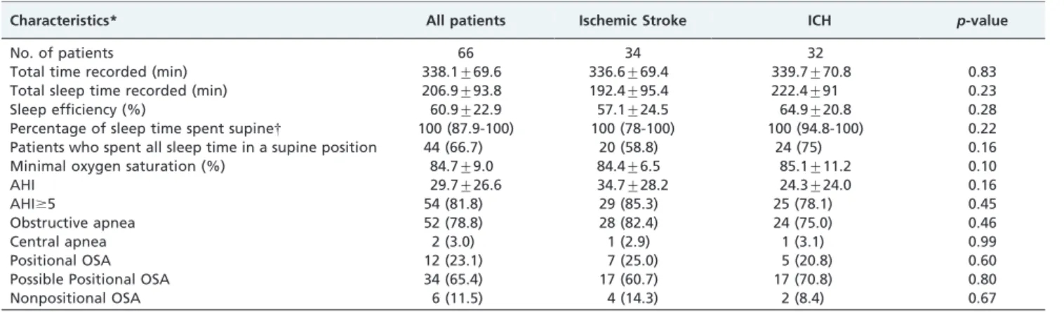
A full night of diagnostic studies was obtained for all subjects. Fifty-two patients (78.8%) exhibited OSA; 42.4% of these patients suffered ischemic stroke, and 36.4% suffered ICH. The mean AHI was 29.7 (¡26.6). Twelve patients
(23.1%) exhibited positional OSA, and 6 patients (11.5%) exhibited nonpositional OSA. The other 34 patients (65.4%) exhibited OSA but spent the entire sleep period in a supine position (i.e., possible positional OSA). No differences were observed between the stroke subtypes and the presence of OSA or positional OSA (Table 2). AHI was significantly reduced when patients changed from a supine to a lateral position (p,0.001 - Table 3).
Table 1 -Demographics and vascular risk factors.
Features* All patients
Ischemic
Stroke ICH p-value
Total patients 66 34 (51.5) 32 (48.5)
Age 57.6¡11.5 58.1¡11.3 57¡11.8 0.69
Sex
Male 54 (81.8) 31 (91.2) 23 (71.9) 0.04
Female 12 (18.2) 3 (8.8) 9 (28.1)
BMI 26.6¡4.9 26.6¡5 26.5¡4.9 0.99
Hypertension 58 (87.9) 26 (76.5) 32 (100) 0.005
Diabetes 16 (24.2) 7 (20.6) 9 (28.1) 0.47
Atrial fibrillation 6 (9.1) 5 (14.7) 1 (3.1) 0.20 Hyperlipidemia 31 (47) 20 (58.8) 12 (37.5) 0.08 Current smoker 36 (54.5) 18 (52.9) 18 (56.2) 0.80 Alcohol
consumption
40 (60.6) 19 (55.9) 21 (65.6) 0.42
NIHSS{
12.5 (7-17) 9.5 (5-15) 15.5 (10.2-19.7) 0.002
ICH: Intracerebral hemorrhage; BMI: body mass index; NIHSS: National Institutes of Health Stroke Scale.*Results are expressed as the means
¡SD or number of patients (%).{
Medians (interquartile ranges). Positional sleep apnea after stroke
Camilo MR et al. CLINICS 2012;67(12):1357-1360
DISCUSSION
We observed high frequencies of obstructive sleep apnea (78.8%) and exclusive supine positioning during sleep (66.7%) in patients with ischemic stroke or ICH. The predominance of supine posture was related to the severity of neurological deficits regardless of the stroke subtype in our patients. Supine sleep may aggravate OSA severity and contribute to hypoxemia, endothelial dysfunction, increased oxidative stress, the activation of the coagulation cascade and inflammation during the acute phase of stroke (13,14). Therefore, prolonged supine positioning during the acute phase of stroke and ICH may potentiate the negative impact of OSA on clinical outcomes.
Positional OSA was confirmed in 23.1% of patients. However, this percentage may be underestimated because the majority of patients spent the entire recorded sleep time in a supine position. The frequency of possible positional OSA increases significantly (up to 65.4%). Similar results were observed in a recent study of ischemic stroke patients in which full polysomnography was performed within the first seven days after symptom onset (9).
Continuous positive airway pressure (CPAP) is the standard treatment for OSA, but its efficacy depends on patient adherence, and stroke patients may have a low tolerance for CPAP (15-17). More definitive results on CPAP treatment in acute stroke patients are required. Therefore, the appropriate positioning of stroke patients is a reasonable and harmless initiative to avoid a prolonged supine decubitus position. Positional OSA may be more frequent during the acute stroke setting, and the incidence may decrease significantly over six months (18). Therefore, the potential for positional therapy to decrease OSA in ischemic stroke patients during the acute phase of stroke requires
further investigation. A recent small, randomized, con-trolled, two-night crossover study demonstrated that posi-tional therapy using a pillow that was designed to prevent supine sleep was adequately tolerated by patients after ischemic stroke and reduced the absolute percentage of time in a supine position by 36% and modestly reduced sleep apnea severity (19).
The limitations of our study include the small sample size and the lack of reliable information on sleep apnea severity prior to stroke. The time spent in a supine position may also be influenced by the availability of local resources and the act of performing the PSG (20). However, other multi-parametric monitoring systems that are routinely used for stroke patients may exert the same influence on sleep position.
In conclusion, positional sleep apnea was detected in one quarter of patients during the acute phase of ischemic stroke and ICH, but the actual frequency was likely under-estimated. The adequate positioning of patients during sleep may be an alternative for reducing the impact of obstructive respiratory events during the acute phase of stroke, especially in patients with a high NIHSS, regardless of their stroke subtype. Further studies are required to examine the efficacy of positional therapy on the outcomes of OSA patients during the acute phase of stroke.
ACKNOWLEDGMENTS
This work received support from Fundac¸a˜o de Amparo a Pesquisa do Estado de Sa˜o Paulo (FAPESP), Conselho Nacional de Desenvolvimento Cientı´fico e Tecnolo´gico (CNPq) and Coordenac¸a˜o de Aperfeic¸oamento de Pessoal de Nı´vel Superior (CAPES).
AUTHOR CONTRIBUTIONS
Camilo MR and Pontes-Neto OM contributed to the data collection and analysis, critical review of the results, and manuscript development. Fernandes RMF and Sander HH contributed to the polysomnography analysis and the manuscript review. Nobre F, Santos-Pontelli T, Dos Santos AC, De Araujo DB, and Leite JP participated in the critical review of the results and the manuscript.
REFERENCES
1. Iranzo A, Santamaria J, Berenguer J, Sanchez M, Chamorro A. Prevalence and clinical importance of sleep apnea in the first night after cerebral infarction. Neurology. 2002;58(6):911-6, http://dx.doi.org/10.1212/ WNL.58.6.911.
Table 2 -Polysomnographic findings.
Characteristics* All patients Ischemic Stroke ICH p-value
No. of patients 66 34 32
Total time recorded (min) 338.1¡69.6 336.6¡69.4 339.7¡70.8 0.83
Total sleep time recorded (min) 206.9¡93.8 192.4¡95.4 222.4¡91 0.23
Sleep efficiency (%) 60.9¡22.9 57.1¡24.5 64.9¡20.8 0.28
Percentage of sleep time spent supine{ 100 (87.9-100) 100 (78-100) 100 (94.8-100) 0.22
Patients who spent all sleep time in a supine position 44 (66.7) 20 (58.8) 24 (75) 0.16
Minimal oxygen saturation (%) 84.7¡9.0 84.4¡6.5 85.1¡11.2 0.10
AHI 29.7¡26.6 34.7¡28.2 24.3¡24.0 0.16
AHI$5 54 (81.8) 29 (85.3) 25 (78.1) 0.45
Obstructive apnea 52 (78.8) 28 (82.4) 24 (75.0) 0.46
Central apnea 2 (3.0) 1 (2.9) 1 (3.1) 0.99
Positional OSA 12 (23.1) 7 (25.0) 5 (20.8) 0.60
Possible Positional OSA 34 (65.4) 17 (60.7) 17 (70.8) 0.80
Nonpositional OSA 6 (11.5) 4 (14.3) 2 (8.4) 0.67
ICH: intracerebral hemorrhage; AHI: apnea–hypopnea index; OSA: obstructive sleep apnea. * Results are expressed as the means¡SD or number of patients (%).{Medians (interquartile ranges).
Table 3 -AHI in supine and lateral positions in patients with positional OSA.
All patients (n = 12)
Ischemic stroke (n = 7)
ICH (n = 5)
Mean AHI supine 36.3¡26.8 38.8¡31.3 32.7¡22
Mean AHI lateral 6.3¡5.8 7.4¡7.4 4.9¡2.5
p-value ,0.001 ,0.001 ,0.001
ICH: intracerebral hemorrhage; AHI: apnea–hypopnea index.
CLINICS 2012;67(12):1357-1360 Positional sleep apnea after stroke
Camilo MR et al.
2. Pontes-Neto OM, Fernandes RM, Sander HH, da Silva LA, Mariano DC, Nobre F, et al. Obstructive sleep apnea is frequent in patients with hypertensive intracerebral hemorrhage and is related to perihematoma edema. Cerebrovasc Dis. 2010;29(1):36-42, http://dx.doi.org/10.1159/000255972. 3. Bassetti CL, Milanova M, Gugger M. Sleep-disordered breathing and
acute ischemic stroke: Diagnosis, risk factors, treatment, evolution, and long-term clinical outcome. Stroke. 2006;37(4):967-72, http://dx.doi.org/ 10.1161/01.STR.0000208215.49243.c3.
4. Sahlin C, Sandberg O, Gustafson Y, Bucht G, Carlberg B, Stenlund H, et al. Obstructive sleep apnea is a risk factor for death in patients with stroke: A 10-year follow-up. Arch Intern Med. 2008;168(3):297-301, http://dx.doi.org/10.1001/archinternmed.2007.70.
5. Parra O, Arboix A, Montserrat JM, Quinto´ L, Bechich S, Garcı´a-Eroles L. Sleep-related breathing disorders: impact on mortality of cerebrovascu-lar disease. Eur Respir J. 2004;24(2):267-72, http://dx.doi.org/10.1183/ 09031936.04.00061503.
6. Oksenberg A, Khamaysi I, Silverberg DS, Tarasiuk A. Association of body position with severity of apneic events in patients with severe nonpositional obstructive sleep apnea. Chest. 2000;118(4):1018-24, http://dx.doi.org/10.1378/chest.118.4.1018.
7. Penzel T, Moller M, Becker HF, Knaack L, Peter JH. Effect of sleep position and sleep stage on the collapsibility of the upper airways in patients with sleep apnea. Sleep. 2001;24(1):90-5.
8. Cartwright RD. Effect of sleep position on sleep apnea severity. Sleep. 1984;7(2):110-4.
9. Brown DL, Lisabeth LD, Zupancic MJ, Concannon M, Martin C, Chervin RD. High prevalence of supine sleep in ischemic stroke patients. Stroke. 2008;39(9):2511-4, http://dx.doi.org/10.1161/STROKEAHA.107.513572. 10. Dziewas R, Hopmann B, Humpert M, Ritter M, Dittrich R, Schabitz WR,
et al. Positional sleep apnea in patients with ischemic stroke. Neurol Res. 2008;30(6):645-8, http://dx.doi.org/10.1179/174313208X289598. 11. Iber C, Ancoli-Israel S, Chesson AL, Quan SF. Respiratory Rules. The
AASM Manual for the Scoring of Sleep And Associated Events: Rules, Terminology And Technical Specifications. Westchester, American Academy of Sleep Medicine, 2007.
12. Cincura C, Pontes-Neto OM, Neville IS, Mendes HF, Menezes DF, Mariano DC, et al. Validation of the national institutes of health stroke scale, modified rankin scale and barthel index in brazil: The role of cultural adaptation and structured interviewing. Cerebrovasc Dis. 2009;27(2):119-22, http://dx.doi.org/10.1159/000177918.
13. Von KR, Loredo JS, Powell FL, Adler KA, Dimsdale JE. Short-term isocapnic hypoxia and coagulation activation in patients with sleep apnea. Clin Hemorheol Microcirc. 2005;33(4):369-77.
14. Kunz AB, Kraus J, Young P, Reuss R, Wipfler P, Oschmann P, et al. Biomarkers of Inflammation and Endothelial Dysfunction in Stroke with and without Sleep Apnea. Cerebrovasc Dis. 2012;33(5):453-60, http://dx. doi.org/10.1159/000336120.
15. Palombini L, Guilleminault C. Stroke and treatment with nasal CPAP. Eur J Neurol. 2006;13(2):198-200, http://dx.doi.org/10.1111/j.1468-1331. 2006.01169.x.
16. Hsu CY, Vennelle M, Li HY, Engleman HM, Dennis MS, Douglas NJ. Sleep-disordered breathing after stroke: A randomised controlled trial of continuous positive airway pressure. J Neurol Neurosurg Psychiatry. 2006;77(10):1143-9, http://dx.doi.org/10.1136/jnnp.2005. 086686.
17. Brown DL, Concannon M, Kaye AB, Zupancic M, Lisabeth LD. Comparison of two headgear systems for sleep apnea treatment of stroke patients. Cerebrovasc Dis. 2009;27(2):183-6, http://dx.doi.org/10. 1159/000185610.
18. Dziewas R, Hopmann B, Humpert M, Ritter M, Dittrich R, Schabitz WR, et al. Positional sleep apnea in patients with ischemic stroke. Neurol Res. 2008;30(6):645-8, http://dx.doi.org/10.1179/174313208X289598. 19. Svatikova A, Chervin RD, Wing JJ, Sanchez BN, Migda EM, Brown DL.
Positional therapy in ischemic stroke patients with obstructive sleep apnea. Sleep Med. 2011;12(3):262-6, http://dx.doi.org/10.1016/j.sleep. 2010.12.008.
20. Metersky ML, Castriotta RJ. The effect of polysomnography on sleep position: Possible implications on the diagnosis of positional obstructive sleep apnea. Respiration. 1996;63(5):283-7, http://dx.doi.org/10.1159/ 000196561.
Positional sleep apnea after stroke
Camilo MR et al. CLINICS 2012;67(12):1357-1360