Follow-up of experimental chronic Chagas’ disease in dogs:
use of polymerase chain reaction (PCR) compared with
parasitological and serological methods
F.M.G. Arau´jo
a, M.T. Bahia
b, N.M. Magalha˜es
c, O.A. Martins-Filho
d,
V.M. Veloso
b, C.M. Carneiro
c, W.L. Tafuri
a, M. Lana
c,*
aNu´cleo de Pesquisas em Cieˆncias Biolo´gicas(NUPEB),Instituto de Cieˆncias Biolo´gicas,Campus Uni6ersita´rio,
Morro do Cruzeiro,Brazil
bDepartamento de Cieˆncias Biolo´gicas,Instituto de Cieˆncias Biolo´gicas,Campus Uni6ersita´rio,Morro do Cruzeiro, Brazil cDepartamento de Ana´lises Clı´nicas,Escola de Farma´cia,Uni6ersidade Federal de Ouro Preto,Rua Costa Sena,171-Centro,
CEP35400-000Ouro Preto,Brazil
dCentro de Pesquisas Rene´ Rachou,FIOCRUZ-BH,A6.Augusto de Lima1715-Barro Preto,CEP30190-002Belo Horizonte,
MG,Brazil
Received 30 March 2001; received in revised form 27 June 2001; accepted 27 August 2001
Abstract
In this study, the polymerase chain reaction (PCR) was compared with parasitological and serological methods to detect the infection in dogs, 5 – 12 years after experimental infection with Trypanosoma cruzi. The ability of parasitological methods to identify a positive animal was 22 and 11% by hemoculture and xenodiagnosis/xenoculture, respectively. On the other hand, the serological tests, including conventional serology and anti-live trypomastigote antibodies (ALTA) were positive in all infected dogs. Despite its low sensitivity, if considering only one reaction, the PCR analysis showed 100% of positivity, demonstrating the presence of parasite kDNA in all infected dogs. To identify a positive dog required at least two blood samples and up to nine repeated reactions using the same sample. Serial blood sample collection, ranging from 1 to 9, revealed that the percentage of dogs with positive PCR ranged from 67 to 100%. These findings suggested that, although the PCR is useful to detect the parasite in infected hosts, it should not be used isolated for the diagnosis of Chagas’ disease and warn for the necessity of serial blood collection and re-tests. Moreover, these data validate once more the dog as a model for Chagas’ disease since they demonstrate the permanence of infection by PCR, parasitological and serological methods, reaching relevant requisites for an ideal model to study this disease. © 2002 Elsevier Science B.V. All rights reserved.
Keywords:Trypanosoma cruzi; Dog; Chronic infection; PCR; Parasitological tests and serology
www.parasitology-online.com
1. Introduction
Dogs have been frequently used as experimental model to study Chagas’ disease. The acute phase * Corresponding author. Tel.: +55-31-3559-1680; fax:
+55-31-559-1628.
E-mail address:[email protected](M. Lana).
F.M.G.Arau´jo et al./Acta Tropica 81 (2002) 21 – 31 22
of the infection has been reproduced with relative facility by different authors, especially in young animals (Andrade, 1984; Pedreira de Castro and Brener, 1985; Lana et al., 1992). However, the development of the different clinical forms of the disease is not easily to be reproduced in this model (Andrade, 1984). The great majority of publications have only recorded the indeterminate form of the disease (Andrade and Andrade, 1980; Andrade et al., 1981) with few exceptions (Laranja et al., 1948; Lana et al., 1988, 1992). Andrade (1984) suggests auto-cure in dogs. Kret-tli et al. (1984) suggest auto-cure in mice. Zeledon et al. (1988) suggest auto-cure in humans. For these reasons, herein is reported a long-term fol-low-up study of dogs, 5 – 12 years after acute
infection with Trypanosoma cruzi (Lana et al.,
1992). The major goal of this study was to
demonstrate the permanence of T. cruzi in these
infected dogs, using different approaches like polymerase chain reaction (PCR), parasitological and serological methods. Since the PCR reaction has been theoretically pointed out as a powerful tool for the diagnosis of Chagas’ disease (Avila et al., 1991; Wincker et al., 1994; Britto et al., 1995a), it was exhaustively performed, every 3 months, during 2 years, in parallel with hemocul-ture, xenodiagnosis/xenoculture and
serology-con-ventional ELISA and detection of anti-live
trypomastigote antibodies (ALTA). The use of these different laboratorial approaches to detect the presence of the parasite in chronically infected dogs could fill a crucial requisite, recommended by WHO (1984), for the establishment of dogs as an experimental model for chronic Chagas’ dis-ease.
2. Material and methods
2.1. Parasite strains
T. cruzi strains were isolated from the patient Berenice, considered the first human case of Chagas’ disease, in 1962 (Be-62) by Salgado et al. (1962) and in 1978 (Be-78) by Lana and Chiari
(1986). Both strains were classified as T. cruzi II
(Lana et al., 1996).
2.2. Animals
Nine out-bred dogs were acutely infected with
Be-62 and Be-78 T. cruzi strains (Lana et al.,
1992), 5 – 12 years before starting this study. Five were infected with Be-62 strain and four infected with Be-78 strain. Animals were inoculated with
2.0×10³ metacyclic trypomastigotes. Two
age-matched uninfected dogs were used as negative controls. All animals were evaluated every 3 months, during 2 years.
2.3. Polymerase chain reaction (PCR)
Ten milliliters of blood were collected with an equal volume of Guanidin – HCl 6 M/EDTA 0.2 M, pH 8.0 solution (Avila et al., 1991). Samples were maintained at room temperature. Seven days later, the samples were boiled during 15 min and stored at 4 °C until processing. DNA extraction was done according to Wincker et al. (1994) pro-tocol with some modifications introduced by Gomes et al. (1998).
PCR conditions were the same described by Gomes et al. (1998) but using the primers: S35
(5%-
AAATAATGTACGGG(T/G)GAGATGCA-TGA-3%) and S36 (5%
-GGGTTCGATTGGGG-TTGGTGT-3%) described by Avila et al. (1990)
that anneal in the conserved microregion of the
parasites kDNA minircicles. Briefly, 2 ml of blood
DNA template was added in 10 mM Tris – HCl
(pH 9.0); 75 mM KCl; 3.5 mM MgCl2; 0.1%
Triton X-100; 0.2 mM of dATP, dCTP, dGTP and dTTP (Sigma Company Ltd., USA); 20 pmol of each primer (S35 and S36, cited above); 1.0
unit of Taq DNA polymerase enzyme (Promega,
Madison, WI, USA) and water up to 20 ml. The
reaction mixture was overlaid with 30 ml of
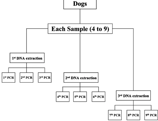
extrac-tions and three PCR reacextrac-tions using each DNA template were performed resulting on up to nine PCR for each blood sample, if necessary. Fig. 1 represents the strategy adopted for each blood sample processed.
2.4. Parasitological methods
2.4.1. Hemoculture
Twenty milliliters of heparinized blood were processed according to Luz et al. (1994). The hemocultures were maintained at 28 °C. Thirty, 60, 90 and 120 days later, each tube was examined for the detection of parasites.
2.4.2. Xenodiagnosis/xenoculture
Fifteen third star nymphs ofTriatoma infestans
were used for each procedure. After blood meal the insects were maintained at 27 °C, 70% of humidity. Forty days later, the insects were exam-ined for the detection of parasites (xenodiagnosis).
Xenoculture was performed according to Bronfen et al. (1989). The xenoculture was maintained at 28 °C and 30, 60, 90 and 120 days later it was examined for detection of parasites.
2.5. Serological methods
2.5.1. Con6entional serology (ELISA)
T. cruzi specific antibodies was detected by the technique described by Voller et al. (1976) modified and adapted by Lana et al. (1991).
ELISA plates were sensitized withT.cruziantigen
prepared by alkaline extraction (Vitor and Chiari, 1987) of Y strain obtained at exponential growth in LIT medium. Antibody binding was detected
by using peroxidase-labeled anti-dog IgG, after
reading in spectrophotometer, using 490 nm filter (BIO-RAD, 3550). The mean absorbance of 10 negative control sera plus two standard deviations was used as a cut-off to discriminate positive and negative results.
F
.
M
.
G
.
Arau
´jo
et
al
.
/
Acta
Tropica
81
(2002)
21
–
31
24
Table 1
Results of PCR of serial blood collection in dogs experimentally infected withT.cruziduring the chronic phase of the infection T.cruzistrain Time of infection (years) Blood sample
Dog Total PCR+(%)
1st 2nd 3rd 4th 5th 6th 7th 8th 9th
Controls
− − −
C1 − − − − − − − − 0
− − − − − − −
C2 − − − − 0
Infected dogs
Be-62 12 − +
1 − + + + + + − 67
Be-62 11 − + + +
2 + + + N N 86
Be-62 11 +
3 − + + + + + N N 86
Be-62 5 + + + +
4 + + + + + 100
Be-62 5 + + + +
5 + + + + + 100
Be-78 10 − + − +
6 + − N N N 50
Be-78 10 + + + + + N
7 N N N 100
Be-78 9 + − − +
8 N N N N N 50
Be-78 6 + + + + + +
9 + + + 100
67 78 67 100
Total (%) 100 86 100 100 75 85
Table 2
Minimal number of DNA extraction and PCR reactions necessary to detect the first positive result in dogs experimentally infected withT.cruziduring the chronic phase of the infection
T.cruzistrain First DNA extraction with PCR+
Time of infection (years) Dog First PCR+
Be-62
5 4 2 5
5 5 Be-62 1 1
Be-78
6 9 2 5
Be-78 2
8 4
9
6
10 Be-78 6 16
Be-78 3
10 7 7
Be-62 6
2 17
11
3
11 Be-62 3 9
Be-62
12 1 5 14
2.5.2. Anti-li6e trypomastigote antibody (ALTA) The immunofluorescence staining to detect ALTA was carried out as described by Martins-Filho et al. (1995), modified for U bottom 96 wells plate as introduced by Cordeiro et al. (2001). Briefly, after incubation with dog sera, the binding of antibodies to trypomastigotes was de-tected using fluorescein isothiocyanate (FITC)
conjugatedanti-dog IgG. The FITC-labeled
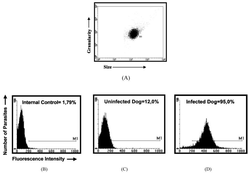
para-sites were fixed before run into cytometer. Flow-cytometric measurements were performed on a
Becton Dickinson FACScalibur. TheCELL-QUEST
software package was used in both data storage and analysis. Trypomastigotes were first identified on the basis of their specific forward (size) and side (granularity) light-scattering properties (Fig. 2A). The relative FITC fluorescence intensity for each parasite preparation after incubation with individual sample was analyzed using a single histogram. A marker was set up on the internal control for unspecific binding (Fig. 2B) and used to determine the percentage of positive fluores-cent parasites (PPFP). The samples were
consid-ered negative when PPFP520% and positive
when PPFP was \20% (Fig. 2C and D).
3. Results
3.1. Polymerase chain reaction (PCR)
PCR results obtained with up to nine reactions
for each blood sample are shown in Table 1. The PCR was found to be positive in 100% of the infected dogs. The identification of a positive dog required at least two blood samples. The percent-age of positive PCR, considering the total blood samples per dog, ranged from 50 to 100%. Only four dogs showed a positive PCR result for all blood samples processed with no association with
the time of infection neither the T. cruzi strain.
Serial blood sample collection, ranging from 1 to 9, revealed that the percentage of dogs with posi-tive PCR ranged from 67 to 100%, depending on the sample processed.
The minimum number of DNA extractions necessary to detect the first positive reaction in ranged from 1 to 6 depending on the animal evaluated (Table 2). The number of PCR
neces-sary to obtain the first positive result
ranged from 1 to 17. Only one infected dog showed a positive result if only one blood sample and the first PCR was taken in account.
No association was observed between the
strain ofT.cruziand the number of DNA
F
.
M
.
G
.
Arau
´jo
et
al
.
/
Acta
Tropica
81
(2002)
21
–
31
26
3.2. Hemoculture, xenodiagnosis and xenoculture
Table 3 summarizes the results of all parasito-logical methods. Serial hemoculture (5 – 9) was
negative in all dogs infected with Be-62 T. cruzi
strain and two dogs infected with Be-78 strain. The total percentage of dogs with positive hemo-culture was 22%. Both, serial xenodiagnosis (5 – 9) and xenoculture were negative in all dogs infected with Be-62 strain and positive in only one dog infected with Be-78 strain. The total percentage of dogs with positive xenodiagnosis/xenoculture was 11%.
3.3. Serology
Immunoenzimatic tests to detect circulating
anti-T.cruziantibodies were performed using 4 – 9
blood samples per animal (Table 4). Results of conventional ELISA are shown in Table 4. De-spite three negative results out of nine reactions
observed for the dog c1, all infected animals
displayed positive ELISA. Repetition of ELISA
confirmed these results. Analysis of ALTA
demonstrated positive results for all samples tested during the 2 years of evaluation. Control animals were always negative in all serological examinations.
4. Discussion
The goal of this work was to demonstrate the
permanency of T. cruzi in dogs during long-term
chronic infection. These dogs were experimentally infected 5 – 12 years prior the beginning of this study. All animals were exhaustively examined during the acute phase, showing positive parasito-logical (fresh blood examination, hemoculture, xenodiagnosis) and serological examinations
(In-direct Immunofluorescence Test (IIT) and
ELISA), commonly used for the diagnosis of Chagas’ disease (Lana et al., 1991, 1992). Many symptoms of the acute phase of the disease and electrocardiographic alterations compatible with Chagas’ disease were also recorded (Lana et al., 1992). Herein they were re-submitted to laborato-rial investigations including well-established para-sitological and serological methods as well as new tools for diagnosing Chagas’ disease, such as ALTA and PCR analysis.
The low performance of parasitological meth-ods observed here is not totally unexpected, since
longitudinal evaluation of these same dogs
showed a lowering positivity of these test during the first 3 years after infection (Lana et al., 1992). The scarcity of positive parasitological examina-tions observed here, mainly in dogs infected with
F.M.G.Arau´jo et al./Acta Tropica 81 (2002) 21 – 31 28
Table 3
Results of hemoculture, xenodiagnosis and xenoculture in dogs experimentally infected withT.cruziduring the chronic phase of the infection
Hc+/Hc total Xd+/Xd total Xc+/Xc total Dog T.cruzistrain
Controls
N N N
C1 –
N N
– N
C2
Infected dogs
0/9
1 Be-62 0/9 0/9
3 Be-62 0/7 0/7 0/7
0/7
4 Be-62 0/7 0/7
0/9 0/9
Be-62 0/9
5
Be-62
6 0/9 0/9 0/9
2/6 1/6
7 Be-78 1/6
1/5 0/5
Be-78 0/5
9
Be-78
10 0/5 0/5 0/5
0/9 0/9
11 Be-78 0/9
22 11 11
Total (%)
N, not preformed.
the Be-62 strain, was previously reported by Lana et al. (1992). Although the time of infection seems to decrease the parasitemia (Castro et al., 1983, 1999) the positivity of hemoculture and
xenodiag-nosis also seems to be dependent on the type ofT.
cruzi strain. Lana et al. (1988), Veloso et al. (2001) easily detected positive hemoculture and xenodiagnosis in dogs experimentally infected with Colombian strain 8 and 15 years after infec-tion which presents a predominance of stout forms in the blood and consequently more resis-tant to lysis mediated by complement (Krettli et al., 1984). Herein, the Be-62 strain, differently from Be-78 strain, which presents a predominance of slender trypomastigotes in blood (Lana and Chiari, 1986) that are more sensitive to comple-ment mediated lysis, resulting on lesser perma-nency on circulating blood, also showed a lower positivity on parasitological methods.
The conventional serological method (ELISA) was positive in all dogs, despite an oscillating result in one of them. Oscillating results in sero-logical tests are also reported in humans (Rassi et al., 1969), and can be the reason why some pa-tients with positive xenodiagnosis/hemoculture and/or PCR present negative serology (Brenie`re et al., 1984; Gomes et al., 1999). However, using the
flow cytometry assay to detect ALTA, all infected dogs were tagged with positive results thoroughly all blood samples. This result confirms the
perma-nency of the T. cruzi in all chronically infected
dogs, since ALTA has been indicated as a good marker for active infection and this method is
Table 4
Serological status of dogs experimentally infected withT.cruzi during the chronic phase of the infection
T.cruzi +ELISA/total
Dog +ALTA/total
Strain
Controls
– 0/9
C1 0/9
– 0/9
C2 0/9
Infected dogs Be-62
1 6/9 9/9
Be-62
3 7/7 7/7
Be-62
4 7/7 7/7
5 Be-62 9/9 9/9
Be-62
6 9/9 9/9
7 Be-78 6/6 6/6
5/5 Be-78 5/5
9
4/4 4/4
10 Be-78
11 Be-78 9/9 9/9
proposed as an new approach to detect cure in Chagas’ disease (Martins-Filho et al., 1995).
Although the PCR was found to be positive in 100% of the infected dogs, to identify a positive dog it was required at least two blood samples and up to nine repeated reactions using the same sample, depending on the blood sample analyzed.
Further investigations, including other PCR
protocols, followed by slot-blot hybridization and analysis of different organs and tissues of these dogs are currently been investigated to
im-prove the detection of T. cruzi during chronic
infection.
The number of PCR necessary to obtain the first positive result ranged from 1 to 17. Only one infected dog showed a positive result if only one blood sample and the first PCR was taken in account. It was interesting to observe that dogs over 10 years of infection required a higher num-ber of PCR reactions and more DNA extractions to show the first positive result.
Britto et al. (1995b), Junqueira et al. (1996) showed only 44.7% and 59.4% of positive PCR, respectively in patients. The most acceptable rea-son to explain the low performance of the PCR to detect DNA in biological material (blood, serum and tissue) is the level of parasitemia pre-sented in the host when the sample is collected (Moser et al., 1989; Gomes et al., 1998). The results presented here do not discard this hypoth-esis, since a low percentage of positivity was observed when parasitological methods were con-duced in parallel to PCR.
However, as the ability of PCR to detect a positive host increases from 67% when one sam-ple was evaluated to 100% when a second samsam-ple was taking in account. The percentage of positive PCR, considering the total blood samples per dog, ranged from 50 to 100%. Then, we do be-lieve that besides considering the low parasitemia of chronic infection as the major restraining fea-ture leading to a negative PCR, the necessity of serial blood collection and re-tests should also be considered. As there are no available methods to test dogs samples for the presence of inhibitor elements during the DNA extraction procedure, we cannot discard that all these repeated negative results of PCR were due to phenol and
chloro-form, which can inhibits the Taq DNA
poly-merase enzyme generating false negative results. In conclusion, the hallmark of this work was:
(1) the evidence of T. cruzi presence in all dogs,
even after a long-chronic period of infection. Then, these results validate the dog as a good experimental model to study the chronic phase of Chagas’ disease and (2) the establishment of the necessity of repeated PCR procedures to assure
the diagnosis of the chronic T. cruzi infection
indicating that this method should not be used isolated as a conclusive tool for diagnosis of Chagas’ disease.
Acknowledgements
We are grateful to Dr Constanc¸a F.C. Britto of the DBBM/Instituto Oswaldo Cruz, Rio de Janeiro, RJ and Drs Egler Chiari and Lu´cia Maria da Cunha Galva˜o of the Departamento de Parasitologia, ICB, UFMG for the scientific sup-port regarding the set-up of PCR. This work was supported by Conselho Nacional de Desenvolvi-mento Cientı´fico e Tecnolo´gico (CNPq) and Fun-dac¸a˜o de Amparo a` Pesquisa do Estado de Minas Gerais (FAPEMIG).
References
Andrade, Z.A., 1984. The canine model of Chagas’ disease. Mem. Inst. Oswaldo Cruz 79 (Suppl.), 77 – 83.
Andrade, Z.A., Andrade, S.G., 1980. A patologia da doenc¸a de Chagas experimental no ca˜o. Mem. Inst. Oswaldo Cruz 75, 77 – 95.
Andrade, S.G., Andrade, Z.A., Sadigursky, M., Maguire, J.H., 1981. Experimental Chagas’ disease in dogs. A pathologic and ECG study of the chronic indeterminate phase of the infection. Arch. Pathol. Lab. Med. 105, 460 – 464. Avila, H., Gonc¸alves, A.M., Nehme, N.S., Morel, C.M.,
Simp-son, L., 1990. Schizodeme analysis ofTrypanosoma cruzi stocks from South and Central America by analysis of PCR-amplified minicircle variable region sequences. Mol. Biochem. Parasitol. 42, 175 – 188.
F.M.G.Arau´jo et al./Acta Tropica 81 (2002) 21 – 31 30
Brenie`re, S.F., Poch, O., Selaes, H., Tibayrenc, M., Lemesre, J., Anteseana, G., Desjeux, P., 1984. Specific humoral depression in chronic patients infected by Trypanosoma cruzi. Rev. Inst. Med. Trop. Sa˜o Paulo 26, 254 – 258. Britto, C., Cardoso, M.A., Monteiro-Vanni, C.M.,
Hass-locher-Moreno, A., Xavier, S.S., Oelemann, W., Santoro, A., Pirmez, C., Wincker, P., 1995a. Polymerase chain reaction detection ofTrypanosoma cruzi in human blood samples as a tool for diagnosis and treatment evaluation. Parasitology 110, 241 – 247.
Britto, C., Cardoso, M.A., Ravel, C., Santoro, A., Borges-Pereira, J., Coura, J.R., Morel, C.M., Wincker, P., 1995b. Trypanosoma cruzi: parasite detection and strain discrimi-nation in chronic chagasic patients from northeastern Brazil using PCR amplification of kinetoplast DNA and nonradioactive hybridization. Exp. Parasitol. 81, 462 – 471. Bronfen, E., Rocha, F.S.A., Machado, G.B.N., Perillo, M.M., Romanha, A.J., Chiari, E., 1989. Isolamento de amostras doTrypanosoma cruzipor xenodiagno´stico e hemocultura de pacientes na fase croˆnica da doenc¸a de Chagas. Mem. Inst. Oswaldo Cruz 84, 237 – 240.
Castro, C.N., Alves, M.T., Macedo, V.O., 1983. Importaˆncia da repetic¸a˜o do xenodiagno´stico para avaliac¸a˜o da para-sitemia na fase croˆnica da doenc¸a de Chagas. Rev. Soc. Bras. Med. Trop. 16, 98 – 103.
Castro, C., Maceˆdo, V., Prata, A., 1999. Aspects of para-sitemia byTrypanosoma cruziin chronic chagasic patients during 13 years. Rev. Bras. Med. Trop. 32, 157 – 165. Cordeiro, F.D., Martins-Filho, O.A., Da Costa Rocha, M.O.,
Adad, S.J., Correa-Oliveira, R., Romanha, A.J., 2001. Anti-Trypanosoma cruziimmunoglobulin G1 can be a use-ful tool for diagnosis and prognosis of human Chagas’ disease. Clin. Diagn. Lab. Immunol. 8, 112 – 118. Gomes, M.L., Macedo, A.M., Vago, A.R., Pena, S.D.J.,
Gal-va˜o, L.M.C., Chiai, E., 1998. Trypanosoma cruzi: opti-mization of polymerase chain reation for detection in human blood. Exp. Parasitol. 88, 28 – 33.
Gomes, M.L., Galva˜o, L.M.C., Macedo, A.M., Pena, S.D.J., Chiari, E., 1999. Chagas’ disease diagnosis: comparative analysis of parasitologic, molecular and serologic methods. Am. J. Trop. Med. Hyg. 60, 205 – 210.
Junqueira, A.C.V., Chiari, E., Wincker, P., 1996. Comparison of the polymerase chain reaction with two calssical para-sitological methods for the diagnosis of Chagas’ disease. Trans. R. Soc. Trop. Med. Hyg. 90, 129 – 132.
Krettli, A.U., Canc¸ado, J.R., Brener, Z., 1984. Criterion of cure of human Chagas’ disease after specific chemoterapy: recent advances. Mem. Inst. Oswaldo Cruz 79, 157 – 164. Lana, M., Chiari, C.A., 1986. Caracterizac¸a˜o biolo´gica
com-parativa das cepas Berenece-78 deTrypanosoma cruzi, iso-ladas da mesma paciente em diferentes perı´odos. Mem. Inst. Oswaldo Cruz 86, 247 – 253.
Lana, M., Tafuri, W.L., Caliari, M.V., Bambirra, E.A., Chiari, C.A., Rios Leite, V.H., Barbosa, A.J.A., Toledo, M.J., Chiari, E., 1988. Fase croˆnica cardı´aca fibrosante da tripanosomı´ase cruzi experimental no ca˜o. Rev. Soc. Bras. Med. Trop. 21, 113 – 121.
Lana, M., Vieira, L.M., Machado-Coelho, J.L.L., Chiari, E., Veloso, V.M., Tafuri, W.L., 1991. H umoral immune response in dogs experimentally infected withTrypanosoma cruzi. Mem. Inst. Oswaldo Cruz 86, 471 – 473.
Lana, M., Chiari, E., Tafuri, W.L., 1992. Experimental Chagas’disease in dogs. Mem. Inst. Oswaldo Cruz 87, 59 – 71.
Lana, M., Chiari, C.A., Chiari, E., Morel, C.M., Gonc¸alves, A.M., Romanha, A.J., 1996. Characterization of two iso-lates of Trypanosoma cruzi obtained from the patient Berenice, the first human case of Chagas’ disease by Carlos Chagas in 1909. Parasitol. Res. 82, 257 – 260.
Laranja, F.S., Dias, J.C., Nobrega, G., 1948. Clı´nica e ter-apeˆutica da doenc¸a de Chagas. Mem. Inst. Oswaldo Cruz 49, 473 – 529.
Luz, Z.M.P., Coutinho, M.G., Canc¸ado, J.R., Krettli, A.U., 1994. Hemocultura: Te´cnica sensı´vel na detecc¸a˜o do Try-panosoma cruziem pacientes chaga´sicos na fase croˆnica da doenc¸a de Chagas. Rev. Soc. Bras. Med. Trop. 27, 143 – 148.
Martins-Filho, O.A., Pereira, M.E.S., Carvalho, J.F., Canc¸ado, J.R., Brener, Z., 1995. Flow cytometry, a new approach to detect anti-live trypomastigote antibodies and monitor the efficacy of specific treatment in human Chagas’ disease. Clin. Diagn. Lab. Immunol. 2, 569 – 573. Moser, D.R., Kirchhoff, L.V., Donelson, J.E., 1989. Detection of Trypanosoma cruzi by DNA amplification using the polymerase chain reaction. J. Clin. Microbiol. 30, 1472 – 1477.
Pedreira de Castro, M.A., Brener, Z., 1985. Estudo parasito-lo´gico e anatomopatoparasito-lo´gico da fase aguda da doenc¸a de Chagas em ca˜es inoculados com duas diferentes cepas do Trypanosoma cruzi. Rev. Soc. Bras. Med. Trop. 18, 223 – 229.
Rassi, A., Amato Neto, V., Siqueira, A.F., 1969. Comporta-mento evolutivo da reac¸a˜o de fixac¸a˜o de compleComporta-mento na fase croˆnica da mole´stia de Chagas. Rev. Inst. Med. Trop. Sa˜o Paulo 11, 430 – 435.
Salgado, J.A., Garcez, P.N., Oliveira, C.A., Galizzi, J., 1962. Revisa˜o clı´nica atual do primeiro caso humano descrito de doenc¸a de Chagas. Rev. Inst. Med. Trop. 4, 330 – 337. Santos, F.R., Pena, S.D.J., Epplen, J.T., 1993. Genetic and
population study of a Y-linked tetranucleotide repeat DNA polymorphism with a simple non-isotopic technique. Hum. Genet. 90, 655 – 656.
Veloso, V.M., Carneiro, C.M., Toledo, M.J.O., Lana, M., Chiari, E., Tafuri, W.L., Bahia, M.T., 2001. Vaiation in susceptibility to benznidazole in isolates derived from Try-panosoma cruzi parental strains. Mem. Inst. Oswaldo Cruz 96, 1005 – 1011.
Vitor, R.W.A., Chiari, E., 1987. Avaliac¸a˜o de antı´genos do Trypanosoma cruzipara a reac¸a˜o de hemaglutinac¸a˜o indi-reta. I. Diferentes extratos antigeˆnicos. Rev. Inst. Med. Trop. 29, 178 – 182.
Wincker, P., Britto, C., Pereira, J.B., Cardoso, M.A., Oele-mann, W.E., Morel, C.M., 1994. Use of a simplified poly-merase chain reaction procedure to detect Trypanosoma cruzi in blood samples patients in a rural endemic area. Am. J. Trop. Med. Hyg. 51, 771 – 777.
WHO — World Health Organization, 1984. Report of the
sci-entific working group on the development and evaluation of animal models for Chagas’ disease. Geneva.