O R I G I N A L A R T I C L E
A potential link among antioxidant enzymes, histopathology and
trace elements in canine visceral leishmaniasis
Carolina C. Souza*, Tatiane de O. Barreto
†, Sydnei M. da Silva
‡, Aldair W. J. Pinto*,
Maria M. Figueiredo*, Olguita G. Ferreira Rocha
§, Silvia D. Canguss
u
¶and Wagner L. Tafuri*
*Departamento de Patologia Geral, Instituto de Ci^encias Biologicas,Universidade Federal de Minas Gerais, Belo Horizonte, Brasil, †Departamento de Bioquımica e Imunologia, Instituto de Ci^encias Biologicas, Universidade Federal de Minas Gerais, Belo Horizonte, Brasil,‡Departamento de Imunologia, Microbiologia e parasitologia, Instituto de Ci^encias Biomedicas, CampusUmuarama, Universidade Federal de Uberl^andia, Uberl^andia,Brasil,§Fundacß~ao Centro Tecnologico de Minas Gerais (CETEC), Belo Horizonte, Brasil and¶Departamento de Ci^encias Biologicas, Instituto de Ci ^encias Exatas e Biologicas, Campus Morro do Cruzeiro, Universidade Federal de Ouro Preto,Ouro Preto, Brasil
INTERNATIONAL
JOURNAL OF
EXPERIMENTAL
PATHOLOGY
doi: 10.1111/iep.12080
Received for publication: 19 September 2013
Accepted for publication: 17 February 2014
Correspondence:
Wagner Luiz Tafuri
Departamento de Patologia Geral Instituto de Ci^encias Biologicas Universidade Federal de Minas Gerais Av. Antonio Carlos 6627^
Belo Horizonte, CEP 31270-901, MG, Brasil
Tel.: +55 31 3409 2889 Fax: +55 31 3409 2879 E-mail: [email protected]
SUMMARY
Canine visceral leishmaniasis (CVL) is a severe and fatal systemic chronic inflamma-tory disease. We investigated the alterations in, and potential associations among, antioxidant enzymes, trace elements and histopathology in CVL. Blood and tissue levels of Cu-Zn superoxide dismutase, catalase and glutathione peroxidase were measured in mixed-breed dogs naturally infected withLeishmania infantum chagasi, symptomatic (n=19) and asymptomatic (n= 11). Serum levels of copper, iron, zinc, selenium and nitric oxide, and plasma lipid peroxidation were measured. Histologi-cal and morphometric analyses were conducted of lesions in liver, spleen and lymph nodes. We found lower blood catalase and glutathione peroxidase activity to be cor-related with lower iron and selenium respectively. However, higher activity of Cu-Zn superoxide dismutase was not correlated with the increase in copper and decreased in zinc observed in infected animals compared to controls. Organ tissue was characterized by lower enzyme activity in infected dogs than in controls, but this was not correlated with trace elements. Lipid peroxidation was higher in symp-tomatic than in asympsymp-tomatic and control dogs and was associated with lesions such as chronic inflammatory reaction, congestion, haemosiderin and fibrosis. Systemic iron deposition was observed primarily in the symptomatic dogs showing a higher tissue parasite load. Dogs with symptomatic CVL displayed enhanced LPO and Fe tissue deposition associated with decreased levels of antioxidant enzymes. These results showed new points in the pathology of CVL and might open new treatment perspectives associated with antioxidants and the role of iron in the pathogenesis of CVL.
Keywords
antioxidant enzymes, canine visceral leishmaniasis, iron, trace elements
Canine visceral leishmaniasis (CVL) is a chronic zoonotic disease in Brazil caused by Leishmania (Leishmania) infan-tum chagasi (syn L. infantum; Mauricio et al. 2000; Shaw 2006) transmitted by the phlebotomine sandfly Lutzomyia longipalpis. Dogs are both natural hosts and the main reservoir of the parasite (Deane & Deane 1955). A typical mononuclear inflammation has been described primarily in organs rich in cells of the mononuclear phagocytic system
such as liver, spleen, bone marrow and lymph nodes (Deane & Deane 1955; Alvaret al.2004).
Macrophages generate highly toxic molecules such as reactive oxygen species (ROS) including superoxide radicals (O
2), hydrogen peroxide (H2O2) and hydroxyl radicals (OH) and reactive nitrogen species including nitric oxide (NO) to combat Leishmania (Mauel et al. 1991; Assrue et al. 1994; Biswas et al. 1997). This can disturb cell
260
structure and function through lipid peroxidation (LPO) which leads to the formation of degradation products, including malonyl dialdehyde (Assrue et al. 1994; Biswas et al. 1997). Leishmania inhibit ROS production and can replicate within macrophages (Cunningham 2002; Paltrinieri et al.2010). However, according to Paltrinieriet al.(2010), an increase in ROS depends on inflammation, rather than directly on the presence ofLeishmania.
To protect against ROS damage, vertebrate hosts possess a variety of antioxidant defences that include metal redistri-bution and antioxidant enzyme systems requiring trace ele-ments such as copper (Cu), zinc (Zn), iron (Fe) and selenium (Se) for their activity. Antioxidant enzymes such as Cu-Zn superoxide dismutase (SOD) remove damaging ROS from the environment by catalysing the dismutation of superoxide radicals to H2O2 and O2. Catalase (CAT) requires iron (Fe), glutathione peroxidase (GSH-Px) requires selenium (Se), and both catalyse the reduction in H2O2 to H2O and O2(Brittiet al.2008).
Although studies of human forms of leishmaniasis (Araujo et al. 2008; Mishra et al. 2010) and experimental animal models (Anstead et al. 2001) show an association between trace element serum levels and oxidative stress, there are only a few reports of trace elements associated with CVL (Nieto et al. 2003; Pasa et al. 2003; Adamama-Moraitou et al. 2005). These studies do not report an association of trace elements and oxidative stress with histological altera-tions, tissue antioxidant enzymes, tissue iron deposition or clinical symptoms in dogs with visceral leishmaniasis.
The aim of this study was to investigate the alterations in, and potential associations among, antioxidant enzymes, his-topathology and trace elements in dogs naturally infected with L. infantumto determine whether imbalance in oxida-tive/antioxidant systems leads to greater oxidative stress and more severe tissue lesions in target organs of infected dogs. These are important factors in understanding the pathology and pathogenesis of CVL and may contribute to possible treatment strategies.
Materials and methods
Dogs
Thirty mixed-breed dogs of unknown age naturally infected with Leishmania (L.) infantum chagasi were obtained from the Control Zoonosis Center of the Munic-ipality of Ribeir~ao das Neves, Belo Horizonte Metropoli-tan Area, Minas Gerais (MG) State, Brazil. The parasite was identified by indirect immunofluorescence antibody testing (IFAT; titre, 1:40 dilution) and enzyme-linked immunosorbent assay (ELISA; optical density, 100; 1:400 dilutions) and subsequently quarantined. Nine uninfected dogs with negative IFAT and ELISA were obtained from a non-endemic geographical area from the Zoonoses Cen-ter of Carandaı, MG, Brazil. Parasitological examinations using bone marrow impression smears, ear biopsies for histology and immunohistochemistry, and conventional
PCR using spleen samples obtained following necropsy were also negative.
Dogs were quarantined for at least 40 days at the experi-mental kennel of the Instituto de Ci^encias Biologicas (ICB), Universidade Federal de Minas Gerais (UFMG) and pro-vided with drinking water and a balanced commercial food (Nero Refeicß~ao, Total Alimentos, Brazil) ad libitumprior to inclusion in the study. Dogs were treated for intestinal helm-inths (Helfine c~aes®, 133 Agener Uni
~
ao, Brazil) and ectopar-asite infestations (Frontline Top Spot®, Merial, Brazil) and vaccinated against rabies (Defensor®, Pfizer Sa
ude Animal, Brazil) and other infectious diseases (Vanguard HTLP 5/ CV-Lâ
, Pfizer Saude Animal, Brazil).
At the end of the quarantine period, infected dogs were physically examined and classified as asymptomatic (n =11) with no clinical signs of disease or symptomatic (n =19) with classical disease symptoms as follows: (i) body condition (weight loss or cachexia and clinical anaemia); (ii) enlarged cervical and popliteal lymph nodes; and (iii) der-matological symptoms (alopecia, dry exfoliative dermatitis or ulcers, onychogryphosis, keratoconjunctivitis; Alvar et al. 1994; Manciantiet al.1998; da Silva 2007; de Amorinet al. 2010). All animals (infected and controls) showed negative serological results forErlichiaandBabesiainfection.
Ethical Approval
All experimental procedure involving animals were per-formed in accordance with National Institute of Health Guide for Care and use of animal and with approval of our institutional ethics committee, which also reviewed and approved this work (CETEA, Universidade Federal de Minas Gerais, protocol no. 213/2007).
Necropsy and parasitological and serological diagnosis of
Leishmania
To confirm the presence of Leishmania after quarantine, dogs were anaesthetized with 2.5% (0.5 ml/kg) intravenous thiopental for obtaining bone marrow aspirates and ear skin tissue (5 mm punch). Smears were air-dried and stained with 10% Giemsa. The entire slide from each bone marrow sample was examined for Leishmania amastigotes by light microscopy at 10009. Control dogs were free of Leish-mania. Ear skin samples obtained from all dogs used in the study were immediately fixed in 10% neutral buffered formalin for immunohistochemistry to detect Leishmania amastigotes (Tafuriet al.2004).
For ELISA determination of anti-Leishmania IgG, soluble Leishmania antigen was derived from L. infantum strain MHOM/BR/1967/BH46 promastigote forms ruptured ultra-sonically (optical density>100>1:400 dilutions). IFAT, employing the sameL. infantumantigen used in ELISA, was used to detectLeishmania antibodies, with titres>1:40 con-sidered positive.
Valencia, CA, USA) used according to the manufacturer’s instructions. DNA amplification was performed using oligonucleotide primers (forward, 50 TGT CGC TTG CAG ACC AGA TG 30and reverse, 50GCA TCG CAG GTG TGA GCA C 30) amplifying a 90 bp fragment of a single-copy-number gene of DNA polymerase of L. infantum(GenBank accession number AF009147; Bretagneet al.2001). The PCR mixture consisted of 15 pmol of DNA template, 15 pmol of each primer, 7ll of Go TaqâGreen Master Mix (Promega, San Luis Obispo, CA, USA) and 3.5ll of nuclease-free water (Go Taqâ
Green Master Mix). Amplification was carried out using an initial denaturation step at 95 °C for 3 min; followed by 30 cycles of annealing at 51°C for 60 s, extension at 72°C for 30 s and denaturation at 94 °C for 60 s; and a final extension step at 72°C for 2 min (da Silvaet al.2009). PCR products were analysed by gel electrophoresis on a 5% non-denaturing polyacrylamide gel in 89 mM Tris–borate buffer (pH 8.0) containing 2 mM EDTA. Fragments were visualized by silver nitrate staining.
Necropsy was conducted immediately after serological and parasitological examination. Dogs were anaesthetized with 0.5 ml/kg thiopental i.v. (2.5%) and killed with thio-pental at 1.0 ml/kg. Samples of liver, spleen and cervical lymph nodes were collected for histology.
Laboratory procedures
SOD, CAT and GSH-Px activity. SOD, CAT and GSH-Px were measured in blood, liver, spleen and lymph node, and GSH-Px was measured in plasma. Samples were lysed for 1 min in an IKA T10 homogenizer and centrifuged for 15 min at 10,000 g. The supernatant was used for biochem-ical assays. Protein concentration in all samples was deter-mined according to Lowry et al.(1951) using bovine serum albumin as standard.
Superoxide dismutase activity was measured according to Giodaet al.(2010). In brief, 40ll of supernatant was added to 50 mM PBS (1 ml, pH 7.8, 37°C) containing 1 mM die-thylenetriaminepentaacetic acid. The reaction was initiated by adding 0.2 mM pyrogallol, and samples were incubated at 37°C for 3 min. The absorbance was determined at 420 nm. Superoxide dismutase activity was calculated as units per mg protein, with one unit constituting the amount of enzyme resulting in 50% inhibition of pyrogallol autoxidation.
Catalase activity was measured according to Giodaet al. (2010). Briefly, 0.3 M H2O2was added as substrate to 0.1 ml of sample supernatant and 2.0 ml of potassium phosphate buffer (50 mM, pH 7.0) to a final H2O2 concentration of 6 mM, and the reaction was run for 1 min at room tempera-ture. Decomposition of H2O2by CAT was measured by the change in absorbance at 240 nm (∆E). Catalase activity was expressed as mM of H2O2decomposed per min per mg protein. Glutathione peroxidase activity was measured from super-natant preserved in ice-cold Tris–HCl buffer (50 mM, pH 7.5, containing 5 mM EDTA and 1 mM dithiothreitol) and centrifuged at 10,000 g for 15 min at 4°C. The experiment was performed using a Cayman Kit (No. 703002) according
to the manufacturer’s instructions and Gioda et al. (2010). Results were expressed in nmol NADPH/min/ml.
Serum levels of Cu, Fe, Zn and Se. Serum was diluted with 0.05% Triton X-100 in 0.01% HNO3 for trace element determination. Cu, Fe and Zn were measured by inductive plasma spectrometry emission coupled with optical emission detection (ICP OES), and Se was assessed by inductively coupled plasma mass spectrometry (ICP MS). All methods were validated according to recommendations of the inter-national and inter-national organizations of the Instituto de Metr-ologia (INMETRO, 2010), International Organization for Standardization (ISO, 1999) and National Association of Testing Authorities (NATA, 1997). The performance param-eters adopted were selectivity, linear range, detection and quantification limits, precision and accuracy at the 95% CI. Recovery was close to 100%, and the accuracy was deter-mined by reference material analysis using Seronorm L1 201405. The quality of results was confirmed by standard reference materials and statistical tools such as media and recovery control charts. All assays were performed in the trace metal laboratory of Fundacß~ao Centro Tecnologico de Minas Gerais (CETEC) in a clean area in ISO 5 and ISO 7 class laminar airflow cabinets using validated methods (Ara-ujoet al.2008; Souzaet al.2013).
TBAR as LPO measures. Lipid peroxidation was deter-mined by measuring the concentration in plasma of thiobar-bituric acid-reactive substance (TBARS; Gioda et al. 2010). Briefly, 200ll of plasma, 1100ll of 1.4% H3PO4 and 500ll of 0.6% thiobarbituric acid were combined and incu-bated for 45 min at 90°C. Subsequently, 200ll of 8.7% sodium dodecyl sulphate was added, and the mixture was centrifuged at 3000 g for 10 min and read in a spectropho-tometer at 532 nm.
Nitric oxide serum levels. Nitrite (NO
2) concentration, an indirect measure of NO, was measured in serum aliquots from all dogs using the Griess reaction (Greenet al. 1982). Twenty-fivell of serum was mixed with 25ll nitrate reduc-tase and incubated overnight or 6 h at 37°C. Subsequently, 50ll of Griess reagent [1% sulphanylamide, 0.1% naph-thylethylene-diamide-dihydrochloride and 2.5% phosphoric acid (Sigma, St. Louis, MO, USA)] was added. The absor-bance was measured at 540 nm using a microplate reader. Results were expressed aslM NO2.
Histology and histomorphometric analysis. Samples of liver, spleen and cervical lymph nodes were fixed in 10% neutral buffered formalin, dehydrated, cleared, embedded in paraffin, cut into 4–5lm sections and stained with haemat-oxylin and eosin (H&E), with Perls’ Prussian blue to detect Fe, Gomori’s ammoniacal silver for collagen detection (Melo et al. 2009) and immunohistochemistry for Leishmania amastigote detection (Tafuriet al.2004).
intralobular granulomas, degenerative hepatocyte lesions (hydropic and steatotic), hypertrophy and hyperplasia of Kupffer cells, haemosiderin deposits and congestion; (ii) spleen: inflammation, hypertrophy and hyperplasia of the red and white pulp; granuloma, congestion and haemosider-in deposits; and (iii) cervical lymph node: thickenhaemosider-ing and chronic inflammatory reaction of the capsule and subcapsu-lar sinus, hyperplasia and hypertrophy of cells (macrophag-es) in the medullar layer (cords and sinus), congestion and haemosiderin deposits. Changes were evaluated semi-quanti-tatively as presence or absence and graded according to intensity over the entire tissue slide: 1, slight (20–30%); 2, moderate (31–60%); and 3, severe (>60%).
Tissue sections were stained with Prussian blue to detect Fe and Gomori’s ammoniacal silver for collagen. Forty-five randomly chosen images of histological sections were exam-ined. Analysis was carried out using an Axiolab light micro-scope (Zeiss) at 4409 and a computer-assisted image analysis system (Kontron Elektronic Image Analyzer, Carl Zeiss, Germany–KS300 software). Using a digital pad, the area stained for Fe or collagen was measured from real images and segmented to generate binary images. The results are expressed in lm2 according to Melo et al. (2009). For detection and assessment of Leishmania amastigotes, para-sites in the entire slide sample were quantified under light microscopy at 10009according to Tafuriet al.(2001).
Statistical analysis
The analyses were performed using GraphPad Prisma 5.0, GraphPad Software, Inc., La Jolla, CA, USA. All analyses were carried out randomly, and dogs were categorized as symptomatic infected, asymptomatic infected or uninfected
(controls). The results obtained for trace elements, enzymes, NO, TBARS, tissue iron deposition and fibrosis were com-pared among groups using one-wayANOVAand Tukey0spost hoc test. The analysis was followed by residual analyses to check for the error distribution and suitability of the normal model. Histological screening results were compared among groups using the Kruskal–Wallis test, as the residuals did not fit the assumptions of homoscedasticity and normality. Spearman’s correlation coefficients were calculated to assess correlation between variables of group. Differences were considered significant whenP≤0.05.
Results
Antioxidant enzyme activity and levels of trace elements
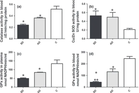
We found significantly lower CAT activity in blood and GSH-Px in plasma (P<0.001 and P<0.01 respectively) of infected dogs, both symptomatic and asymptomatic, than in controls (Figure 1a,c). GSH-Px activity in blood was also lower in symptomatic (P<0.0001) and asymptomatic dogs (P <0.001) than in controls. Symptomatic dogs showed sig-nificantly lower (P<0.01) GSH-Px activity compared to asymptomatic (Figure 1d). Infected dogs showed significantly higher SOD activity than controls (P<0.01; Figure 1b).
Liver, spleen and lymph node CAT, GSH-Px and SOD were significantly lower (P<0.05) in infected dogs than in controls, with no difference between asymptomatic and symptomatic (Table 1).
Fe and Zn were significantly lower in blood of symptom-atic dogs than in either asymptomsymptom-atic or controls (P<0.01; Figure 2a,c). In contrast, serum Cu concentrations were higher in symptomatic dogs than in asymptomatic and
(a) (b)
(c) (d)
control dogs (P<0.01 and P<0.001 respectively; Fig-ure 2b). The Se concentration was significantly lower in infected dogs than in controls (P<0.001 and P<0.01 respectively) and lower in symptomatic than in asymptom-atic dogs (P<0.01; Figure 2d).
We observed significant positive correlation of GSH-Px with Se in blood (r=0.758 andP =0.001) and plasma (r=0.793 andP=0.01) and significant positive correlation of CAT with
Fe (r=0.658 andP=0.001) of infected dogs but no correla-tion between SOD and Cu, Zn or serum trace elements with liver, spleen and lymph node antioxidant enzyme activity.
Lipid peroxidation
Lipid peroxidation was higher in plasma of all infected (symptomatic and asymptomatic) dogs than in controls Table 1Antioxidant enzyme activity and iron deposition in organs of uninfected dogs and those with canine visceral leishmaniasis. Infected dogs: symptomatic (SD) and asymptomatic (AD) compared to uninfected control dogs (C)
Parameters
Liver Spleen
SD AD C SD AD C
CAT 0.2220.01* 0.2800.02* 0.37990.05 0.2390.01* 0.3120.02* 0.4750.03 GSH-Px 66.78.0* 88.38.3* 174.46.2 60.846.5* 89.13.3* 127.417.9 SOD 0.2100.02* 0.2720.04* 0.5180.05 0.1780.03* 0.2660.02* 0.3640.04 Fe 546.760.8* ** 303.863* 101.725.3 2356529.7* ** 791.1215.1* 483.557.9
(continued)
Parameters
Lymph node
SD AD C
CAT 0.2190.01* 0.3340.02* 0.4530.03 GSH-Px 58.35.7* 83.81.3* 128.118.6 SOD 0.1320.01* 0.3060.03* 0.3730.03 Fe 1237216.7* ** 358.5148.6* 22663.13
Values are shown as meanstandard deviation. Significant differences (P≤0.05) relative to control are represented by*and relative to AD by**P<0.01 for all. Parameters: CAT, catalase; GSH-Px, glutathione peroxidase; SOD, Cu-Zn superoxide dismutase; Fe, iron.
(a) (b)
(c) (d)
(P<0.0001 and P<0.01) and higher in symptomatic than in asymptomatic dogs (P<0.001; Figure 3b).
Histopathological and parasitological aspects
Liver. Liver of infected dogs showed severe chronic inflam-mation involving the capsule, portal area and hepatic lob-ules with intralobular granulomas comprising macrophages (epithelioid cells), lymphocytes and plasma cells (Table 2). Congestion and haemosiderin pigment deposits were present (Figure 4a). Swelling and vacuolization of hepatocytes, iden-tified as hydropic degeneration and steatosis, were found (Figure 4b). Semi-quantitative analysis showed higher score lesions (P <0.05) in infected dogs than the control group (Table 2). The chronic inflammatory reaction (P<0.04) was more intense in symptomatic dogs than asymptomatic (Figure 4b). In contrast, we observed higher numbers of hepatic intralobular granulomas in asymptomatic than symptomatic dogs (P =0.01). Widespread haemosiderin pig-ment deposits mainly in hepatic sinusoids (haemosiderosis) were significantly greater in symptomatic than in asymptom-atic and control dogs (P=0.04; P<0.02 respectively). Fibrosis was more widely distributed in symptomatic than in asymptomatic and controls dogs (P<0.05 and P<0.1
respectively; Table 2). Tissue parasite load was higher in symptomatic dogs (P=0.006; Table 2).
Spleen. Semi-quantitative analysis showed that all tissue changes had more frequent presence and higher grading in infected dogs than in the control group (P<0.05; Figure 4e and Table 3). Inflammation was more intense in symptom-atic dogs than in asymptomsymptom-atic (P=0.01; Table 3). Virchow’s granulomas were more frequently present and of higher grade in symptomatic than in asymptomatic dogs (P =0.001 and P=0.01). Hyperplasia and hypertrophy of macrophages of red pulp, with and withoutLeishmania am-astigotes, were also found, with no differences observed between symptomatic and asymptomatic dogs. As in liver, the parasite load in spleen was higher in symptomatic ani-mals (P=0.0269; Table 3). Higher presence and grade of haemosiderin deposits and congestion was observed in red pulp of symptomatic than of asymptomatic dogs (P=0.01 and P<0.01; Figure 4f and Table 3). Splenic fibrosis was more widely distributed in symptomatic dogs than in asymp-tomatic and controls (P <0.01 andP<0.001; Table 3).
Lymph nodes. Cervical lymph nodes showed chronic lymphadenitis in all infected dogs. However, chronic
inflam-(a) (b)
Figure 3 Nitrite and lipid peroxidation (LPO) in uninfected dogs and dogs infected withL. infantum. NO in serum (a), LPO in plasma (b) of symptomatic (SD) and asymptomatic (AD) dogs compared to uninfected control dogs (c). Significant differences (P<0.05) from control are represented by*from AD by**.
Table 2Histopathological and parasitological evaluation of liver in canine visceral leishmaniasis. Analyses of presence quantification (P) and grading (G) of tissues abnormalities following the distribution: slight (S), moderate (M) and severe (SV)
Symptomatic
n=19
Asymptomatic
n=11
Control
n=9
Histopathology
Grading aP≤0.05 Grading aP≤0.05 Grading
S M SV G/P S M SV G/P S M SV
Inflammation 8 5 3 */* ** 5 2 2 */* 0 0 0
Hypertrophy/hyperplasia of Kupffer 5 10 3 */* 3 6 2 */* 0 0 0 Intralobular granulomas 5 3 1 */* ** 3 3 2 */* 0 0 0
Degeneration 5 8 6 */* 2 6 3 */* 2 0 0
Congestion 4 10 2 */* 5 3 3 */* 2 0 0
Haemosiderin 5 3 0 * **/* ** 2 0 0 */* 0 0 0
Fibrosis (lm2)b 2751866*,** 2039597 1567303 Parasitological diagnosisb 26.45** 10.62
aSignificant differences (P≤0.05) relative to control are represented by * and relative to asymptomatic by **. bValues are shown as
(a) (e)
(b) (f)
(c) (g)
(d) (h)
Figure 4 Liver, spleen and lymph node sections of symptomatic dogs naturally infected withL. infantum. Liver (a, b, c, d): (a) Note congestion of sinusoid vessels (S). Inflammation and capsule thickness (Cap). (b) Intense hydropic degeneration (HD), (black arrow heads) granuloma (GR) and congestion. (c) In the centre of the micrograph is an intralobular granuloma (GR) comprising epithelioid cells with an elongated or oval nucleus (large white arrow), plasma cells (white arrow heads) and lymphocytes (white small arrows). Haemosiderin deposition as circular brown structures can be noted (black arrows). (d) Perls’ Prussian blue staining confirmed haemosiderin deposition inside macrophages of granulomas (*) where it has been visualized as homogeneous circular blue structures. (e, f) Spleen sections: (e) Note capsule (Cap) thickening (black bar) and congestion of the red pulp sinusoid vessels (S). Haemosiderin deposition as brown (black arrows). (f) Perls’ Prussian blue staining confirming Virchow’s granuloma (VG) haemosiderin deposition inside cells of the red pulp (macrophages*) visualized as homogeneous circular blue structures. (g, h) Lymph node sections: (g) Haemosiderin in macrophages (black arrows) of medullar and sinus cords. (h) Perls’ Prussian blue staining confirming haemosiderin deposition inside foam macrophages (*). H&E (a, b, c, e and g); Perls’ Prussia blue. (d, f, h); a, e (Bar=32lm), and b, c, d, f, g, h
matory reaction (P<0.01) was higher in symptomatic dogs (Table 4) than in asymptomatic and control dogs. The pri-mary alterations in all cases were hyperplasia and hypertro-phy of macrophages of the medullary cords and sinuses. Congestion was intense (P <0.05) in all infected dogs (Table 4). Haemosiderin pigment deposition (Figure 4g) as confirmed by Prussian blue staining (Figure 4h) was more intense (P<0.01;P=0.01) in symptomatic than in asymp-tomatic dogs. The tissue parasite load was greater in symptomatic dogs (P=0.03). Fibrosis was more widely dis-tributed in symptomatic dogs than in controls (P<0.001; Table 2).
A significant negative correlation was found between tis-sue antioxidant enzyme activity and the inflammatory response in all three studied organs. However, pathologies such as congestion and/or haemosiderosis were only corre-lated to tissue antioxidant enzymes in spleen. Fibrosis, although present in all studied organs, was only correlated
tissue antioxidant enzymes in liver and spleen. Lymph nodes showed correlation of inflammation with CAT only (Table 5).
Nitric oxide production
Nitric oxide was higher in all infected dogs than in controls (P <0.001) and higher in asymptomatic than in symptom-atic dogs (P=0.001; Figure 3a).
Iron in tissue
Iron deposition was observed in liver, spleen and lymph node, accompanied by decreased serum levels without signif-icant cell necrosis. Prussian blue staining showed Fe inside Kupffer cells and hepatocytes (Figure 4d). More prominent deposits of Fe in the liver of symptomatic dogs were observed than in the asymptomatic group and controls Table 3Histopathological and parasitological characteristics of the spleen in cases of canine visceral leishmaniasis. Analyses of presence quantification (P) and grading (G) of tissue abnormalities following the distribution: slight (S), moderate (M) and severe (SV)
Symptomatic
n=19
Asymptomatic
n=11 Controln=9
Histopathology
Grading
aP≤0.05 Grading aP≤0.05 Grading
S M SV G/P S M SV G/P S M SV
Inflammation 6 5 3 */* ** 2 1 1 */* 0 0 0
Granuloma de Virchow 12 4 2 ** */* ** 3 1 0 */* 0 0 0 Hypertrophy/hyperplasia white/red pulp 5 5 0 */* 2 6 1 */* 0 0 0
Haemosiderin 2 5 3 ** */* ** 2 1 0 */* 0 0 0
Congestion 7 6 6 ** */* 4 5 0 */* 0 0 0
Fibrosis (lm2)b 3064881* ** 2022385 1516345 Parasitological diagnosisb 32
6** 8.42
aSignificant differences (P≤0.05) relative to control are represented by * and relative to asymptomatic by**. bValues are shown as
meanstandard deviation.
Table 4Histopathological and parasitological evaluation of the cervical lymph nodes in cases of canine visceral leishmaniasis. Analyses of presence quantification (P) and grading (G) of tissue abnormalities following the distribution: slight (S), moderate (M) and severe (SV)
Symptomatic
n=19
Asymptomatic
n=11 Controln=9
Histopathology
Grading
aP≤0.05 Grading aP≤0.05 Grading
S M SV G/P S M SV G/P S M SV
Inflammation 7 9 3 * **/* ** 4 3 0 */* 0 0 0
Macrophages hyperplasic 2 12 2 */* 2 8 1 */* 0 0 0
Hypertrophy/hyperplasic of nodules (lymphatic follicles) 2 4 4 */* 4 2 2 */* 0 0 0 Hypertrophy/hyperplasia medullar sinus/cords 3 11 5 */* 3 8 0 */* 0 0 0
Haemosiderin 8 3 4 *.**/* ** 1 1 1 */* 0 0 0
Congestion 3 5 2 */* 2 4 2 */* 0 0 0
Fibrosis (lm2)b 1943392* 1596261 12803217
Parasitological diagnosisb 28
3** 6.72
a
(Figure 4d and Table 1). In spleen, Fe deposition was found in red pulp sinusoids, but not in the white pulp (Figure 4f). Symptomatic dogs showed higher Fe deposition than asymp-tomatic and controls (Table 1). In cervical lymph nodes (Figure 4h), Fe deposition was higher in symptomatic dogs than in asymptomatic and controls (Table 1).
In infected animals, a significant positive correlation was seen between LPO and iron deposition in liver (P=0.003, r =0.659), spleen (P<0.002,r=0.728) and cervical lymph node (P<0.01,r=0.765). A significant positive correlation was observed in infected animals between Fe and tissue damage in (i) liver: capsule inflammation, portal inflamma-tion, haemosiderin, hepatocyte degeneration; (ii) spleen: cap-sule inflammation, congestion, haemosiderin; and (iii) cervical lymph node: haemosiderin (Table 5).
Discussion
We observed low antioxidant enzyme activity and altered oxidant/antioxidant balance and trace elements associated with tissue iron deposition and damage in CVL.
The body’s ability to counteract oxidative stress depends on the status and activity of the antioxidant system includ-ing CAT, SOD and GSH-Px (de Luna et al. 2000; Heidar-pour et al. 2012; Samanta et al. 2012). The importance of Zn, Cu, Se and Fe on the immune response to CVL, and their redistribution during the course of infection is well documented (Nieto et al. 2003; Pasa et al. 2003; Heidar-pouret al.2012).
In our study, we found higher SOD activity in blood of infected dogs than in controls, along with lower CAT and GSH-Px activity. These results were also reported by Britti et al. (2008) in CVL and Dimri et al. (2012) in dogs infected with Dirofilaria immitis. In addition, Dimri et al. (2012) suggest that SOD activity increase might be attrib-uted to upregulation of its synthesis to counteract free radi-cals. As a consequence, there is an increase in H2O2 production accompanied by a decrease GSH-Px and CAT, which can result in tissue damage. We found decreased activity of the antioxidant enzymes SOD, CAT and GSH-Px, which have not been previously studied in CVL.
As these antioxidant enzymes require trace elements for activity, we evaluated those elements in serum. Lower serum levels of Zn, Fe and Se and higher serum Cu were found in
all infected dogs, especially those symptomatic, compared to controls. Changes in trace mineral metabolism are an inte-gral aspect of acute-phase response and have been shown in CVL (Nieto et al. 2003; Pasa et al. 2003), possibly related to host susceptibility (Faryadi & Mohebali 2003; Weyen-berghet al. 2004). We did not observe a correlation of Cu or Zn serum levels with SOD activity in any group. How-ever, GSH-Px activity was correlated with lower Se plasma/ blood levels, and CAT activity with lower Fe blood levels, in all infected dogs. We did not observe a correlation of these elements with low enzyme activity in liver, spleen or lymph node tissue.
Classical histological alterations in CVL have been gener-ally described as an intense chronic inflammatory reaction, fibrosis and degeneration of organs such as liver, spleen, lymph node, kidney and lung (Alvaret al.2004; Alexandre-Pires et al. 2006; Melo et al. 2009). Our results were in accordance with previous reports and symptomatic dogs exhibiting a more pronounced chronic inflammatory reac-tion than controls. This indicates that severity of the disease is a consequence of histological alterations directly associ-ated with increasing utilization or redistribution of trace ele-ments and deficiency in antioxidant enzyme activity.
Tissue changes observed may result from ROS but also from NO production by inflammatory cells (Mauel et al. 1991; Assrue et al. 1994; Kocyigit et al. 2005). Research has shown that symptomatic dogs show a heavier parasite load and lower serum levels of NO than do asymptomatic (Zafraet al.2008). In the present study, low NO levels with high parasite load in symptomatic dogs seemed to be related to Fe tissue deposition and severe lesions. We found sys-temic iron deposition in all infected dogs, with widely dis-tributed haemosiderin deposits in symptomatic dogs. Prussian blue staining showed intracytoplasmatic granules especially inside macrophage inflammatory exudate or gran-ulomas in liver, spleen and lymph node. Iron level was posi-tively correlated with grade of inflammation, haemosiderin, and fibrous connective tissue in all organs of symptomatic dogs and negatively correlated with tissue antioxidant enzymes. Iron is an important component of biological pro-cesses and has been associated with ROS production, ery-throcatheresis, localized tissue lesions and stimulating fibrinogenesis, probably due to oxygen free radical forma-tion (Arthur 1996; Biswas et al. 1997; Britti et al. 2008). Table 5Correlation between presence of histopathology and tissue antioxidant enzyme or iron in canine visceral leishmaniasis
Tissue histopathology
Liver Spleen Lymph node
CAT SOD GSH-Px Fe CAT SOD GSH-Px Fe CAT Fe
Inflammation 0.735 0.768 0.781 0.659 0.723 0.584 0.787 0.780 0.729
Degeneration 0.758
Haemosiderin 0.597 0.659 0.653 0.783 0.677 0.729
Congestion – 0.696 0.624 0.774 0.723
Fibrosis 0.677 0.598 0.699 0.598 0.648 0.685 0.598 0.678 0.726
Although Fe2+and Fe3+ may provoke LPO with subsequent cell membrane damage (Huynh and Andrew 2008), studies of lesions associated with chronic tissue Fe increase in CVL were not considered until this work.
Although bone marrow, potentially involved in iron metabolism, was not evaluated, our results may provoke dis-cussion of increase in tissue iron deposition and cell damage in association with low antioxidant enzyme activity in CVL that could explain differences in clinical signs and factors associated with severity.
Organ tissue Fe deposits may result from multiple distinct but overlapping conditions: (i) Systemic oxidative stress may decrease RBC lifespan (senescent erythrocytes; de Lunaet al. 2000; Samanta et al. 2012) and increase LPO levels in plasma of infected dogs, as demonstrated by higher LPO levels together with a positive correlation of LPO with Fe tissue deposits. Senescent erythrocytes are taken up through erythrophagocytosis and destroyed within macrophages, and Fe is released from haemoglobin and rapidly shifted to reu-tilization by incorporation into iron proteins or iron storage within ferritin (Weiss et al. 1999). (ii) CVL is a chronic infection characterized by a systemic inflammatory reaction in organs rich in cells of the mononuclear phagocyte system (Alvaret al. 2004). Tissue damage or inflammation in liver and spleen of infected dogs showed a positive correlation with LPO in plasma and Fe in tissue and negative correlation with antioxidant enzymes. (iii) According to Al-exandre-Pires et al. (2006) and Melo et al. (2009), major abnormalities in the splenic microvascular system and liver architecture result in changes to circulatory dynamics, bring-ing about a reduction in blood flow directly related to congestion and haemosiderosis. (iv) Moreover, intracellular Fe overload may block the transcription of inducible NO synthase as described by Weiss et al. (1999). Thus, the lower NO formation in symptomatic dogs might be due to higher tissue Fe deposits.
In addition, we have shown evidence that dogs with symptomatic VL display enhanced LPO and Fe tissue depo-sition associated with decreased levels of antioxidant enzymes. These alterations in oxidant balance may further aggravate infection-associated pathology in canine VL as observed by Faryadi and Mohebali (2003) and Weyenbergh et al. (2004) in human VL. Together these results suggest a functional derangement in infected animals that may be responsible for the clinical features and open new approaches for treatment perspectives associated with an-tioxidants and the role of iron in the pathogenesis of CVL.
Acknowledgements
The authors thank Dr Marco Antonio Alves Carneiro for statistical advice; Miss V^ania Aparecida N. Silva and Olin-da D. Rodrigues for histological technical support and Mr Weder Gomes de Oliveira for animal care. This study was supported by research grants from Fundacß~ao de Ampar-o a Pesquisa do Estado de Minas Gerais (FAPEMIG) (APQ00068-08; APQ-01355-09), Conselho Nacional de
Desenvolvimento Cientıfico e Tecnologico (CNPq) (473601/ 2009-5) and Pro-reitoria de Pesquisa da Universidade Fed-eral de Minas Gerais (PRPq-UFMG). We also thank Lucidus Consultancy ([email protected]) for correction of English.
References
Adamama-Moraitou K.K., Saridomichelakis M.N., Polizopoulou Z., Kritsepi M., Tsompanakou A. & Koutinas A.F. (2005) Short-term exogenous glucocorticosteroidal effect on iron and copper status in canine leishmaniosis (Leishmania infantum). Can. J. Vet. Res. 69, 287–292.
Alexandre-Pires G., Pais D., Correia M. & Pina J.A. (2006) Leish-maniosis: a report about the microvascular and cellular architec-ture of the infected spleen inCanis familiaris.Microsc. Res. Tech. 69, 227–235.
Alvar J., Molina R., San Andres M.et al.(1994) Canine leishmania-sis: clinical, parasitological and entomological follow-up after chemotherapy.Ann. Trop. Med. Parasitol.88, 371–378.
Alvar J., Ca~navate C., Molina R., Moreno J. & Nieto J. (2004) Canine leishmaniasis.Adv. Parasitol.57, 1–88.
de Amorin I.F.G., Freitas E., Alves C.F. et al. (2010) Humoral immunological profile and parasitological statuses of Leishmuneâ
vaccinated and visceral leishmaniasis infected dogs from an ende-mic area.Vet. Parasitol.173, 55–63.
Anstead G.M., Chandrasekar B., Zhao W., Yang J., Perez L. & Melby P. (2001) Malnutrition alters the innate immune response and increases early visceralization followingLeishmania donovani
infection.Infect. Immun.69, 4709–4718.
Araujo A.P., Mayrink W. & Machado-Coelho G.L. (2008) The influ-ence of copper, selenium and zinc on the response to Montenegro skin test in subjects vaccinated against American cutaneous leish-maniasis.Trans. R. Soc. Trop. Med. Hyg.102, 64–69.
Arthur M.J. (1996) Iron overload and liver fibrosis.J. Gastroenter-ol. HepatGastroenter-ol.11, 1124–1129.
Assrue J., Cunha F.Q. & Epperlein M. (1994) Production of nitric oxide and superoxide by activated macrophages and killing of
Leishmania major.Eur. J. Immunol.24, 672–676.
Biswas T., Ghosh D.K., Mukherjee N. & Ghosal J. (1997) Lipid peroxidation of erythrocytes in visceral leishmaniasis.J. Parasitol. 83, 151–152.
Bretagne S., Durand R., Olivi M.et al.(2001) Real-time PCR as a new tool for quantifyingLeishmania infantumin liver in infected mice.Clin. Diagn. Lab. Immunol.4, 828–831.
Britti D., Sconza S., Morittu V.M., Santori D. & Boari A. (2008) Superoxide dismutase and Glutathione peroxidase in the blood of dogs with Leishmaniasis.Vet. Res. Commun.32, S251–S254. Cunningham A.C. (2002) Parasitic adaptive mechanisms in infection
byLeishmania.Exp. Mol. Pathol.72, 132–141.
Deane L.M. & Deane M.P. (1955) Sobre a biologia do Phleboto-mus longipalpis, transmissor da leishmaniose visceral em zona end^emica do Estado do Ceara. I. Distribuicß~ao, predomin^ancia e variacß~ao estacional.Rev. Bras. Biol.15, 83–95.
Dimri U., Singh S.K., Sharma M.C., Behera S.K., Kumar D. & Tiwari P. (2012) Oxidant/antioxidant balance, minerals status and apoptosis in peripheral blood of dogs naturally infected with
Dirofilaria immitis.Res. Vet. Sci.93, 296–299.
Gioda C.R., de Oliveira-Barreto T., Prımola-Gomes T.N. et al.
(2010) Cardiac oxidative stress is involved in heart failure induced by thiamine deprivation in rat. Am. J. Physiol. Heart Circ. Physiol.298, H2039–H2045.
Green L.C., Wagner D.A., Glogowski J., Skipper P.L. & Wishnok J.S. (1982) Analysis of nitrate, nitrite, and nitrate in biological flu-ids.Anal. Biochem.126, 131–138.
Heidarpour M., Soltani S., Mohri M. & Khoshnegah J. (2012) Canine visceral leishmaniasis: relationships between oxidative stress, liver and kidney variables, trace elements, and clinical sta-tus.Parasitol. Res.111, 1491–1496.
Huynh C. & Andrews N.W. (2008) Iron acquisition within host cells and the pathogenicity of Leishmania. Cell Microbiol. 10, 293–300.
Instituto Nacional de Metrologia Normalizalizacß~ao e Qualidade Industrial – INMETRO (2010) Orientacß~oes sobre validacß~ao de metodos de ensaios quımicos-DOQ-CGCRE-008. Rio de Janeiro. 2. ed. 33 p.
International Organization for Standardization–ISO (1999) Part 1: Water quality–Calibration and evaluation of analytical methods and estimation of performance characteristics. 8466-1 Switzer-land, 11 pp.
Kocyigit A., Keles H., Selek S., Guzel S., Celik H. & Erel O. (2005) Increased DNA damage and oxidative stress in patients with cuta-neous leishmaniasis.Mutat. Res.585, 71–78.
Lowry O.H., Rosenbrough H.J., Farra L. & Randall R.J. (1951) Protein measurement with folin phenol reagent. J. Biol. Chem. 193, 265–275.
de Luna R., Ferrante M., Severino L.et al. (2000) Decreased lipid fluidity of the erythrocyte membrane in dogs with leishmaniasis associated anaemia.J. Comp. Pathol.122, 213–216.
Mancianti F., Gramiccia M., Gradoni L. & Pieri S. (1998) Studies on canine leishmaniasis control. I. Evolution of infection of differ-ent clinical forms of canine leishmaniasis following antimonial treatment.Trans. R. Soc. Trop. Med. Hyg.82, 566–567. Mauel J., Ransijn A. & Buchmuller-Rouiller Y. (1991) Killing of
Leishmania parasites in activated murine macrophages is based on n L-arginine-dependent process that produces nitrogen deriva-tives.J. Leukoc. Biol.49, 73–82.
Mauricio I.L., Stothard J.R. & Miles M.A. (2000) The strange case ofLeishmania chagasi.Parasitol. Today16, 188–189.
Melo F.A., Moura E.P., Ribeiro R.R.et al.(2009) Hepatic extracel-lular matrix alterations in dogs naturally infected with Leish-mania (LeishLeish-mania) chagasi.Int. J. Exp. Pathol.90, 538–548. Mishra J., Carpenter S. & Singh S. (2010) Low serum zinc levels in
an endemic area of visceral leishmaniasis in Bihar, India.Indian J. Med. Res.131, 793–798.
National Association of Testing Authorities –NATA (1997) (Aus-tralia), Format and content of test methods and procedures for validation and verification of chemical test methods, technical note. 8.
Nieto J., Alvar J., Mullen A.B. et al. (2003) Pharmacokinetics, toxicities, and efficacies of sodium stibogluconate formulations
after intravenous administration in animals.Antimicrob. Agents Chemother.47, 2781–2787.
Paltrinieri S., Ravicini S., Rossi G. & Roura X. (2010) Serum con-centrations of the derivatives of reactive oxygen metabolites (d-ROMs) in dogs with leishmaniosis.Vet J.186, 393–395. Pasa S., Kargin F., Bildik A., Seyrek K., Ozbel Y. & Ozensoy S.
(2003) Serum and hair levels of zinc and other elements in dogs with visceral leishmaniasis.Biol. Trace Elem. Res.94, 141–147. Samanta S., Ghoshal A., Bhattacharya K., Saha B., Walden P. &
Mandal C. (2012) Sialoglycosylation of RBC in visceral leishman-iasis leads to enhanced oxidative stress, calpain-induced fragmen-tation of spectrin and hemolysis.PLoS ONE7, 1–10.
Shaw J.J. (2006) Further thoughts on the use of the name Leish-mania(Leishmania)infantum chagasifor the aetiological agent of American visceral leishmaniasis.Mem. Inst. Oswaldo Cruz101, 577–579.
da Silva S.M. (2007)Avaliacß~ao clınica e laboratorial de c~aes natu-ralmente infectados por Leishmania (Leishmania) chagasi
(CUNHA & CHAGAS, 1937) submetidos a um protocolo te-rap^eutico em clınica veterinaria de Belo Horizonte. Belo Horizon-te: Universidade Federal de Minas Gerais.
da Silva S.M., Ribeiro V.M., Ribeiro R.R., Tafuri W.L., Melo M.N. & Michalick M.S. (2009) First report of vertical transmission of
Leishmania (Leishmania) infantumin a naturally infected bitch from Brazil.Vet. Parasitol.166, 159–162.
Souza C.C., Fabrino J.H.F., Beinner M.A. et al. (2013) Develop-ment and validation of methods for the determination of copper and iron in serum of dogs with canine visceral leishmaniasis using multivariate optimization and GFAAS. Anal. Methods5, 3129–
3135.
Tafuri W.L., Santos R.L., Arantesa R.M.et al. (2004) An alterna-tive immunohistochemical method for detecting Leishmania am-astigotes in paraffin-embedded canine tissues. J. Immunol. Methods292, 17–23.
Tafuri W.L., De Oliveira M.R. & Melo M.N. (2001) Canine vis-ceral leishmaniasis: a 726 remarkable histopathological picture of one case reported from Brazil.Vet. Parasitol.96, 203–212. Weiss G., Umlauft F., Urbanek M. et al. (1999) Associations
between cellular immune effector function, iron metabolism and disease activity in patients with chronic hepatitis C virus infec-tion.J. Infect. Dis.180, 1452–1458.
Weyenbergh J.V., Santana G., D’Oliveira A. Jr et al. (2004) Zinc/ copper imbalance reflects immune dysfunction in human leishmaniasis: anex vivoandin vitrostudy.BMC Infect. Dis.4, 1–17.