361
Corresponding author: Dra Márcia Dalastra Laurenti. e-mail: [email protected]
Received 19 November 2015 Accepted 29 February 2016
Canine antibody response to
Lutzomyia longipalpis
saliva in endemic area of visceral leishmaniasis.
Luís Fábio da Silva Batista
[1],[2],
Vânia Lúcia Ribeiro da Matta
[1],
Thaise Yumie Tomokane
[1],
Acácio Duarte Pacheco
[3],
Fernando Tobias Silveira
[4],[5],
Claudio Nazaretian Rossi
[6],
Mary Marcondes
[3]and
Márcia Dalastra Laurenti
[1][1]. Laboratório de Patologia de Moléstias Infecciosas, Faculdade de Medicina, Universidade de São Paulo, São Paulo, São Paulo, Brasil. [2]. Departamento de Patologia, Faculdade de Medicina Veterinária e Zootecnia, Universidade de São Paulo, São Paulo, São Paulo, Brasil. [3]. Departamento de Clínica, Cirurgia e Reprodução Animal, Faculdade de Medicina Veterinária, Universidade Estadual de São Paulo, Araçatuba, São Paulo, Brasil. [4]. Instituto Evandro Chagas, Ministério da Saúde, Ananindeua, Pará, Brasil. [5]. Núcleo de Medicina Tropical, Universidade Federal do Pará, Belém, Pará, Brasil.
[6]. Instituto de Ciências da Saúde, Universidade Paulista, Campinas, São Paulo, Brasil.
Abstract
Introduction: Canine exposure to Lutzomyia longipalpis bites and the potential of Leishmania infantum transmissibility for the vector were evaluated. Methods: Immunoglobulin G (IgG) anti-Lu longipalpis saliva and -L. infantum, and blood parasite load were determined in dogs from endemic areas of visceral leishmaniasis. Results: Blood parasitism was similar between symptomatic and asymptomatic dogs. IgG anti-L. infantum was higher in symptomatic dogs, but IgG anti-Lu. longipalpis saliva was mostly observed in higher titers in asymptomatic dogs, indicating vector preference for feeding on asymptomatic dogs.
Conclusions: Our data suggest a pivotal role of asymptomatic dogs in L. infantum transmission in endemic areas.
Keywords: Canine leishmaniasis. Anti-Lutzomyia longipalpis saliva antibody.Transmissibility.
In Brazil, visceral leishmaniasis (VL) is considered a zoonotic disease caused by the protozoan Leishmania infantum
[syn. Leishmania (Leishmania) chagasi] that is mainly transmitted by the sand fly Lutzomyia longipalpis. Dogs
(Canis familiaris) are considered the main domestic reservoir
for VL transmission and have been the target of control actions, because they form part of the transmission cycle of L. infantum(1).
Previous studies concerning the potential transmission of VL from infected dogs are controversial. Whilst some authors reported that asymptomatic dogs were non-infective to vectors(2),
others stated that they are indeed infective vectors, but to a lesser extension than symptomatic dogs(3). Recently, our group
demonstrated that sand ly infection can occur independently
of a dog’s skin parasitism, and that asymptomatic dogs were more likely than symptomatic dogs to transmit L. infantum to
the sand ly, thus promoting a higher infection rate of sand lies
feeding on asymptomatic dogs(4).
In accordance with this life cycle, during the blood meal
the female sand ly injects its saliva into the hosts’ skin which
contains proteins that have anti-hemostatic, immunomodulatory, and antigenic properties(5). The saliva induces antibody
production in repeatedly bitten hosts that are highly speciic to the sand ly species Lu. longipalpis, and the antibody level
relects the intensity of vector exposure(6). Therefore, antibodies
against sand ly saliva have been used in epidemiological studies
as an exposure marker to the vector bite, as well as to estimate the likely risk of Leishmania transmission(7).
The phlebotomine sand fly could be found in the peridomiciliary zone in urban and rural areas, since the transmission of the infection is associated to precarious living
conditions and environmental modiications, modulated by the
intense urbanization process, which favoured the adaptation of the vector to the urban environment.(3) Usually, the sand ly feeding in
the crepuscular period and dogs who lives in the street or houses backyard are exposed. Apparently, the ears skin represent the most frequently exposed areas, likely due to its lower hair density and cerumen attractiveness, leading to increased exposure to the insect vector’s bite(8).
Thus, the main objective of this study was to evaluate
the level of immunoglobulin G (IgG) anti-Lu. longipalpis in dogs living in urban endemic areas for VL to determine the vector exposure of symptomatic and asymptomatic dogs, verify whether clinical condition affects female attractionfor hematophagy, and correlate the data with anti-L. infantum
antibody and parasite burden in canine whole blood, the main
source of feeding for sand lies.
Rev Soc Bras Med Trop 49(3):361-364, May-June, 2016 doi: 10.1590/0037-8682-0360-2015
362
Batista LFS et al. - Antibody against Lu. longipalpis in saliva of Canines
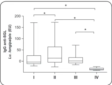
FIGURE 1 - Boxplot showing median and interquartile range of IgG anti-Lu. longipalpis salivary gland lysate in EU in the sera of dogs from endemic and non-endemic area for visceral leishmaniasis. I. Symptomatic dogs. II. Asymptomatic dogs. III. Control dogs from endemic area. IV. Control dogs from non-endemic area. IgG: immunoglobulin G; anti-SGL: anti-salivary gland lysate; Lu: Lutzomyia; EU: ELISA Units; ELISA: enzyme linked immunosorbent assay. *p < 0.05.
I II III IV
0 50 100 150
200
*
*
*
*
IgG anti-SGL
Lu. longipalpis
(EU)
Upon clinical examination, whole blood was collected from
the jugular or cephalic vein, and the serum was stored at -20°C
until its use for determination of IgG anti-Lu. longipalpis saliva by enzyme linked immunosorbent assay (ELISA) according to the methodology described by Rohousova et al., 2005(9).
Lu. longipalpis captured in Cametá municipality, Pará state,
Brasil were used as a source of antigens for ELISA. Its salivary glands were collected in cold phosphate buffered saline (PBS) from 5- to 7-day-old F1 laboratory-reared females. Just before use, salivary gland lysate (SGL) was prepared by disruption of glands with 3 freeze-thaw cycles. Proteins were measured by the Bradford reaction(10). Briely, 96-well microtiter plates
were coated with SGL (60ng of protein per well) in 0.01M
carbonate-bicarbonate buffer (pH 9.5) overnight at 4°C. After
automated PBS washing for three times, 10% skimmed milk
powder in PBS for 45 min at 37°C was used to block unspeciic
binding sites. After another washing, the canine serum was diluted 1:400 times in PBS containing 0.05% Tween 20 and
incubated in duplicates for 90 min at 37°C, followed by
another incubation with alkaline-phosphatase anti-canine IgG
(Bethyl, USA) at 1/20,000 dilution for 45 min at 37°C.
Para-nitrophenyl phosphate - pNPP (Sigma-Aldrich, EUA) was used as a substrate solution for 30 min at 25oC and 2N sodium hydroxide
(50µL per well) was used to block the reaction. The absorbance was measured using the Multiskan reader (Labsystems, USA) at 405nm. The titers of IgG were expressed as ELISA units (EU). Another ELISA using crude L. infantum antigen (MHOM/BR/72/
strain46) and anti-canine IgG (A40-123AP) conjugated to alkaline
phosphatase (Bethyl, USA) was performed according to Laurenti et al., 2014(11) to determine the level of IgG anti-L. infantum.
Samples of whole blood were also used for parasite-load determination by real time polymerase chain reaction (PCR). This assay was performed using primers targeting a 120-bp sequence found in kinetoplast DNA (kDNA)(12). The
ampliication was performed in a inal volume of 15µL that
consisted of 5µL of total DNA diluted in deionized water to 10ng/µL, 7.5µL of Kapa SYBR Fast Universal 2× qPCR Master Mix (Kapa Biosystems, USA), 0.5µL of each primer
at a inal concentration of 300nM, and 1.5µL of deionized
water. A standard curve was generated using serial dilutions of
L. infantum DNA from 106 to 10-3 parasites/µL. The standard
curve was considered acceptable when the slopes ranged
between -3.1 and -3.4 with correlation coeficients ≥0.98. The ampliications were performed in duplicate for each sample,
each concentration of parasites, and negative control. The parasite load was obtained by plotting the cycle threshold (Ct) values against the standardized parasite concentrations. Prism 5.00 for Windows (GraphPad software Inc., USA) was used to perform all statistical analysis and graphics. Differences among the clinical groups were analyzed by nonparametric
Kruskal-Wallis test and p value < 0.05 was considered signiicant.
In the current study, the IgG anti-Lu. longipalpis salivary gland lysate was found in many of the dogs in the endemic areas. From a total of 185 sera evaluated, 134 (72%) were positive. The highest proportion of dogs with detectable anti-saliva antibodies were found in the asymptomatic group (64/73; 88%), followed by the uninfected controls from the endemic
area (15/20; 75%) and symptomatic dogs (55/92; 60%). Levels of anti-salivaantibodies (median, interquartile range
25% to 75%) were signiicantly higher in asymptomatic dogs
(13.7, -4.53 to 63.53 EU) in comparison with the symptomatic
ones (-3.76, -10.99 to 25.07 EU) (p ≤ 0.05) (Figure 1).
Figure 1 shows that the levels of anti-saliva IgG were higher in asymptomatic dogs, which suggests that the state
of being asymptomatic inluences the attraction of the female sand ly and inluences their preference for feeding. These indings are consistent with a previous study carried out on
experimental conditions of xenodiagnoses, when we showed a larger number of engorged females after blood meal conducted in asymptomatic dogs compared to symptomatic dogs(4). As early
as 1931, Adler and Theodor warned that ulcerated skin lesion infected by bacteria and fungus of symptomatic animals would
be less attractive to the sand ly vector, which could explain our indings(13). Skin involvement is one of the main clinical
indings in dogs with VL that usually present with alopecia,
exfoliative dermatitis, and nodular and ulcerated lesions(14). In
the present study 63% of symptomatic dogs had the skin lesions aforementioned. It is important to consider that the anemic status of symptomatic dogs is not a limiting factor for vector feeding in the host(4), as the symptomatic dogs with (35%) and without
anemia (65%) did not differ in the number of positive cases for anti-Lu. longipalpis saliva IgG as well as in their titer.
Dogs infected by L. infantum have detectable levels of
anti-Leishmania antibody in the serum, which increases with the
363 Rev Soc Bras Med Trop 49(3):361-364, May-June, 2016
I II III IV
0 50 100 150
*
*
*
*
*
IgG anti
-L. (-L.) infantum
(EU)
FIGURE 2 - Boxplot showing median and interquartile range of IgG
anti-L. infantum in EU in the sera of dogs from endemic and non-endemic area for visceral leishmaniasis. I. Symptomatic dogs. II. Asymptomatic dogs. III. Control dogs from endemic area. IV. Control dogs from non-endemic area. IgG: immunoglobulin G; anti-L. infantum: anti-Leishmania infantum; EU: ELISA units; ELISA: enzyme linked immunosorbent assay. *p < 0.05.
Asymptomatic Symptomatic 0
20 40 60 80 100 120 140 1,000 1,200
Blood parasite loa
d
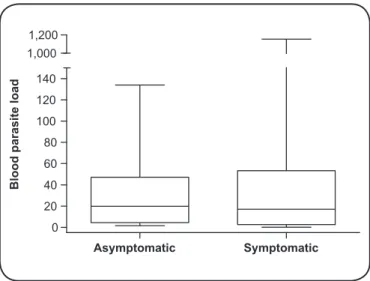
FIGURE 3 - Box plot showing median and interquartile range of parasitic load in the blood of symptomatic and asymptomatic dogs from endemic areas of visceral leishmaniasis.
in symptomatic dogs showing that they have a high immune competency to produce antibodies against foreign antigens.
The skin parasitism found in both symptomatic and asymptomatic animals has been implicated as an important
source of infection for sand lies(3). However, our previous
report showed that vector infection can occur independently of dogs’ skin parasitism(4). Thus, parasite loads in the blood,
the main source of feeding for Lu. longipalpis females, was also investigated in the present study. The parasite load was measured in the buffy coat by quantitative real-time PCR
and no signiicant difference (p = 0.55) was observed in the blood parasitism between symptomatic and asymptomatic dogs (Figure 3). Although peripheral blood is not the favorite tissue for the proliferation and maintenance of L. infantum;
and a luctuation of parasitemia may occur during the course
of infection(15), the lack of signiicance in parasite load between
clinical groups suggests asymptomatic and symptomatic dogs likely have similar potential of transmissibility.
In summary, besides asymptomatic dogs being competent for
infecting phlebotomine sand lies(3) (4), our present study showed
that asymptomatic dogs have blood parasitism comparable to symptomatic dogs and were preferably bitten by Lu. longipalpis.
The higher attraction of female sand lies to asymptomatic
dogs may signify their contribution in parasitic transmission
for vectors. Together, these indings suggest that asymptomatic
dogs play a pivotal role in the maintenance of the L. infantum
cycle and spread of parasite in endemic areas of VL. Moreover,
to the best of our knowledge, this is the irst study to show
the relationship between the Lu. longipalpis anti-sand ly saliva
antibodies, and blood parasite load, main source of vector feeding, and clinical status of dogs naturally infected by L. infantum.
ETHICAL CONSIDERATIONS
In the present study, 185 breed dogs (Rottweiler n = 50, Labrador Retriever n = 72 and Shepherd dogs n = 63) domiciled or resident for at least 18 months in high endemic areas for VL were used. According to the clinical signs, biochemical laboratory features, and infection status (presence/absence of
Leishmania deoxyribonucleic acid (DNA) in lymph nodes),
dogs were grouped into symptomatic (n = 92), asymptomatic (n = 73), and control (n = 20) from endemic area of São Paulo state, Brazil. Blood serum of uninfected dogs (n = 20) from non-endemic area was used as the negative control. This study was approved by the Ethics Committee for the use of animals in the Veterinary School of the University of São Paulo, under protocol 2391/2011. All procedures were performed in accordance with the guidelines of Brazilian College of Animal Experimentation (COBEA), under the owners’ signed consent.
Acknowledgments
We wish to thank Raissa de Andrade Dias and Thais Bruna Ferreira da Silva for their technical assistance.
Conlict of interest
The authors declare that there is no conlict of interest.
Financial Support
Grant #2012/50285-9 from the São Paulo Research Foundation (Fundação de Amparo a Pesquisa do Estado de São Paulo - FAPESP), Grant #301549/2011-7 from Research National Council (Conselho Nacional de Desenvolvimento
Cientíico e Tecnológico - CNPq) and Laboratório de Investigação Médica
do Hospital das Clínicas da Faculdade de Medicina da Universidade de São
364
REFERENCES
1. Dantas-Torres F, Brandão-Filho SP. Visceral leishmaniasis in Brazil: Revisiting paradigms of epidemiology and control. Rev Inst Med Trop Sao Paulo 2006; 48:151-156.
2. Verçosa BLA, Lemos CM, Mendonça IL, Silva SMMS, de Carvalho
SM, Goto H, et al. Transmission potential, skin inlammatory
response, and parasitism of symptomatic and asymptomatic dogs with visceral leishmaniasis. BMC Vet Res 2008; 4:45.
3. Soares MRA, de Mendonça IL, do Bonim JM, Rodrigues JA, Werneck GL, Costa CHN. Canine visceral leishmaniasis in Teresina, Brazil: relationship between clinical features and
infectivity for sand lies. Acta Trop 2011; 117:6-9.
4. Laurenti MD, Rossi CN, da Matta VL, Tomokane TY, Corbett CE, Secundino NF, et al. Asymptomatic dogs are highly competent to transmit Leishmania (Leishmania) infantum chagasi to the natural vector. Vet Parasitol 2013; 196:296-300.
5. Kamhawi S. The biological and immunomodulatory properties
of sand ly saliva and its role in the establishment of Leishmania infections. Microbes Infect 2000; 2:1765-1773.
6. Hostomska J, Rohousova I, Volfova V, Stanneck D, Mencke N, Volf
P. Kinetics of canine antibody response to saliva of the sand ly
Lutzomyia longipalpis. Vector Borne Zoonotic Dis 2008; 8:443-450.
7. Vlkova M, Rohousova I, Drahota J, Stanneck D, Kruedewagen EM, Mencke N, et al. Canine antibody response to Phlebotomus perniciosus bites negatively correlates with the risk of Leishmania infantum transmission. PLoS Negl Trop Dis 2011; 5:e1344.
8. Travi BL, Tabares CJ, Cadena H, Ferro C, Osorio Y. Canine visceral leishmaniasis in Colombia: Relationship between clinical
and parasitologic status and infectivity for sand lies. Am J Trop
Med Hyg 2001; 64:119-124.
9. Rohousova I, Ozensoy S, Ozbel Y, Volf P. Detection of
species-speciic antibody response of humans and mice bitten by sand lies.
Parasitology 2005; 130:493-499.
10. Laurenti MD, Silveira VMS, Secundino NFC, Corbett CEP, Pimenta PPF. Saliva of laboratory-reared Lutzomyia longipalpis exacerbates Leishmania (Leishmania) amazonensis infection more potently than saliva of wild-caught Lutzomyia longipalpis. Parasitol Int 2009; 58:220-226.
11. Laurenti MD, de Santana Leandro Jr MV, Tomokane TY, De Lucca HRL, Aschar M, Souza CSF, et al. Comparative evaluation of the DPP® CVL rapid test for canine serodiagnosis in area of visceral leishmaniasis. Vet Parasitol 2014; 205:444-450.
12. Francino O, Altet L, Sánchez-Robert E, Rodriguez A, Solano-Gallego L, Alberola J, et al. Advantages of real-time PCR assay for diagnosis and monitoring of canine leishmaniosis. Vet Parasitol 2006; 137:214-221.
13. Adler S, Theodor O. Skin infection in canine visceral leishmaniasis. Brit Med J 1931; 2:1179.
14. Feitosa MM, Ikeda FA, Luvizotto MCR, Perri SHV. Aspectos clínicos de cães com leishmaniose visceral no município de Araçatuba – São Paulo (Brasil). Clin Vet 2000; 28:36-44.
15. Gramiccia M, Di Muccio T, Fiorentino E, Scalone A, Bongiorno G, Cappiello S, et al. Longitudinal study on the detection of canine Leishmania infections by conjunctival swab analysis and correlation with entomological parameters. Vet Parasitol 2010; 171:223-228.