Evaluatio n o f radio induce d dam age
and re pair capacity in blo o d
lym pho cyte s o f bre ast cance r
patie nts
Supervisão de Radiobiologia, Departamento de Bioengenharia, Instituto de Pesquisas Energéticas e Nucleares, CNEN/SP, São Paulo, SP, Brasil
P.A. Nascimento, M.A. da Silva, E.M. Oliveira, M.F. Suzuki and K. O kazaki
Abstract
Genetic damage caused by ionizing radiation and repair capacity of blood lymphocytes from 3 breast cancer patients and 3 healthy donors were investigated using the comet assay. The comets were analyzed by two parameters: comet tail length and visual classification. Blood samples from the donors were irradiated in vitro with a 60Co source at
a dose rate of 0.722 Gy/min, with a dose range of 0.2 to 4.0 Gy and analyzed immediately after the procedure and 3 and 24 h later. The basal level of damage and the radioinduced damage were higher in lymphocytes from breast cancer patients than in lymphocytes from healthy donors. The radioinduced damage showed that the two groups had a similar response when analyzed immediately after the irradia-tions. Therefore, while the healthy donors presented a considerable reduction of damage after 3 h, the patients had a higher residual damage even 24 h after exposure. The repair capacity of blood lymphocytes from the patients was slower than that of lymphocytes from healthy donors. The possible influence of age, disease stage and mutations in the BRCA1 and BRCA2 genes are discussed. Both parameters adopted proved to be sensitive and reproducible: the dose-response curves for DNA migration can be used not only for the analysis of cellular response but also for monitoring therapeutic interventions. Lymphocytes from the breast cancer patients presented an initial radiosensitivity similar to that of healthy subjects but a deficient repair mechanism made them more vulnerable to the genotoxic action of ionizing radiation. However, since lymphocytes from only 3 patients and 3 normal subjects were analyzed in the present paper, additional donors will be necessary for a more accurate evaluation.
Co rre spo nde nce
K. O kazaki Travessa R, 400 05508-900 São Paulo, SP Brasil
Fax: + 55-11-3816-9232 E-mail: kokazaki@ net.ipen.br
Research supported by CNPq. Publication supported by FAPESP.
Received June 12, 2000 Accepted November 29, 2000
Ke y wo rds
·Comet assay
·DNA damage
·DNA repair
·Ionizing radiation
·Breast cancer
·Blood lymphocytes
Intro ductio n
Breast cancer has attracted increasing at-tention over the last years because of its high incidence among women. In industrialized countries one in ten women develops this
about 20% have a family history including a 5% rate of autosomal dominant mutations of high penetrance (2).
Alterations of the tumor suppressor genes BRCA1 and BRCA2 confer a high risk of breast cancer. Mutations in the BRCA1 gene are involved in about 50% of families with a high incidence of breast cancer and in at least 80% of families with a high rate of early breast and ovarian cancers (3). Similarly, mutations of the BRCA2 gene cause early breast cancer in women and also a higher risk in men (4).
Although breast and ovarian cancers are types of neoplastic disease with a strong familial component, many uncertainties ex-ist about the role of hereditary predisposi-tion because of many factors that impair analysis. These include the existence of spo-radic cancer and mutations of variable pen-etrance, in addition to the heterogeneity of the disease as revealed by epidemiological evidence and molecular analysis (1).
The deficiency in repair has been pointed out as a factor of susceptibility to cancer development. The biological importance of the DNA repair mechanism in cancer devel-opment is illustrated by recessive autosomal disease such as chromosome fragility syn-drome. Affected individuals, i.e., subjects with xeroderma pigmentosum, Fanconis anemia, or ataxia telangiectasia, are defi-cient in a type of repair and are at high risk for malignancy when exposed to specific mutagens such as UV light, alkylating agents and ionizing radiation (5). There are many cell repair mechanisms and, depending on the type of DNA lesion, a specific repair mechanism is initiated. Recent molecular studies have shown that the BRCA1 and BRCA2 genes are involved in DNA repair. Both protein products act directly or indi-rectly with yeast Rad51 protein homologues or Escherichiacoli RecA protein which par-ticipates in DNA double-strand break or re-combinant repair (2,6).
In addition to these genes, other tumor
suppressors such as the p53 and AT genes, which are involved in breast cancer, partici-pate in cell repair processes (7,8). On this basis, the analysis of the association be-tween cell repair capacity and genotoxic ex-posure to environmental agents in breast cancer patients would be of great interest.
There are few studies on this subject and some data reported in the literature are con-tradictory. Some investigators found a high sensitivity and a reduced repair capacity in peripheral blood lymphocytes from breast cancer patients when exposed to X-rays, gamma and UV light, as evaluated by the determination of chromosome aberrations (9-11) and by the micronucleus test (12). While Hsu et al. (13), using bleomycin in a mutagenic test, did not find a significant difference between blood cells from breast cancer patients and healthy subjects in terms of number of chromatid breaks per cell, Jaloszynski et al. (14) reported a high sensi-tivity and reduced repair capacity in periph-eral blood cells from breast cancer patients using the comet assay. Rothfuss et al. (15) reported a higher sensitivity to gamma radia-tion and oxygen peroxide in patients with BRCA1 mutation than in healthy subjects using the micronucleus assay but not when using the comet assay.
In the present study, we investigated the damage induced by 60Co gamma radiation
and the repair capacity of 3 breast cancer patients (2 with cancer cases in their fami-lies) by analyzing peripheral blood lympho-cytes immediately and 3 and 24 h after irra-diation by the comet assay. The objective was to determine if the irradiated cells of these patients present the same radiosensi-tivity and repair the damage with the same intensity as observed in healthy individuals.
Mate rial and Me tho ds
D o no rs
and from 3 patients with breast cancer were obtained for analysis from Instituto de Radioterapia de São Paulo (São Paulo, SP, Brazil), by written authorization. This project was evaluated and approved by the Ethics Committee on Research of IPEN-CNEN/SP (No. 006/CEP).
Healthy donors. The group of healthy do-nors consisted of 2 women (donor A, 49 years and donor B, 40 years) and 1 man (donor C, 45 years), all of them nonsmokers who were not consuming alcohol or taking any medication at the time of blood collection. None of them had cases of cancer in the family.
Breast cancer patients. This group con-sisted of 3 women with ductal breast carci-noma (donor D, 47 years; E, 72 years; F, 65 years) in the primary stage and with no me-tastasis. All patients had been mastectomized but none had been submitted to chemotherapy or radiotherapy at the time of blood sam-pling. Donor D had an uncle with intestinal cancer, an aunt with breast cancer and a brother with skin cancer. Donor F had a brother with prostate cancer and a daughter with ovarian cancer. Donor E had no cancer case in her family.
All donors completed a written question-naire to obtain information related to their life style, such as dietary habits, medical history and exposure to chemical and physi-cal agents.
Blo o d sample co lle ctio n and irradiatio n
About 5 ml of blood was collected from each donor by venipuncture into heparinized tubes. The samples were then fractionated in Eppendorf tubes in equal volumes of 0.8 ml each and irradiated with a panoramic 60Co
source at 0.2, 0.6, 1.0, 2.0 and 4.0 Gy (dose rate of 0.722 Gy/min) at room temperature. One tube was maintained as control (0 Gy).
Co m e t assay
The alkaline version described by Singh
et al. (16) was used, with some modifica-tions. After irradiation, all samples were kept on ice to avoid repair of radioinduced dam-age (no incubation period) for the evaluation of initial damage.
For repair evaluation, blood samples were incubated at 37oC for 3 and 24 h after
irradia-tion. These periods were chosen based on literature data about the repair kinetics of induced damage evaluated over periods rang-ing from minutes to some hours after expo-sure to low linear energy transfer (LET) radiation (16-18). The period of 24 h was adopted because it was considered sufficient to analyze non-repaired damage.
For each fractionated blood sample, irra-diated or not, fully frosted microscope slides (in duplicate) were covered with 300 µl of normal melting agarose (Sigma Chemical Co., St. Louis, MO, USA; 0.75% Ca2+ and
Mg2+ free PBS at 65oC) and maintained at
4oC for 5-10 min for gel solidification. On
this gel layer, 5 µl of blood was dissolved in 90 µl of low melting agarose (Sigma; 0.5% Ca2+ and Mg2+ free PBS at 37oC). After
solidification at 4oC, another 90-µl layer of
low melting agarose was laid.
The slides were then placed vertically in a cuvette containing lysis solution (2.5 M NaCl, 100 mM EDTA, 10 mM Tris, 1% sodium sarcosinate, 1% Triton X-100, and 10% DMSO) for 2 h at 4oC to remove
pro-teins. After lysis, the slides were placed side-by-side in a horizontal electrophoresis tank (10 x 20 cm, Permatron) and immersed in alkaline buffer, pH >12 (1 mM EDTA and 300 mM NaOH) for 30 min to allow DNA damage expression, and electrophoresis was performed at 25 V, 300 mA (Pharmacia, Uppsala, Sweden) for 30 min at 4oC.
additional damage to the DNA.
Micro sco pe slide analysis
The slides were analyzed with a fluores-cent microscope (Carl Zeiss) at 200X, with a 515-560-nm exciting filter and a 590-nm barrier filter. Approximately 50-70 comets were analyzed for each radiation dose. All comets were photographed using black and white ASA 400 TMAX Kodak film.
D amage and re pair e valuatio n
The radioinduced damage was evaluated in two ways:
Comet measurement. The analysis was done on the negative by projecting the comet image as a photo slide (Zoom Cabin). DNA damage was quantified for each cell by meas-uring the total length (head and tail) accord-ing to the criteria adopted by McKelvey-Martin et al. (19).
Damage category. Damage was assigned to 5 classes (0-4) based on the visual aspect of the comets, considering the extent of DNA migration according to the criteria estab-lished by Visvardis et al. (20). Comets with a bright head and no tail were classified as class 0 (cells with no DNA migration) and comets with a small head and a long diffuse tail were classified as class 4 (severely dam-aged cells). Comets with intermediate char-acteristics were assigned to classes 1, 2 and 3. Radioinduced DNA damage (DD) and repair capacity (R) were estimated quantita-tively 3 and 24 h after irradiation using the equations described by Jaloszynski et al. (14). DD values ranged from 0 to 400 arbi-trary units (au), corresponding to situations ranging from no damaged comets to all com-ets extremely damaged.
DD = (n1 + 2n2 + 3n3 + 4n4)/(S/100)
R = (DD immediately after exposure - DD after exposure at time 3 or 24 h)/(DD
imme-diately after exposure - DD0) 100%, where,
DD: DNA damage (au), n1-n4: number of
class 1 to 4 comets, S: total number of scored comets, including class 0, and DD0: DNA
damage in nonirradiated samples.
Statistical analysis
The radioinduced damage and repair ca-pacity 3 and 24 h after irradiation were com-pared between groups by nonparametric two-way ANOVA. Dose-response curves were fitted according to nonlinear regression:
Y = A.e(-kD) + B
where, Y: tail length (µm) or DNA damage (au); A, B, k: constants; D: radiation dose (Gy).
The statistical analyses were performed using the Graph Pad Prism Software.
Re sults
In this study, radioinduced damage and DNA repair capacity of blood lymphocytes from breast cancer patients and healthy sub-jects were compared by the comet assay 3 and 24 h after 60Co irradiation. Nonirradiated
cells showed a nuclear matrix with a fluores-cent halo formed by DNA filaments limited to the original nuclear area (nucleoid or nucleus-like structure) (19). The nucleoid diameter was about 28-29.5 µm. The irradi-ated cells formed images similar to comets with a head and tail, and tail length increased with radiation dose.
Evaluatio n o f no nirradiate d ce lls
Comets of nonirradiated cells from both groups were analyzed by measuring tail length and DNA damage 3 and 24 h after irradiation (Table 1).
ten-dency to an increase was observed in the cancer group. In contrast, significant differ-ences were detected by tail length measure-ment (P<0.05).
No significant difference was observed in either parameter with respect to time of incubation (P>0.05).
Evaluatio n o f radio induce d damage
The results for blood cells irradiated with
60Co gamma radiation from healthy donors
(A, B, C) and from breast cancer patients (D, E, F) analyzed at different times after irradia-tion are presented in Tables 2 and 3.
In both groups, total length and tail length increased with dose, but the values were smaller at 24 h, and head diameter tended to decrease with dose.
As expected, the percentage of affected cells (comets with tails) increased with dose in both groups. Three hours later the fre-quency of affected cells was higher in
pa-tients at all radiation doses.
The quantitative estimation of radioin-duced DNA damage based on visual classifi-cation is presented in Table 4. In both groups the DD values increased as a function of dose, with a tendency to decrease with time.
Figure 1 shows the dose-response curve for DNA migration (tail length and DNA damage) fitted by nonlinear regression. There
Table 2 - M ean values of comet length (head and tail) obtained for blood samples from 3 healthy donors, processed immediately or 3 and 24 h after
in vitro exposure to 60Co.
Tail length = Total length - head length.
Time after Dose Number of Total length Head length Tail length ± SEM Number of cells Number of cells
irradiation (Gy) cells (µm) (µm) (µm) w ithout a tail (% ) w ith a tail (% )
0 h 0.0 168 30.16 29.02 1.14 ± 0.11 151 (89.9) 17 (10.1)
0.2 187 46.14 28.67 17.47 ± 3.80 53 (28.3) 134 (71.7)
0.6 163 54.84 28.10 26.74 ± 8.60 34 (20.9) 129 (79.1)
1.0 155 65.54 29.16 36.38 ± 5.83 22 (14.2) 133 (85.8)
2.0 163 72.10 28.74 43.36 ± 4.27 4 (2.5) 159 (97.5)
4.0 177 104.40 27.75 76.65 ± 11.21 0 (0.0) 177 (100.0)
3 h 0.0 171 31.49 30.17 1.32 ± 0.52 160 (93.6) 11 (6.4)
0.2 202 32.34 29.58 2.76 ± 0.63 171 (84.7) 31 (15.3)
0.6 193 35.46 30.34 5.12 ± 0.93 143 (74.1) 50 (25.9)
1.0 180 35.19 30.04 5.15 ± 0.63 124 (68.9) 56 (31.1)
2.0 211 41.23 29.43 11.80 ± 1.85 113 (53.6) 98 (46.4)
4.0 223 44.46 28.04 16.42 ± 2.05 92 (41.3) 131 (58.7)
24 h 0.0 163 30.60 29.39 1.21 ± 0.57 152 (93.3) 11 (6.7)
0.2 195 33.33 28.60 4.73 ± 2.02 149 (76.4) 46 (23.6)
0.6 170 36.69 27.44 9.25 ± 1.71 110 (64.7) 60 (35.3)
1.0 152 35.03 27.18 7.85 ± 1.74 94 (61.8) 58 (38.2)
2.0 198 40.97 27.09 13.88 ± 0.56 106 (53.5) 92 (46.5)
4.0 208 47.07 26.16 20.91 ± 1.59 85 (40.9) 123 (59.1)
Table 1 - Basal values obtained by the comet assay for blood samples from 3 healthy donors and 3 breast cancer patients.
Data are reported as means ± SEM .
Group Time after irradiation Tail length (µm) DNA damage (au)
Healthy 0 h 1.14 ± 0.11 13.33 ± 1.76
3 h 1.32 ± 0.52 10.00 ± 9.26
24 h 1.21 ± 0.57 12.89 ± 2.56
Patient 0 h 2.07 ± 0.19 15.85 ± 1.89
3 h 2.39 ± 1.38 14.03 ± 1.46
Table 3 - M ean values of comet length (head and tail) obtained for blood samples from 3 breast cancer patients, processed immediately or 3 and 24 h after in vitro exposure to 60Co.
Tail length = Total length - head length.
Time after Dose Number of Total length Head length Tail length ± SEM Number of cells Number of cells
irradiation (Gy) cells (µm) (µm) (µm) w ithout a tail (% ) w ith a tail (% )
0 h 0.0 198 29.98 27.91 2.07 ± 0.19 182 (91.9) 16 (8.1)
0.2 169 34.72 27.47 7.25 ± 1.48 111 (65.7) 58 (34.3)
0.6 191 46.31 26.88 19.43 ± 2.20 62 (32.5) 129 (67.5)
1.0 180 58.07 25.77 32.30 ± 1.62 10 (5.6) 170 (94.4)
2.0 186 71.60 25.97 45.63 ± 2.00 1 (0.5) 185 (99.5)
4.0 167 91.79 25.18 66.61 ± 3.47 0 (0.0) 167 (100.0)
3 h 0.0 206 30.28 27.89 2.39 ± 1.38 193 (93.7) 13 (6.3)
0.2 197 36.32 27.55 8.77 ± 1.73 129 (65.5) 68 (34.5)
0.6 161 43.50 26.55 16.95 ± 2.92 78 (48.4) 83 (51.6)
1.0 170 45.36 26.45 18.91 ± 3.13 92 (54.1) 78 (45.9)
2.0 219 49.71 26.70 23.01 ± 2.45 86 (39.3) 133 (60.7)
4.0 171 67.28 26.03 41.25 ± 8.39 36 (21.1) 135 (78.9)
24 h 0.0 183 32.10 28.10 4.00 ± 0.66 158 (86.3) 25 (13.7)
0.2 179 33.37 27.79 5.58 ± 1.18 141 (78.8) 38 (21.2)
0.6 163 37.02 27.01 10.01 ± 1.89 118 (72.4) 45 (27.6)
1.0 186 42.03 26.94 15.09 ± 5.67 115 (61.8) 71 (38.2)
2.0 186 45.46 26.40 19.06 ± 4.32 91 (48.9) 95 (51.1)
4.0 185 52.72 26.30 26.42 ± 5.12 72 (38.9) 113 (61.1)
Table 4 - M ean values of DNA damage obtained for blood samples from 3 healthy donors and 3 breast cancer patients, processed immediately or 3 and 24 h after in vitro exposure to 60Co.
Healthy Patient
Time after irradiation Dose (Gy) Number of cells DNA damage (au) Time after irradiation Dose (Gy) Number of cells DNA damage (au)
0 h 0.0 150 13.33 ± 3.06 0 h 0.0 177 15.85 ± 3.27
0.2 154 103.38 ± 16.30 0.2 163 62.36 ± 11.12
0.6 153 164.09 ± 29.21 0.6 152 125.31 ± 25.20
1.0 154 207.90 ± 37.54 1.0 152 158.05 ± 53.84
2.0 154 232.46 ± 11.55 2.0 152 232.18 ± 16.44
4.0 151 290.59 ± 18.34 4.0 152 290.82 ± 20.10
3 h 0.0 150 10.00 ± 3.46 3 h 0.0 194 14.03 ± 2.54
0.2 150 31.33 ± 16.04 0.2 160 62.15 ± 11.36
0.6 152 57.00 ± 16.64 0.6 139 89.06 ± 16.08
1.0 152 55.36 ± 14.45 1.0 149 84.82 ± 19.44
2.0 150 82.67 ± 11.72 2.0 160 162.17 ± 55.34
4.0 154 123.97 ± 11.12 4.0 152 230.28 ± 78.26
24 h 0.0 148 12.89 ± 4.44 24 h 0.0 179 14.93 ± 7.74
0.2 150 31.33 ± 16.29 0.2 164 33.71 ± 6.83
0.6 152 48.17 ± 19.80 0.6 157 53.53 ± 28.15
1.0 155 49.44 ± 21.10 1.0 149 76.57 ± 9.93
2.0 154 81.72 ± 40.10 2.0 181 110.97 ± 9.33
was a significant increase in DNA migration as a function of dose at all 3 periods of time analyzed.
Both groups showed a similar response when analyzed immediately after exposure. Analysis of variance showed that there were no differences in comet length between groups (P>0.05), but visual classification showed a significant difference (P<0.05).
Both tail length and DD values showed a considerable reduction after 3 and 24 h when compared with the values obtained immediately after exposure, with significant differences between groups after 3 h (P<0.01) and 24 h (P<0.05). The breast cancer group presented a greater quantity of dam-age than controls.
Evaluatio n o f ce ll re pair
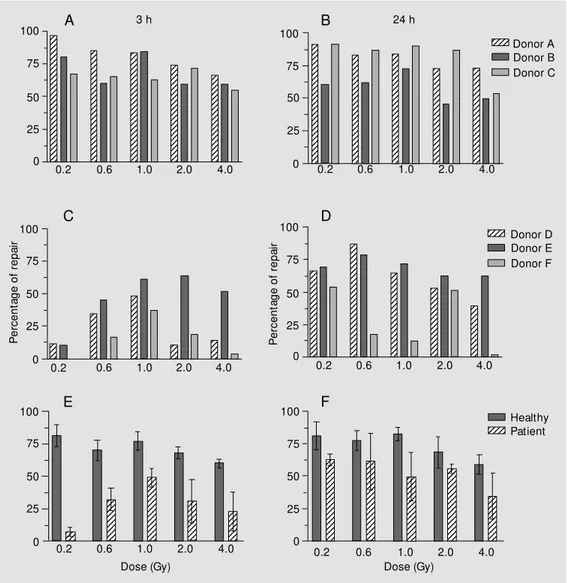
Repair capacity of radioinduced damage in blood cells was estimated by the formula described by Jaloszynski et al. (14). The individual values and means at 3 and 24 h are illustrated in Figure 2.
There were interindividual differences in repair capacity of blood lymphocytes in both groups, but this variability was more marked among patients.
Figure 2 shows that radioinduced dam-age was less efficiently repaired among pa-tients than among controls after all doses at both times of analysis. Among patients, the blood cells of donor E showed a better
effi-Figure 1 - Dose-response curves of DNA migration obtained for blood samples from 3 healthy donors (full lines) and 3 breast cancer patients (broken lines), processed immediately (A, D) or 3 (B, E) and 24 h (C, F) after in vitro exposure to 60Co.
Ta
il
le
ng
th
(µ
m
)
100
D
N
A
d
am
ag
e
(a
u)
75
50
25
0
100
75
50
25
0
100
75
50
25
0
400
300
200
100
0
400
300
200
100
0
0 1 2 3 4 0 1 2 3 4
0 1 2 3 4
0 1 2 3 4
Dose (Gy)
0 1 2 3 4
0 1 2 3 4
Dose (Gy) 400
300
200
100
0
A D
B E
C F
0 h 0 h
3 h 3 h
ciency in repair capacity. After 24 h, only donor E had a performance equal to controls. Among controls, donor C had a less effi-cient repair after 3 h, but presented the best efficiency after 24 h. We observed a ten-dency to a decrease in repair capacity of blood lymphocytes with increasing doses in both groups at 3 and 24 h. The repair ob-served in peripheral blood cells from healthy donors after 24 h did not differ from that observed after 3 h, whereas this difference was evident among patients. Statistical anal-ysis showed that the repair capacity of the groups differed both 3 h (P<0.001) and 24 h (P<0.05) after irradiation.
D iscussio n
Ionizing radiation is an etiologic agent known to act on the induction of breast cancer (21), but on the other hand it is a therapeutic modality used in cancer treat-ment. Therefore, a better knowledge of the action of ionizing radiation on the cellular response is of importance both clinically and therapeutically.
The head diameters obtained here for cells without DNA migration agreed with data reported by other laboratories (20,22).
A significant difference in relation to basal values was observed between groups
P er ce nt ag e of r ep ai r 100 Donor A 1234 1234 1234 Donor B Donor C 12 12 12 12 12 12 12 12 12 12 12 12 12 12 12 12 12 12 12 12 12 12 12 12 12 12 12 12 12 12 12 12 12 12 12 12 12 12 12 12 12 12 12 12 12 12 12 12 12 12 12 12 12 12 12 12 12 12 12 12 12 12 12 12 12 12 12 12 12 12 12 12 12 12 12 12 12 12 12 12 12 12 12 12 12 12 12 12 12 12 12 12 12 12 12 12 12 12 12 12 12 12 12 12 12 12 12 12 12 12 75 50 25 0
0.2 0.6 1.0 2.0 4.0
100 75 50 25 0 12 12 12 12 12 12 12 12 12 12 12 12 12 12 12 12 12 12 12 12 12 12 12 12 12 12 12 12 12 12 12 12 12 12 12 12 12 12 12 12 12 12 12 12 12 12 12 12 12 12 12 12 12 12 12 12 12 12 12 12 12 12 12 12 12 12 12 12 12 12 12 12 12 12 12 12 12 12 12 12 12 12 12 12 12 12 12 12 12 12 12 12 12 12 12 12 12 12 12 12 12 12 12 12 12 12 12 12 12 Donor D 1234 1234 1234 Donor E Donor F
0.2 0.6 1.0 2.0 4.0
3 h 24 h
A B 12 12 12 12 12 12 12 12 12 12 12 12 12 12 12 12 12 12 12 12 12 12 12 12 12 12 12 12 12 12 12 12 12 12 12 12 12
0.2 0.6 1.0 2.0 4.0
100 75 50 25 0 123 123 123 123 123 123 123 123 123 123 123 123 123 123 123 123 123 123 123 123 123 123 123 123 123 123 123 123 123 123 123 123 123 123 123 123 123 123 123 123 123 123 123 123 123 123 123 123 123 123 123 123 123 123 123 123 123 123 123 123 123 123 123 123 123 123 123 123 123 123 123 123 123 123 123 123 123 123 123 123 123 123 123 123 123
0.2 0.6 1.0 2.0 4.0
P er ce nt ag e of r ep ai r 100 75 50 25 0 C D 123 123 123 123 123 123 123 123 123 123 123 123 123 123 123 123 123 123 123 123 123 123 123 123 123 123 123 123 123 123 123 123 123 123 123 123 123 123 123 123 123 123
0.2 0.6 1.0 2.0 4.0
Dose (Gy) 12 12 12 12 12 12 12 12 12 12 12 12 12 12 12 12 12 12 12 12 12 12 12 12 12 12 12 12 12 12 12 12 12 12 12 12 12 12 12 12 12 12 12 12 12 12 12 12 12 12 12 12 12 12 12 12 12 12 12 12 12 12 12 12 12 12 12 12 12 12 12 12 12 Healthy 1234 1234 1234Patient
0.2 0.6 1.0 2.0 4.0
100 75 50 25 0 Dose (Gy) 100 75 50 25 0 E F
by tail measurement, but not by visual classi-fication. This incongruent result may be ex-plained by the fact that cells with little dam-age barely differ from cells with no damdam-age so that visual analysis may not permit a distinction between them.
Statistical analysis showed no differences in results after 3 and 24 h of incubation. We conclude that the maintenance of blood samples for 24 h does not cause additional DNA lesions.
About the radioinduced damages, the dose-response curves for DNA migration (tail length) obtained just after irradiation showed an increase in tail length as a func-tion of radiafunc-tion dose. The increasing dose caused an increase in DNA lesions and so damaged filaments migrated to the anode during the electrophoretic run, giving origin to the comet tails. Statistical analysis showed no difference between groups, whereas the difference determined by visual classifica-tion (DD), although small, was considered significant. This result may be explained as a consequence of the slight difference between class 0 and class 1.
In this context, we conclude that both groups had an analogous response when an-alyzed immediately after exposure. The ini-tial radioinduced damage may have been quantitatively similar.
The quantitative estimation of repair ca-pacity in blood lymphocytes showed that most of the radioinduced damage in the healthy group was repaired within 3 h while patients had more lesions even after 24 h. These data show that the breast cancer do-nors analyzed in this study had deficient radioinduced damage repair. These data do not agree with those reported by Hsu et al. (13) who used bleomycin as a mutagenic test in peripheral blood lymphocytes from pa-tients with various cancer types. The authors found a higher frequency of chromatid breaks per cell in patients with lung, colon and upper digestive tract cancer but not in breast cancer patients. They suggested that
sensi-tivity to the mutagenic agents may play an important role in the carcinogenesis of tis-sues and organs in contact with the external environment, a fact that does not occur with mammary tissue.
In contrast, Jaloszynski et al. (14) re-ported that breast cancer patients were more sensitive to bleomycin by expressing higher level of DNA damage than controls, as ob-served in the comet assay.
The less efficient repair observed in blood lymphocytes in the present patients may ex-plain the high basal level of damage.
Since carcinogenesis may originate from defects in DNA repair (14,23), we may raise various hypotheses. The reduced repair ca-pacity observed in these patients may be a consequence of mutations in genes involved in cell cycle control and DNA repair regula-tion. The tumor suppressor genes BRCA1, BRCA2, p53 or ATM are fundamental fac-tors for the maintenance of genome integrity (2,24).
Rothfuss et al. (15) observed that familial breast cancer patients with a mutated BRCA1 gene show deficiency in radioinduced dam-age repair as detected by the micronucleus test. These data show a direct relation be-tween BRCA1 mutation and radioinduced damage repair. Therefore, molecular analy-sis of the genes revealed that somatic muta-tions of either BRCA1 or BRCA2 are rare events in the majority of sporadic cases of breast and ovarian cancer (2,6,23,25). Then, some authors such as Futreal et al. (26) raised the hypothesis that sporadic and fa-milial tumors have different etiologies, sug-gesting that BRCA1 and BRCA2 may not play a critical role in the genesis of sporadic cancer cases.
relatives with ovarian and breast cancer, be-sides other types of cancer in the families. There seems to be a predisposition to cancer development in their families, although we cannot rule out the hypothesis of exposure of the entire family to the same carcinogenic agent.
Other genes may be involved but further studies are necessary to test this possibility. One of the possible genes involved is p53, which has been described in almost all hu-man cancers, including breast cancer (27), and plays a role in the repair mechanism of radioinduced damage (9). There is evidence that p53 and rad51 are involved in vivo (28). We cannot rule out the possibility of an ATM gene action since this gene acts on BRCA1 phosphorylation after exposure to ionizing radiation as an initial signal for the recombinational repair process of DNA double-strand breaks (24). The sensitivity to ionizing radiation is also observed in ataxia telangiectasia patients who are defective in DNA strand break repair. The disease is related to ATM gene mutation and heterozy-gotes are 0.5-1% of the population more predisposed to breast cancer (2).
These observations suggest the occur-rence of other genes that can act in the same way as suppressor genes. On this basis, vari-ous other molecular targets for sporadic tu-mors may exist (26).
In relation to controls, the blood cells from donor C showed the most efficient repair after 24 h, suggesting a slower repair mechanism than in the other donors.
Patient age was not an important factor because the oldest one, donor E, showed the best repair. The control group ranged in age from 40-50 years. A possible influence of age may have affected the comparison be-tween groups.
The relation between age and damage susceptibility in somatic cells had been stud-ied but data are contradictory. Duffaud et al. (29) and Peace and Succop (30) observed an increase in the frequency of affected cells
with aging. This could be attributed to the accumulation of genetic damage in cells (31) or to the aging process, such as altered cellu-lar metabolism and/or decrease in the effi-ciency of DNA repair (32). Other authors, however, did not find any association be-tween age and genetic damage in cells (33,34).
The influence of gender is not significant for the biological response to environmental genotoxic agents (29,30), although some evidence shows that women are more sus-ceptible to some carcinogenic agents (35).
In addition to these considerations, there is the possibility that the difference between groups was a consequence of the pathologi-cal stage (malignancy) of the tumors. Other factors such as immunological, metabolic and dietary ones, also considering that life style is influenced by various environmental agents that can act synergistically, have been suggested by several authors (29,33). There-fore, nothing that could explain the differ-ence between patients and healthy donors was observed in the replies to the question-naire.
Ionizing radiation is known to induce multiple DNA lesions (17). Although the comet assay does not permit the direct iden-tification of the damage or repair mechanism involved, it is known that the pH of the electrophoresis buffer is a fundamental fac-tor: under neutral conditions, double-strand breaks are the type of damage most fre-quently detected, while under alkaline con-ditions (pH >12), as used in the present study, single- and double-strand breaks as well as alkali-labile lesions make a signifi-cant contribution to the DNA quantity found in comet tails (16,18).
2 h (16-18). Olive (36) described a slower repair system for more complex lesions, which was completed only after 30-40 h.
Based on this information, we suggest that the discordant results obtained by Rothfuss et al. (15) may have been due to the period of time used to check the breast can-cer cell repair. The authors did not find a significant difference in repair induced by 2 Gy 60Co analyzed one hour later by the comet
assay, but did find a difference by the micro-nucleus test used to evaluate the non-re-paired damage.
Double-strand breaks are among the most difficult radioinduced lesions to repair. Non-repaired damage can result in cell death, aging and cancer.
Both parameters adopted in the present study, i.e., comet measurement and visual classification, proved to be safe and sensi-tive and therefore could be used as an alter-native method for the evaluation of cellular response.
The results showed that cellular
radio-sensitivity depends not only on the amount of imposed initial damage, but also on the cell repair capacity, among other factors.
Since in the present study only a small number of patients and controls was ana-lyzed, it is necessary to investigate a larger number of donors including other family members, with an equal distribution of pa-tients and healthy donors with respect to age, gender and other risk factors of increased DNA fragility, and to perform molecular analysis of possible genes involved in breast cancer susceptibility for a more accurate evaluation.
Ackno wle dgm e nts
The authors are indebted to Dr. Regina Godoy Lopes, Instituto de Radioterapia de São Paulo, São Paulo, SP, Brazil, for supply-ing the blood samples from the patients, and to Dr. Maria Aparecida Pires Camillo for the statistical analysis.
Re fe re nce s
1. Sankaranarayanan K & Chakraborty R (1995). Cancer predisposition, radiosensi-tivity and the risk of radiation-induced can-cers. I. Background. Radiation Research,
143: 121-143.
2. Venkitaraman AR (1999). Breast cancer genes and DNA repair. Science, 286: 1100-1102.
3. Easton DF, Bishop DT, Ford D & Crockford GP (1993). Genetic linkage analysis in fa-milial breast and ovarian cancer: results from 214 families. The breast cancer link-age consortium. American Journal of Hu-man Genetics, 52: 678-701.
4. Rahman N & Stratton M R (1998). The ge-netics of breast cancer susceptibility. An-nual Review of Genetics, 32: 95-121. 5. M achado CR & M enck CFM (1997).
Hu-man DNA repair diseases: from genome instability to cancer. Brazilian Journal of Genetics, 20: 755-762.
6. Scully R, Chen J, Plug A, Xiao Y, Weaver D, Feunteun J, Ashley T & Livingston DM (1997). Association of BRCA1 w ith Rad51
in mitotic and meiotic cells. Cell, 88: 265-275.
7. Smith M L, Chen I-T, Zhan Q, Bae I, Chen C-Y, Gilmer TM , Kastan M B, O’Connor PM & Fornace Jr AJ (1994). Interaction of the p53-regulated protein Gadd45 w ith proliferating cell nuclear antigen. Science,
266: 1376-1380.
8. M allya SM & Sikpi M O (1998). Evidence of the involvement of p53 in gamma-ra-diation-induced DNA repair in human lym-phoblasts. International Journal of Radia-tion Biology, 74: 231-238.
9. Rigaud O, Guedeney G, Duranton I, Leroy A, Doloy M T & M agdelenat H (1990). Genotoxic effects of radiotherapy and chemotherapy on the circulating lympho-cytes of breast cancer patients. I. Chro-mosome aberrations induced in vivo. M u-tation Research, 242: 17-23.
10. Helzlsouer KJ, Harris EL, Parshad R, Fogel S, Bigbee WL & Sanford KK (1995). Famil-ial clustering of breast cancer: possible interaction betw een DNA repair
profi-ciency and radiation exposure in the de-velopment of breast cancer. International Journal of Cancer, 64: 14-17.
11. Parshad R, Price FM , Bohr VA, Cow ans KH, Zujew ski JÁ & Sanford KK (1996). Deficient DNA repair capacity, a predis-posing factor in breast cancer. British Journal of Cancer, 74: 1-5.
12. Scott D, Barber JBP, Levine EL, Burrill W & Roberts SA (1998). Radiation-induced micronucleus induction in lymphocytes identifies a high frequency of radiosensi-tive cases among breast cancer patients: a test for predisposition? British Journal of Cancer, 77: 614-620.
13. Hsu TC, Johnston DA, Cherry LM , Ram-kissoon D, Schantz SP, Jessup JM , Winn RJ, Shirley L & Furlong C (1989). Sensitiv-ity to genotoxic effects of bleomycin in humans: possible relationship to environ-mental carcinogenesis. International Jour-nal of Cancer, 43: 403-409.
(1997). Bleomycin-induced DNA damage and its removal in lymphocytes of breast cancer patients studied by comet assay.
M utation Research, 385: 223-233. 15. Rothfuss A, Schutz P, Bochum S, Volm T,
Eberhardt E, Kreienberg R, Vogel W & Speit G (2000). Induced micronucleus fre-quencies in peripheral lymphocytes as a screening test for carriers of a BRCA1 mutation in breast cancer families. Can-cer Research, 60: 390-394.
16. Singh NP, M cCoy M T, Tice RR & Schnei-der EL (1988). A simple technique for quantitation of low levels of DNA damage in individual cells. Experimental Cell Re-search, 175: 184-191.
17. Price A (1993). The repair of ionising ra-diation-induced damage to DNA. Cancer Biology, 4: 61-71.
18. Tice RR (1995). The single cell gel/comet assay: a microgel electrophoretic tech-nique for the detection of DNA damage and repair in individual cells. In: Phillis DH & Venitt S (Editors), Environmental M u-tagenesis. Bioscientific, Oxford, 315-339. 19. M cKelvey-M artin VJ, Green M HL, Schme-zer P, Poolzobel BL, De M éo M P & Collins A (1993). The single cell gel electrophosis assay (comet assay): A European re-view . M utation Research, 288: 47-63. 20. Visvardis E-E, Tassiou AM & Piperakis SM
(1997). Study of DNA damage induction and repair capacity of fresh and cryopre-served lymphocytes exposed to H2O2 and
g-irradiation w ith the alkaline comet as-say. M utation Research, 383: 71-80. 21. van Bekkum DW & Broerse JJ (1991).
Induction of mammary tumors by ionizing
radiation. Radiation and Environmental Biophysics, 30: 217-220.
22. Singh NP & Stephens RE (1997). M icrogel electrophoresis: sensitivity, mechanisms, and DNA electrostretching. M utation Re-search, 383: 167-175.
23. Kinzier KW & Vogelst ein B (1997). Gatekeepers and caretakers. Nature, 386:
761-763.
24. Cortez D, Wang Y, Qin J & Elledge SJ (1999). Requirement of ATM -dependent phosphorylation of BRCA1 in the DNA dam age response t o double-st rand breaks. Science, 286: 1162-1166. 25. Sharan SK, M orimatsu M , Albrecht U, Lim
D-S, Regel E, Dinh C, Sands A, Eichele G, Hasty P & Bradley A (1997). Embryonic lethality and radiation hypersensitivity m ediated by Rad51 in m ice lacking BRCA2. Nature, 386: 804-810.
26. Futreal PA, Liu Q, Shattuck-Eidens D, Cochran C, Harshman K, Tavtigian S, Ben-nett LM , Haugen-Strano A, Sw ensen J, M iki Y, Eddington K, M cClure M , Frye C, Weaver-Feldhaus J, Ding W, Gholami Z, Söderkvist P, Terry L, Jhanw ar S, Ber-chuck A, Iglehart JD, M arks J, Ballinger DG, Barrett JC, Skolnick M H, Kamb A & Wiseman R (1994). BRCA1 mutations in primary breast and ovarian carcinomas.
Science, 266: 120-122.
27. Hollstein M , Sidransky D, Vogelstein B & Harris CC (1991). p53 mutations in human cancers. Science, 253: 49-53.
28. Sturzbecher HW, Donzelmann B, Henning W, Knippschild U & Buchhop S (1996). p53 is linked directly to homologous re-combination processes via RAD51/RecA
protein interaction. EM BO Journal, 15: 1992-2002.
29. Duffaud F, Orsière T, Villani P, Pelissier AL, Volot F, Favre R & Botta A (1997). Com-parison betw een micronucleated lympho-cyte rates observed in healthy subjects and cancer patients. M utagenesis, 12: 227-231.
30. Peace BE & Succop P (1999). Spontane-ous micronucleus frequency and age: w hat are normal values? M utation Re-search, 425: 225-230.
31. Fenech M & M orley AA (1989). Kineto-chore detection in micronuclei: an alterna-tive method for measuring chromosome loss. M utagenesis, 4: 98-104.
32. Ganguly BB (1993). Cell division, chromo-somal damage and micronucleus forma-tion in peripheral lymphocytes of healthy donors: related to donor’s age. M utation Research, 295: 135-148.
33. Sarto F, Finotto S, Giacomelli L, M azzotti D, Tomanin R & Levis AG (1987). The micronucleus assay in exfoliated cells of the human buccal mucosa. M utagenesis, 2: 11-17.
34. Gantenberg H-W, Wuttke K, Streffer C & M uller W-U (1991). M icronuclei in human lymphocytes irradiated in vitro or in vivo.
Radiation Research, 128: 276-281. 35. Perera FP (1997). Environment and
can-cer: w ho are susceptible? Science, 278: 1068-1073.