251 Magnetic resonance dacryocystography
Radiol Bras. 2008 Jul/Ago;41(4):251–254 Original Article • Artigo Original
Magnetic resonance dacryocystography: comparison
between conventional surface coils and microscopic
coils*
Ressonância magnética das vias lacrimais: estudo comparativo entre bobinas de superfície convencionais e microscópicas
Luiz de Abreu Junior1, Angela Maria Borri Wolosker1, Maria Lucia Borri1, Mário de Melo Galvão Filho1, Giuseppe D’Ippolito2, Luiz Guilherme de Carvalho Hartmann3, Cláudio Campi de Castro4
OBJECTIVE: Magnetic resonance imaging has been utilized in the evaluation of the lacrimal apparatus with some advantages over conventional dacryocystography. The present study was aimed at acquiring high-resolution images utilizing microscopic coils for evaluating typical structures of the lacrimal apparatus as compared with the findings observed with conventional surface coils. MATERIALS AND METHODS: Five asymptomatic volunteers with no history of epiphora were submitted to high-field magnetic resonance imaging with microscopic and conventional surface coils, and STIR sequence after instillation of saline solution. The definition of normal anatomic structures of lacrimal apparatuses was compared utilizing conventional and microscopic surface coils. Based on a consensual scoring system, the mean values for each structure were calculated by two observers. RESULTS: In 90% of cases, higher scores were attributed to images acquired with the microscopic coil. On average, a 1.17 point increase was observed in the scoring of anatomic structures imaged with the microscopic coil. Additionally, a subjective improvement was observed in the signal-to-noise ratio with the microscopic coil. CONCLUSION: Magnetic resonance dacryocystography with microscopic coils is the appropriate method for evaluating the lacrimal apparatus, providing images with better quality as compared with those acquired with conventional surface coils.
Keywords: Magnetic resonance imaging; Lacrimal apparatus; Dacryocystography; Microscopic coil.
OBJETIVO: A ressonância magnética tem sido utilizada para avaliar as vias lacrimais, com vantagens em relação à dacriocistografia por raios-X. O objetivo deste trabalho é obter imagens de alta resolução utilizando bobinas de superfície microscópicas para avaliação de estruturas normais das vias lacrimais, comparando com o aspecto observado utilizando-se bobinas de superfície convencionais. MATERIAIS E MÉTODOS: Cinco voluntários assintomáticos, sem histórico de lacrimejamento, submeteram-se a ressonância magnética de alto campo, com bobinas de superfície (convencional e microscópica), com seqüência STIR após instilação de soro fisiológico. A identificação das estruturas anatômicas normais das vias lacrimais foi comparada uti-lizando-se as duas bobinas. Mediante uso de um sistema de escore, um valor médio de cada estrutura foi calculado por dois examinadores, consensualmente. RESULTADOS: Em 90% das vezes houve aumento do escore, atribuído à estrutura anatômica no estudo com a bobina microscópica. Em média, houve aumento de 1,17 ponto no escore, por estrutura anatômica visualizada, quando se utilizou a bobina microscópica. Ob-servou-se, ainda, melhora subjetiva da relação sinal-ruído ao se utilizar a bobina microscópica. CONCLU-SÃO: A dacriocistografia por ressonância magnética com bobinas microscópicas é um método adequado para o estudo das vias lacrimais, resultando em imagens de melhor qualidade quando comparada ao uso de bobinas de superfície convencionais.
Unitermos: Imagem por ressonância magnética; Vias lacrimais; Dacriocistografia; Bobina microscópica. Abstract
Resumo
* Study developed at Scopo Diagnóstico, Unit of US, CT and MRI, Hospital São Luiz, São Paulo, SP, Brazil.
1. PhDs, MDs, Radiologists at the Unit of US/CT/MRI, Hospi-tal e Maternidade São Luiz, São Paulo, SP, Brazil.
2. PhD, Associate Professor, Department of Imaging Diagno-sis, Universidade Federal de São Paulo/Escola Paulista de Me-dicina (Unifesp/EPM), São Paulo, SP, MD, Responsible for the Unit of US/CT/MRI, Hospital e Maternidade São Luiz, São Paulo, SP, Brazil.
INTRODUCTION
Lacrimal drainage system disorders present an estimated prevalence of 3%(1),
representing a relatively frequent complaint among ophthalmological patients. Epi-phora – an overflow of tears due to obstruc-Abreu Jr L, Wolosker AMB, Borri ML, Galvão Filho MM, D’Ippolito G, Hartmann LGC, Castro CC. Magnetic resonance dacryocystography: comparison between conventional surface coils and microscopic coils. Radiol Bras. 2008;41(4): 251–254.
0100-3984 © Colégio Brasileiro de Radiologia e Diagnóstico por Imagem
3. MD, Radiologist at the Unit of US/CT/MRI, Hospital e Ma-ternidade São Luiz, São Paulo, SP, Brazil.
4. Private Docent, Head for the Unit of Magnetic Resonance Imaging, Instituto do Coração (InCor), São Paulo, SP, Brazil.
Mailing address: Dr. Luiz de Abreu Junior. Rua Doutor Alceu de Campos Rodrigues, 95/143, 1º subsolo. São Paulo, SP, Bra-zil, 04544-000. E-mail: [email protected]
252
Abreu Jr L et al.
Radiol Bras. 2008 Jul/Ago;41(4):251–254 tive causes – may be caused by
inflamma-tion, tumors, trauma, surgery, radiotherapy or congenital alterations. The evaluation of the lacrimal pathways includes clinical evidences (intubation and irrigation) and imaging studies.
Imaging methods available for evaluat-ing the lacrimal pathways include: dacryo-cystography with conventional radiogra-phy, dacryoscintigraphy (nuclear medi-cine), computed tomography dacryocysto-graphy, and magnetic resonance imaging (MRI) dacryocystography.
MRI dacryocystography is currently an excellent diagnostic method for evaluating the lacrimal pathways, with potential ad-vantages, considering the absence of ion-izing radiation which minimizes the risk for development of cataract. MRI provides high contrast resolution for soft tissues imaging, with instillation of saline solution or diluted gadolinium, and combining the non-invasiveness with the capability of evaluating functional alterations. Tradition-ally, surface coils have been utilized for acquisition of diagnostic images. However, with the introduction of the so called mi-croscopic coils, i.e., surface coils with re-duced diameter, allowing a better resolu-tion of superficial structures and, conse-quently, an improvement in the signal-to-noise ratio, the authors considered study-ing normal structures of the lacrimal sys-tem in asymptomatic individuals, compar-ing the results with those obtained with conventional surface coils.
MATERIALS AND METHODS
A prospective study was developed with five asymptomatic volunteers (ten lacrimal pathways) – two women and three men with ages ranging between 29 and 33 years (mean, 31.2 years). These individuals were submitted to magnetic resonance imaging in an Intera 1.5T (Philips Medical Systems; Best, The Netherlands) with conventional surface coils (C1 – 160 mm) and micro-scopic coils (47 mm). Initially, images were acquired with C1 surface coils utilized in the majority of clinical applications which require imaging of superficial structures. Afterwards, images were acquired with another type of surface coil called “micro-scopic coil”, characterized by reduced
in-ner diameter (< 5.0 cm). Because of this features, microscopic coils result in a sig-nificant increase in the signal of superficial structures, with improvement of the sinal-to-noise ratio, providing lower voxels with-out affecting the images quality. The coil utilized in the present study (microscopic coil –Philips Medical Systems; Best, The Netherlands) has a 47 mm inner diameter. STIR (TR = 3800 ms / TE = 90 ms / TI = 180 ms / 1.8 mm thickness) T2-weighted sequences were performed after instillation of two drops of saline solution every minute into both conjunctival sacs during five minutes. The same sequence was per-formed with the two different coils at five-minute and ten-five-minute acquisition inter-vals. The anatomical definition of the dif-ferent structures included in the lacrimal system (superior lacrimal canaliculi, lacri-mal sac, and nasolacrilacri-mal duct) was sub-jectively evaluated by consensus between two observers (a radiologist specialized in
head and neck, and a general radiologist) and rated according to a scoring system. The scoring system utilizes the following visualization rating scale: 0 – nonvisu-alization; 1 – poor visunonvisu-alization; 2 – good visualization; 3 – optimum visualization of the structure.
RESULTS
The radiologists’ consensual evaluation is summarized on Table 1.
In 36 of the 40 (90%) structures evalu-ated, a mean 1.17 point increase was ob-served in the score assigned to the images in the study of each anatomical structure with the microscopic coil as compared with the C1 surface coil. Only 10% (four ana-tomical structures – two nasolacrimal ducts in case 2 and two lacrimal sacs in case 5) of the whole sample did not present any increase in the score assigned to images acquired with the microscopic coil. A
sub-Table 1 Consensual scoring attributed by two observers to the identification of lacrimal pathways, ac-cording to the coil utilized.
Superior lacrimal duct Inferior lacrimal duct Lacrimal sac Nasolacrimal duct
Case 1 Case 2 Case 3 Case 4 Case 5
C1 M C1 M C1 M C1 M C1 M
R 2 2 2 2 L 1 1 2 2 R 3 3 3 3 L 2 2 3 3 R 0 0 2 2 L 0 1 2 2 R 2 2 3 2 L 3 3 3 3 R 3 3 3 2 L 2 2 3 2 R 1 2 3 1 L 1 1 3 1 R 2 2 3 2 L 0 0 2 0 R 0 1 2 1 L 3 3 3 2 R 3 3 3 3 L 2 2 3 2 L 1 1 2 1 R 1 1 2 1
C1, conventional surface coil; M, microscopic surface coil; R, right; L, left.
253 Magnetic resonance dacryocystography
Radiol Bras. 2008 Jul/Ago;41(4):251–254
offices (about 3% of ambulatory visits). Data about level and type of obstruction play a significant role in the definition of the best therapeutic strategy to be adopted (dacryocystorhinostomy, dacryocysto-plasty, nasolacrimal stent or antibiotics ir-rigation). Additionally to the clinical evalu-ation, an imaging method should be uti-lized for this purpose.
The principles of dacryocystography with conventional radiography were first described by Ewing in 1909 and, up to re-cently, this was the method most frequently utilized in the evaluation of the lacrimal system. Despite providing an excellent anatomical delineation, this technique is relatively invasive and involves certain risks such as iatrogenic trauma and post-catheterization stenosis, besides the harm-ful effects of the ionizing radiation on the crystalline lens, without offering any func-tional information, considering that the contrast agent is administered under pres-sure by a syringe/catheter.
Dacryoscintigraphy is an alternative method for evaluating functional alter-ations of the lacrimal pathways. However, the following disadvantages of this method should be considered: the low spatial reso-lution for orbital soft tissues imaging and crystalline lens exposure to radiation.
Despite representing an important alter-native in the study of lacrimal pathways, computed tomography dacryocystography also presents relative disadvantages con-nected with the utilization of ionizing ra-diation and the potential conjunctival irri-tation caused by the iodinated contrast agent.
MRI has been reported in the literature as a feasible method for evaluating the lac-rimal pathways. The main advantages in re-lation to the other methods are: non-inva-siveness, absence of ionizing radiation, (in-stillation rather than catheterization), ana-tomical and functional evaluation, and fea-sibility of simultaneous bilateral evalua-tion. Either T1- or T2-weighted sequences can be utilized for images acquisition(2–7).
In the first case, a gadolinium solution acts as a contrast agent; on T2-weighted se-quences, likewise in the present study, the 0.9% saline solution is the medium that allows the identification of the lacrimal pathways.
jective improvement was observed in the signal-to-noise ratio for the whole sample in the comparison between images ac-quired with microscopic coil and with con-ventional surface coil.
DISCUSSION
Epiphora (abnormal overflow of tears due to obstruction of lacrimal pathways) is a frequent complaint(1) in ophthalmologic
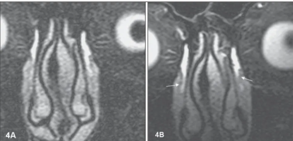
Figure 2.Case 1. A better definition of superior and inferior lacrimal ducts as well as lacrimal sac is observed on images acquired with microscopic coil (2B), as compared with those acquired with the con-ventional surface coil (2A).
254
Abreu Jr L et al.
Radiol Bras. 2008 Jul/Ago;41(4):251–254 Surface coils allow high-quality
imag-ing of superficial anatomical structures like the normal lacrimal pathways structures. Conventional surface coils, with larger di-ameters, can provide images with a non-op-timum sinal-to-noise ratio, utilizing very thin slices. In the present protocol, 1.8 mm-thick slices presented a poor signal-to-noise ratio with the conventional coil. Despite the fact that the majority of structures could be visualized with this coil, their visualization was rated as non-optimum.
Several studies in the literature report an improvement of the signal-to-noise ratio with the utilization of microscopic coils for
evaluating several structures of different tissues such as knee ligaments and me-nisci(8) and pathological cervical lymph
nodes(9). The present study is compatible
with these reports, considering that all the lacrimal system structures were better char-acterized in the evaluation with the micro-scopic coil, even in the presence of artifacts (motion artifacts, for example). This aspect is particularly relevant if the identification of lacrimal ducts is considered. Consider-ing their small caliber, sometimes the visu-alization of these structures with conven-tional surface coils is limited(2). In the
present study, the lacrimal ducts
character-ization improved with the utilcharacter-ization of the microscopic coil. This finding allows the authors to consider the possibility of im-provement in the diagnosis of lacrimal pathways obstruction, especially high (pre-saccular) obstructions. However, further studies with symptomatic individuals would be necessary to confirm this hypoth-esis.
CONCLUSION
Magnetic resonance imaging dacryo-cystography utilizing microscopic coils is the appropriate method for identifying nor-mal structures of the lacrinor-mal pathways, resulting in higher-quality images as com-pared with images acquired with conven-tional surface coils.
REFERENCES
1. Linberg JV, McCormick SA. Primary acquired na-solacrimal duct obstruction. A clinicopathologic report and biopsy technique. Ophthalmology. 1986;93:1055–63.
2. Caldemeyer KS, Stockberger SM Jr, Broderick LS. Topical contrast-enhanced CT and MR dacryocystography: imaging the lacrimal drain-age apparatus of healthy volunteers. AJR Am J Roentgenol. 1998;171:1501–4.
3. Kirchhof K, Hähnel S, Jansen O, et al. Gado-linium-enhanced magnetic resonance dacryo-cystography in patients with epiphora. J Comput Assist Tomogr. 2000;24:327–31.
4. Yoshikawa T, Hirota S, Sugimura K. Topical con-trast-enhanced magnetic resonance dacryocysto-graphy. Radiat Med. 2000;18:355–62. 5. Takehara Y, Isoda H, Kurihashi K, et al. Dynamic
MR dacryocystography: a new method for evalu-ating nasolacrimal duct obstructions. AJR Am J Roentgenol. 2000;175:469–73.
6. Manfrè L, de Maria M, Todaro E, et al. MR dac-ryocystography: comparison with dacryocysto-graphy and CT dacryocystodacryocysto-graphy. AJNR Am J Neuroradiol. 2000;21:1145–50.
7. Hoffmann KT, Hosten N, Anders N, et al. High-resolution conjunctival contrast-enhanced MRI dacryocystography. Neuroradiology. 1999;41: 208–13.
8. Niitsu M, Ikeda K. Magnetic resonance micro-scopic images with 50-mm field-of-view of the medial aspect of the knee. Acta Radiol. 2004;45: 760–8.
9. Sumi M, Van Cauteren M, Nakamura T. MR microimaging of benign and malignant nodes in the neck. AJR Am J Roentgenol. 2006;186:749– 57.
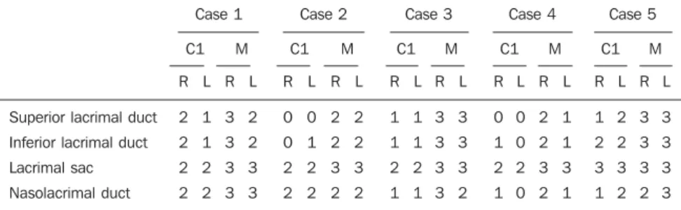
Figure 6. Additionally to the better signal-to-noise ratio, the study with microscopic coil (6B), has al-lowed a better characterization of lacrimal ducts at right (arrows).