304
INTRODUCTION
Sclerosing encapsulating peritonitis (SEP) is a rare disease entity characterized by total or partial encasement of small bowel by a thick fibrous sac, resulting in intestinal obstruction. SEP is classified as primary (idiopathic) or secondary based on the aetiological factors. This condition was first described in 1907 as “peritonitis chronic fibrosa incapsulata” by Owtschinnikow.1 Idiopathic cases which lack
any identifiable aetiology have been described as‘abdominal cocoon syndrome’.1
Infection of the fallopian tubes or retrograde menstruation with subclinical peritoneal infection may be related to the disease onset and progression. The idiopathic form primarily affects young women from tropical countries.2
The secondary form has been associated with
long term peritoneal dialysis, β blocker therapy,
abdominal tuberculosis, previous abdominal surgery, peritoneal shunts and gastrointestinal malignancies.3
Presentation in a middle aged man, as in our case is rare. There is a need for awareness regarding this relatively rare entity as a cause of intestinal obstruction, which can present as an emergency, as it did in our case.
CASE REPORT
A 45-year-old man presented to our emergency room with abdominal pain, distension without passage of stools or flatus for the last 3 days. He had two episodes of abdominal pain in the past few months with spontaneous symptomatic relief. He had no past history of abdominal
Case Report:
Abdominal cocoon: a rare cause of acute intestinal obstruction
Silpa Kadiyala,1 Sivaramakrishna Gavini,2 Rashmi Patnayak,3 B. Vijayalakshmi Devi,1 S. Sarala,1
A.Y. Lakshmi1
Departments of 1Radiology, 2Surgical Gastroenterology, 3Pathology,
Sri Venkateswara Institute of Medical Sciences, Tirupati
ABSTRACT
Sclerosing encapsulating peritonitis (SEP) is a relatively rare cause of intestinal obstruction resulting from encasement of variable lengths of bowel by dense fibro-collagenous membrane. The idiopathic cases of SEP, which lack any identifiable cause from clinical, radiological and histopathological findings, are also reported under the descriptive term “abdominal cocoon syndrome”. Patient with SEP present with intestinal obstruction. Persistent untreated SEP may advance to bowel gangrene or intestinal perforation, which are life threatening conditions.
We report the rare occurrence of SEP in a 45-year-old male presenting with signs of intestinal obstruction. Imaging findings revealed abdominal cocoon with bowel gangrene leading to perforation and the same was confirmed at surgery. Surgical excision of the fibrotic sac encasing the bowel, resection of gangrenous bowel segment and end ileostomy were performed. Histopathology of the excised membrane confirmed the diagnosis of SEP. Radiologists should be aware of this relatively rare cause of intestinal obstruction, its imaging findings and complications, as an accurate preoperative diagnosis will prevent diagnostic delay and aid the surgeon in planning treatment.
Key words:Intestinal Perforation, Intestinal obstruction, Peritoneal fibrosis
Kadiyala S, Gavini S, Patnayak R, Vijayalakshmi Devi B, Sarala S, Lakshmi AY. Abdominal cocoon: a rare cause of acute intestinal obstruction. J Clin Sci Res 2015;4:304-7. DOI: http://dx.doi.org/10.15380/2277-5706.JCSR.14.068
Corresponding author: Dr Silpa Kadiyala, Assistant Professor, Department of Radiology, Sr i Ven kateswara In stitute of Medical Sciences, Tirupati, India.
e-mail: silpakadiyala@gmail.com
Received: December 12, 2014; Revised manuscript received: May 24, 2015; Accepted: May 28, 2015.
Abdominal cocoon: a rare cause of acute intestinal obstruction Silpa Kadiyala et al
Online access
http://svimstpt.ap.nic.in/jcsr/Oct-dec15_files/4cr15.pdf
305 surgery, tuberculosis or hepatic disease. Physical examination revealed distension, abdominal blood pressure 110/60 mm Hg, pulse:130 beats per minute, temperate 100o F,
diffuse tenderness all over the abdomen and decreased bowel sounds. Per rectal examination was unremarkable. Laboratory investigations showed elevated total leucocyte count 16,800/ mm3 with neutrophils 92%. Erect radiograph
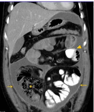
of abdomen (Figure 1) revealed dilated small bowel loops in left lower quadrant (arrow) and multiple air-fluid levels in right iliac fossa (arrow head). Abdominal ultrasonography showed clustering of dilated fluid-filled small bowel loops with intervening fluid between them in mid-abdomen. Contrast-enhanced computed tomography (CECT) (Figure 2) showed a thin soft tissue density membrane encasing small bowel loops in midline giving the appearance of cocoon (arrow), few dilated small bowel loops within, dilated distal ileal loop showing thinned out wall with intramural air and small bowel faeces sign (asterisk), loculated fluid surrounding the loops within the cocoon (arrow head).
After obtaining informed consent, emergency exploratory laparotomy was performed for stoma, which showed a thick shiny whitish membrane encapsulating the small bowel loops (Figure 3). On incising the membrane, gangrenous small bowel loops (Figure 3) and 700 mL of serosangvinous free fluid were noted within the cocoon which was sent for culture. Twenty cm of terminal ileum, five cm proximal to ileocaecal valve was found to be gangrenous. Resection of gangrenous bowel with end ileostomy was performed. Post-operative recovery was uneventful.
Histopathological examination of the excised peritoneal capsule showed proliferation of the fibroconnective tissue with signs of non-specific inflammatory reaction and no signs of tuberculosis/malignancy (Figure 4), suggestive of SEP. Histopathology of excised gangrenous bowel suggested acute ischaemic pathology.
Culture of the peritoneal fluid showed no growth.
DISCUSSION
Abdominal cocoon syndrome, also known as idiopathic SEP, is a rare cause of acute intestinal obstruction in which the small bowel becomes encased (o r co cooned) by a dense fibrocollagenous membrane. While some patients with SEP remain asymptomatic, majority present with recurrent attacks of acute, sub-acute or chronic intestinal obstruction, weight loss, loss of appetite, palpable abdominal mass or ascites. Gastrointestinal perforation is a relatively rare complication of SEP.4 Our patient with primary SEP had
presented with perforation at distal ileum with gangrene of small bowel, in a case of primary SEP. To our knowledge, only two cases of SEP related perforation have been reported.5 One of
them had documented a high jejunal perforation in a case of SEP secondary to tuberculosis 5
and the other with primary SEP with co-existing
Figure 1: Erect plain radiograph of abdomen showing dilated small bowel loops in left lower quadrant (arrow) and multiple air fluid levels in right iliac fossa (arrow heads)
306 incarcerated meckel’s diverticulum in inguinal hernia presented with ileal perforation.5
Imaging plays an important role in preoperative diagnosis and further disease management.
Erect abdominal radiograph, dilated bowel loops and air-fluid levels suggestive of small bowel obstruction. On a barium meal follow-through study classical findings include a serpentine or concertina-like configuration of dilated small bowel loops in a fixed “U shaped” cluster giving a “cauliflower appearance”.6
Delayed transit time has been considered diagnostic of the condition. Characteristic ultra sonographic findings include altered peristalsis, adherence of the bowel to the anterior abdominal wall, intra peritoneal echogenic strands and membrane formation during the late stages of the disease. Classic computed tomography (CT) findings include small bowel loops congregated in a single area or in the midline encased by a soft tissue density envelope. Other CT findings include ascites, loculat ed fluid collect ion thickening, enhancement or calcification of peritoneum, peritoneal deposists, thickening of omentum bowel wall, tethering or fixation of bowel loops and abdominal lymphadenopathy. Presence of these may suggest secondary form of SEP due to causes like TB or malignancy. Prior to the era of cross-sectional imaging, definitive diagnosis was usually made at the time of surgery. Currently, CECT is the investigation of choice as it gives more accurate information on the degree of obstruction, types of bowel loops involved and associated complications.8
Figure 4: Photomicrograph showing areas of sclerosis (arrow head) with foreign body giant cells (arrow) (Haematoxylin and eosin, 400)
Abdominal cocoon: a rare cause of acute intestinal obstruction Silpa Kadiyala et al
Figure 2: Coronal CECT showing thin soft tissue density membrane encasing small bowel loops in midline giving the appearance of cocoon (arrow), few dilated small bowel loops within, dilated distal ileal loop showing thinned out wall with intramural air and small bowel faeces sign (asterisk), loculated fluid surrounding loops within cocoon (arrow head)
307 Internal hernia is a close differential diagnosis which can be differentiated by the absence of membrane like sac encasing the dilated bowel loops.9
Surgical intervention is the treatment of choice. It includes freeing of adhesions, total excision of the membrane, or partial intestinal resection when necessary10 as was done in our case.
In our pat ient , clinical, labo rato ry and radiographic findings were concordant with abdominal cocoon. Because of lack of clinical and radiological findings suggestive of other mimics like TB of peritonitis, peritoneal carcinomatosis, previous abdominal surgery, dialysis. Further, as a negative history regarding there was a long term use of practolol, or chronic disease and non contributory laboratory test results, a diagnosis of idiopathic abdominal cocoon was presumed. Preoperative diagnosis was made on CECT, which showed classic findings and raised the suspicion of complications of gangrene and perforation there by facilitating early emergency surgical intervention in this patient.
Abdominal cocoon presenting as a cause of acute intest inal obstructio n with bo wel gangrene and perforation is rare. Radiologists should be aware of this relatively rare cause of intestinal obstruction, its imaging findings and complications, as preoperative diagnosis will prevent delay and aid in appropriate treatment planning by the surgeon. Identification of soft tissue density membrane encasing congregated small bowel loops into a single area on CECT gives diagnostic clue. Surgical excision of sac, release of bowel loops and adhesions with
partial intestinal resection when necessary can be life saving.
REFERENCES
1. Foo KT, Ng KC, Rauff A, Foong WC, Sinniah R. Unusual small intestinal obstruction in adolescent gir ls: th e abdomin al cocoon . Br J Surg 1978;65:427-30.
2. Rastogi R. Abdominal cocoon secondary to tuberculosis. Gastroenterol 2008;14:139-41. 3. Sh ar ma D, Nair RP, Dan i T, Sh etty P.
Abdominal cocoon-a rare cause of intestinal
obstruction. Int J Surg Case Rep 2013;4:955-7. 4. Akbulut S, Yagmur Y, Babur M. Coexistence of
abdominal cocoon, intestinal perforation and incarcerated Meckel’s diverticulum in an inguinal h er n ia: a troublesome condition. Wor ld J Gasrtrointest Surg 2014;27:51-4.
5. Bani-Hani MG, Al-Nowfal A, Gould S. High jejunal perforation complicating tuberculous abdominal cocoon: a rare presentation in immune-competent male patient. J Gastrointest Surg 2009;13:1373-5.
6. Sieck JO, Cowgill R, Larkworthy W. Peritoneal encapsulation and abdominal cocoon. Case reports and a review of the literature. Gastroenterology 1983;84:1597-1601.
7. Maguire D, Srinivasan P, O’Grady J, Rela M, Heaton ND Scler osin g en capsulatin g
peritonitis after orthotopic liver transplantation.
Am J Surg 2001;182:151-4.
8. Gupta S, Shirahatti RG, Anand J. CT findings of an abdomin al cocoon . An J Roen tgen ol 2004;183:1658-60.
9. Kaur R, Ch auh an D, Dalal U, Kh ur an a U. Abdominal cocoon with small bowel obstruction: two case reports. Abdom Imaging 2012;37:275-8. 10. Serter A, Kocakoç E, Çipe G. Supposed to be rare cause of intestinal obstruction; abdominal cocoon: report of two cases. Clinical Imaging 2013;37:586-9.

