Abstract
Submitted: February 12, 2016
Accepted: September 23, 2016
Role of
Candida
species from HIV
infected children in enamel caries
lesions: an
in vit ro
study
Objectives: This study analyzed the capacity of Candida spp. from dental
in vitro by Transversal Microhardness (TMH), Polarized Light Microscopy (PLM)
and the quantity of calcium ions (Ca2+) released from the enamel. Material and
Methods: Candida
C. albicans
formed by C. albicans and C. t ropicalis C.
albicans and C. parapsilosis C. albicans,
C. parapsilosis and C. glabrat a C. albicans ATCC
(Group 5) and medium without Candida
the TMH and images of enamel were analyzed by PLM. The quantity of Ca2+
released, from Groups 1 and 6, was determined using an Atomic Absorption
Spectrophotometer. The SPSS program was used for statistical analysis and the
hardness (p<0.05) from the 1st to 15th
in porosity. C. albicans caused the release of Ca2+
formation. Conclusion: Candida
cause demineralization of primary enamel in vit ro.
Ke yw or ds: Candida albicans. Dental caries. HIV infections. Child. Hardness. Senda CHARONE1,2
Maristela Barbosa PORTELA3
Karol de Oliveira MARTINS3
Rosangela Maria SOARES4
Gloria Fernanda CASTRO1
Original Article http://dx.doi.org/10.1590/1678-77572016-0021
1Universidade Federal do Rio de Janeiro, Faculdade de Odontologia, Departamento de
Odontopediatria e Ortodontia, Rio de Janeiro, RJ, Brasil.
2Universidade de Brasília, Faculdade de Odontologia, Departamento de Cariologia,
Brasília, DF, Brasil.
3Universidade Federal Fluminense, Faculdade de Odontologia, Departamento de Clínica e
Odontopediatria, Niterói, RJ, Brasil.
4Universidade Federal do Rio de Janeiro, Instituto de Microbiologia Paulo de Góes,
Departamento de Microbiologia, Rio de Janeiro, RJ, Brasil.
Corresponding address: Gloria Fernanda Barbosa de Araújo Castro Rua Araguaia, 994, bl. 2, apt 304 - Freguesia -
Introduction
The most common oral lesion of HIV infected
children is candidiasis25 and the major etiologic
agent of this oral lesion is C. albicans26. However,
various studies have evidenced the presence of other
species with pathogenic features such as C. t ropicalis,
C. par apsilosis, C. glabr at a, C. gu iller m on dii and
C. du blin ien sis22,24. Studies in the literature have
associated the presence of Candida spp. in the oral
cavity with the development of caries lesions7,30.
15 (1994) suggested the possibility
C. albicans have increased
cariogenicity, because this yeast can produce lactic
acid through the fermentation of carbohydrates and degenerate the dental hydroxyapatite structure. Also,
C. albicans can produce enzymes with collagenolytic
activity (aspartic proteases) and dental collagen
hydrolysate17.
Children infected with HIV tend to present a higher
prevalence of caries compared with noninfected
children7,19. The most probable hypotheses that
support this are: the ingestion of a hypercaloric
diet, a high sucrose content in medicines20, low
immunosuppression4 and unsatisfactory oral hygiene23.
Considering these possible roles of Candida in the
development of caries and the fact that HIV infected children present a high prevalence of these fungi
and caries lesions, further studies are needed. Also,
other possible factors involved in the beginning and
development of caries disease in these patients should be investigated.
In this study, we hypothesized that, in vit ro, Candida
(HIV+) children is able to demineralize primary molar enamel. Thus, strains of C. albicans and non- albicans
in vit ro capacity of these Candida
spp. samples to demineralize primary molar enamel was analyzed by Transversal Microhardness (TMH)
and Polarized Light Microscopy (PLM) and furthermore
the quantity of calcium ion (Ca2+) released from the
enamel was determined.
Material and methods
Microorganisms
The Candida spp. samples were isolated from the
23 on the dental surfaces of
according to the CDC criteria6. The HIV+ children were
selected by convenience over an 8-month period.
These children attended the Pediatric AIDS Outpatient
Clinic at the Pediatric Hospital of the Federal University
of Rio de Janeiro (IPPMG-UFRJ) on a regular basis and were assisted by the School of Dentistry of the same
institution. Children who had been under antifungal
therapy in the least three months or used topic oropharyngeal antimicrobial drugs were not included
in the study. Other medical data were obtained from
their medical records.
were rubbed against the easiest accessed dental
surface that allowed relative isolation of saliva. The
material collected was transferred to Eppendorffs
under refrigeration until analysis. For the analysis,
100 mL of these suspensions was seeded onto Petri
plates containing CHROMagar Can d id a® medium
and incubated at 37°C for 72 hours18. Colonies were
analyzed by using biochemical tests of fermentation
and sugar assimilation (API 20C System, Biomerieux,
Marcy L’Étoile, Lyon, France). Also, green colonies
were inoculated in Sabouraud dextrose agar to screen their ability to grow at 45°C in 48 hours and thus
differentiate between C. albicans and C. dubliniensis8.
This study was approved by the Ethics Committee
of the UFRJ and informed consent form was obtained from the caregivers of the children.
Preparation of specimens of human primary
teeth
Forty (40) sound primary molars without any visible alteration (stereoscopic microscope, 40x, Astro Optics
Division, Montpelier, Vermont, USA) were selected.
They were cut by a double sided diamond disc, forming
5 all the
areas were protected by nail varnish (two layers, with
24 hours for drying each layer) except for a circular
area with 6.25 mm2 This
area was exposed to Can dida biofilms, while the
protected area served as its own control since it was
not exposed to the
Candida
spp., we observed that 45 patients presented positive
three presented positive growth of C. albicans and C.
parapsilosis simultaneously; one, positive growth of
both C. albicans and C. t ropicalis; and one, positive growth of C. albicans, C. parapsilosis and C. glabrat a
simultaneously. C. albicans
were randomly selected from five (5) different
patients, who had just presented this species. Also, isolates of C. albicans and non-albicans pertaining to
patients who simultaneously presented isolates of: C.
albicans + C. parapsilosis; C. albicans + C. t ropicalis; and C. albicans + C. parapsilosis + C. glabrat a (one
isolate of each species) were selected. One reference
isolate of C. albicans (ATCC 24433) was used.
1
well plates containing YCB-agar culture medium
(Yeast Carbon Base, Difco, Trenton, New Jersey,
USA), with 1% BSA (Bovine Serum Albumin)8. Ten
divided into six groups: Group 1 – enamel exposed
to C. albican s ;
C. albicans and C.
t ropicalis (n=14; one plate);
formed by C. albicans and C. par apsilosis (n=14;
one plate); C.
albicans, C. parapsilosis and C. glabrat a (n=14; one
plate); Group 5 – C. albicans (ATCC 24433)
(n=14; one plate); Group 6 (control group) – absence
of Candida
suspensions containing 105 yeasts/mL sowed in YCB
medium supplemented with BSA 1% under mixing for 48 hours at 37°C were inoculated in each well, except
incubation at 37°C.
plate after the 1st, 3rd, 5th, 8th, 10th, 12th and 15th
day, and inserted in Falcon tubes containing 5 mL of
cell viability was assessed through the Trypan blue
a soft brush with water and pumice paste and then
future analysis.
Transversal Microhardness (TMH)
The enamel TMH was measured using a Knoop
indenter with a 50 gram load for 15 seconds8,10. The
equatorial region by a precision sectioning cutter
double sided diamond disc, forming two enamel
resin and its enamel surface was polished with mesh sandpaper (1000 grit, 1200 grit and 2400 grit) (3M,
Sumaré, São Paulo, Brazil) for 10 minutes. Then, a
Ltda; São Paulo, São Paulo, Brazil) and abrasive
alumina (1 and 0.3 μm; South Bay Technology Inc.;
San Clemente, California, USA) were used until the
surfaces were plain and smooth. Indentations were made at distances of 12.5, 25, 37.5, 50, 62.5, 75,
87.5, 100, 112.5, 125, 137.5, 150 μm from the
external surface into the dentin-enamel junction
(DEJ)28. All the previously patterned indentations had
as well as in those protected areas.
Polarized Light Microscopy (PLM) analysis
For the qualitative analysis, 14 enamel fragmentsselected. They were examined through a 100x
accessories. The fragments were prepared by hand using increasing grades of water sandpaper (1000 grit,
1200 grit and 2400 grit). After that, they were polished
with felt polishing paste until a 100 μm longitudinal
section was observed. This measure was obtained by using a digital micrometer.
Determination of Ca
2+released
To determine the amount of calcium ion (Ca2+)
at the following operating conditions: lamp current 3
(selected randomly) of Group 1 (C. albicans, n=14) and from the plate of Group 6 (control group, n=14)
was collected from each well on the 1st, 3rd, 5th, 8th, 10th,
12th and 15th day. After centrifugation (3000 xg, for 3
minutes, at 4°C) the supernatant portions were stored
in plastic tubes with screw caps containing 250 μL of
nitric acid 65% (HNO3
was put into 3 mL of 53 mM La (NO3)3 and then 50
readings of the calcium ion release were compared
with a standard curve obtained from readings of the
standard solutions. Hence, the calcium release was
calculated on the 1st, 3rd, 5th, 8th, 10th, 12th and 15th day.
Statistical analysis
In order to determine the TMH, the sum of
the indentation values was made, followed by the arithmetic mean. Data were analyzed by SPSS
Statistics Program 20. Parametric statistical tests were
(p<0.05). The means of TMH, on the different days,
in each group, were compared using the Analysis of
Variance (ANOVA). Comparisons between the TMH
values of the exposed and nonexposed areas were made for Group 1 using the T-Student test. For the
other groups, only a descriptive analysis was made
due to the small sample size. Also, the results of PLM
and Ca2+ releases were descriptively analyzed.
Results
For the in vit ro experiments, from the 45 patients that presented positive growth only for C. albicans in
C albicans isolates
patients that presented positive growth of C. albicans
and other species simultaneously were very few. Thus,
one isolate of C. albicans and one of non-albicans, pertaining to patients who simultaneously presented
these isolates, were selected [Group 2: C. albicans +
C. parapsilosis; Group 3: C. albicans + C. t ropicalis;
Group 4: C. albicans + C. parapsilosis + C. glabrat a (one isolate of each specie)], resulting in 14 dental
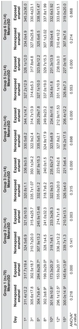
The results of TMH are shown in Table 1. A gradual decrease (p<0.05; ANOVA) of the TMH values in areas
the 1st to 15th day) in all groups exposed to Candida
Group 6 (no Candida
difference in TMH values.
The TMH values of exposed and nonexposed areas
were daily compared only in the group with C. albicans
Group 1 (n=70)
Mean±
SD
Group 2 (n=14)
Mean±
SD
Group 3 (n=14)
Mean±
SD
Group 4 (n=14)
Mean±
SD
Group 5 (n=14)
Mean±
SD
Group 6 (n=14)
Mean±
SD
Day
N
onexposed enamel Exposed enamel
Nonexposed
enamel
Exposed enamel
Nonexposed
enamel
Exposed enamel
Nonexposed
enamel
Exposed enamel
Nonexposed
enamel
Exposed enamel
Nonexposed
enamel
Exposed enamel
1 st 31 1.4±12.2 3 17.7±19.4 3 24.5±6.9 3 21.0±10.8 3 20.8±1.1 336.9±3.6 3 30.5±8.2 344.1±14.7 327.2±12.9 326.1±12.0 3 34.0±4.9 345.2±36.0 3 rd 305.6±17.8 302.5±18.7 310.7±20.0 315.4±25.7 335.7±1.07 350.1±34.2 322.9±2.4 3 14.7±13.9 314.6±5.3 317.8±4.8 3 27.0±25.8 3 30.4±23.1 5 th 304.7±9.4 A 284.8±28.7 A 307.9±13.2 245.9±13.46 3 32.6±1.2 287.9±15.3 335.5±5.7 2 55.29±7.8 3 27.1±10.0 3 23.8±12.2 3 25.6±6.5 338.5±1.47 8 th 302.3±8.9 B 199.3±29.0 B 328.1±4.5 2 41.0±32.3 3 18.7±6.2 240.0±1.5 324.9±17.9 249.9±23.2 337.0±19.0 3 29.7±15.3 3 07.1±3.02 337.8±1.99 10 th 301.9±10.8 C 173.3±20.3 C 318.7±8.1 2 33.3±26.3 3 22.9±3.3 235.82±7.2 323.8±8.9 234.8±1 1.6 334.6±1.8 2 31.3±13.9 3 09.0±4.6 332.6±3.92 12 th 296.1±12.5 D 153.9±20.1 D 336.2±12.2 214.7±1.8 335.0±20.0 2 23.6±2.6 327.8±2.1 2 12.9±1 1 .53 3 31.1±10.9 2 38.0±3.0 310.4±10.2 333.6±13.1 15 th 300.0±14.9 E 145.6±13.6 E 339.7±1.3 2 10.5±13.1 3 26.5±4.0 221.5±6.4 316.2±17.5 206.4±12.2 326.8±7.3 2 27.0±18.1 3 07.9±7.6 319.0±20.0 p value 0.218 0.000 0.141 0.003 0 .315 0.000 0.698 0 .000 0.553 0.000 0 .214 0.868 C. albicans C. albicans and C. tropicalis C. albicans and C.parapsilosis C. albicans , C. parapsilosis and C. glabrata C. albicans T a ble
Mean values of transversal microhardness (TMH) of primary enamel according to the groups and duration (in days) of the experim
Figure 1- Polarized light microscopy (PLM) images of the enamel blocks from Group 1 (C. albicans
C. albicans on 1st, 5th, 8th and 15th day respectively.
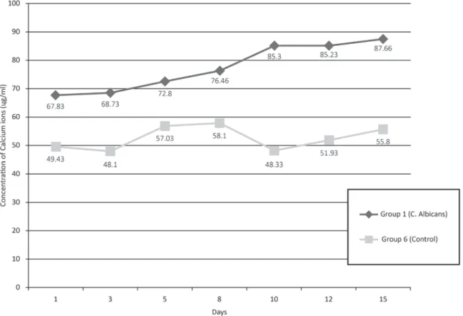
Figure 2- Release of calcium ions (μg/mL) during C. albicans
1st and 3rd day, but from the 5th
difference (p<0.05; Student’s t-test) was observed.
results show a similar behavior in Groups 2, 3 and 4.
In Group 5 the decrease in the TMH values may be
observed as of the 10th day, however, in Group 6, TMH
values were similar throughout the experiment (Table 1). However, statistical test for these comparisons
because of the reduced sample size in each day, in these groups.
Mineral losses observed using PLM showed that
enamel areas exposed to C. albicans
refraction) and lesions in the sub-surface zone with
positive birefringence (double refraction), indicating
an increase in porosity. This porosity increase along
the enamel-dentin junction, increased during the experiment. In Figure 1, the images 2A, 2C, 2E and 2G
show the enamel areas exposed to C. albicans during
the 1st, 5th, 8th and 15th days respectively. The images
2B, 2D, 2F and 2H show their respective controls (protected areas).
The quantity of Ca2+ ions released into the
medium during the Candida albicans
experiment is shown in Figure 2. There was an increase
in the quantity of Ca2+ ions released over the days of
the experiment in Group 1 (C. albicans
Discussion
16 (2003) observed that C. albicans
presents cariogenic potential and has the ability to
dissolve hydroxyapatite crystals in large proportions. According to these authors, this species has
regular and denatured forms by different mechanisms, which could contribute to the persistence of the yeast
on the dissolved hydroxyapatite surface14,21. Our
results show that Candida
not, has a potential to cause enamel demineralization in vit ro, since the microhardness analysis of enamel
Also, we noted that in the C. albicans
from the 5th day on, the TMH values of exposed and
nonexposed areas were different. We hypothesized
that this may have been due to the fact that yeasts
present a more intense metabolism after the 5th day
or this result could be associated with an increase
of lactic acid production along the days, as an
accumulative effect. In addition, a recent study from
C. albicans
can be observed as of the 5th day, under the same
experimental conditions (data not shown).
Secreted aspartyl proteinases (Saps) are among
the most important virulence factors of Candida spp. Their relationship to the development of candidiasis
by adhesion to human tissues, degradation of
extracellular matrix and other important proteins
Recently, Li, et al.13 (2014) demonstrated the role of
Candida albicans-Saps in severe early childhood caries.
The authors observed by enzymatic activity that the
early childhood caries were statically higher producers
of Saps than the C. albicans isolates of caries free
group. Moreover, different members of the Sap family might be differentially expressed depending on the
environment and host conditions. The gene expression
levels of Sap1–5 are the predominant protease genes
expressed in C. albicans
may play an important role in the development of
severe early childhood caries13. Additionally, Brighenti,
et al.3 (2014) also observed caries associated with
other virulence factors (production of acid, extracellular polysaccharides, proteins and metabolic activity) of C.
albicans
C.
albicans
presented a greater caries-associated virulence than
isolates from healthy children.
A limitation of our study was the small sample
by C. albicans and non- albicans, which did not allow
a quantitative analysis between TMH values of the
exposed versus nonexposed areas. Therefore, these results must be discussed with caution. On the other
hand, our descriptive analysis shows that the values of
by C. albicans and non-albicans species presented a C. albicans, but
with a smaller decrease. Some competition between
the Candida species may be occurring in the mixed
C. albicans species in the system29. Future investigations
Candida
On the other hand, although some results were
descriptively analyzed, the comparability of our
results with those in the literature shows some
consistency8,21,30. Although this in vit ro study shows a
the in vit ro demineralization potential of Candida spp.
processes. Besides, there are studies that have already
demonstrated a potential synergism among Candida
albicans and St rept ococcus m ut ans9,12. However, we
did not consider S. m ut ans in our experiments in order
to only compare the demineralization potentials of
Candida albicans and non-albicans, since our objective
these yeasts. Additionally, the importance of S. m ut ans
in the development of caries lesions is
in the current literature. However, novel investigations
regarding the synergistic demineralization potential
Candida spp., isolated from
oral cavity of HIV infected children, and S. m ut ans
are been initiated to better understand the biological
The small variation of microhardness values on
the enamel nonexposed to Can d id a spp. biofilm
(Groups 1 to 5), as well as the whole of Group 6
(control group), shows that the pH variation of the
mineral loss. Moreover, our results showing the loss
of calcium ions by atomic force absorption and the
found in microhardness of the enamel exposed to
C albicans
However, on some days, in the control group (Group
6), the calcium concentration in the suspension reduced instead of increasing gradually. This could be explained
calcium release
was performed in only one isolate per day, randomly selected, and the isolate used may have been one of
those that causes less mineral loss.
Although various studies have already shown the
demineralization ability of Candida species isolated
from other oral cavity sites11,30,the literature is very
scarce in studies about strains from HIV-infected
children, who have high caries prevalence. In parallel,
these yeasts have characteristics that are strain
environment. Arzmi, et al.2 (2015) showed that
different strains of C. albicans
of coaggregation with A. naeslundii and S. m ut ans
These data reinforce the
importance of this study.
Conclusions
Candida spp.
cause in vit ro enamel demineralization.
and elucidating the exact participation of Candida spp. in the development of caries will contribute to caries
disease control in HIV infected patients.
This study was supported by FAPERJ – Rio de
Janeiro Research Foundation , Brazil.
References
1- Amaechi BT, Higham SM, Edgar WM. Efficacy of sterilization methods and their effect on enamel demineralisation. Caries Res.
1998;32:441-6.
2- Arzmi MH, Dashper S, Catmull D, Cirillo N, Reynolds EC, McCullough
M Coaggregation of Candida albicans, Act inom y ces naeslundii and
St r ept ococcus m ut ans is Candida albicans strain dependent. FEMS
Yeast Res. 2015;15:fov038.
3- Brighenti FL, Medeiros AC, Matos BM, Ribeiro ZE, Koga-Ito CY.
Candida
albicans
anemia. J Appl Oral Sci. 2014;22:484-9.
to cariogenic bacteria in HIV-positive children and its correlation
with caries prevalence and levels of cariogenic microorganisms. Oral Microbiol Immunol. 2004;19:281-8.
5- Ccahuana-Vásquez RA, Tabchoury CPM, Tenuta LM, Del Bel Cury AA, Vale GC, Cury JA. Effect of frequency of sucrose exposure on dental
6- Center for Disease Control and Prevention - CDC. Revised
children less than 13 years of age. MMWR. 1994;43:1-10.
7- Cerqueira DF, Portela MB, Pomarico L, Soares RM, Souza IP, Castro
GF. Examining dentinal carious lesions as a predisposing factor for the oral prevalence of Candida ssp in HIV-infected children. J Dent Child
(Chic). 2007;74:98-103.
8- Charone S, Portela MB, Chagas MS, Araújo Soares RM, Araújo Castro
Candida albicans from oral cavity of an HIV-infected child: challenge on enamel microhardness. Oral Surg Oral Med Oral Pathol
Oral Radiol. 2013;115:500-4.
9- Falsetta ML, Klein MI, Colonne PM, Scott-Anne K, Gregoire S, Pai
CH, et al. Symbiotic relationship between St rept ococcus m ut ans and
Candida albicans in vivo. Infect
Immun. 2014;82:1968-81.
M, et al. First-time isolation of Candida dubliniensis from plaque and carious dentine of primary teeth. Eur Arch Paediatr Dent.
2015;16:365-70.
12- Koo H, Bowen WH. Candida albicans and St rept ococcus m ut ans: a
potential synergistic alliance to cause virulent tooth decay in children. Future Microbiol. 2014;9:1295-7
13- Li W, Yu D, Gao S, Lin J, Chen Z, Zhao W. Role of Candida albicans -secreted aspartyl proteinases (Saps) in severe early childhood caries.
Int J Mol Sci. 2014;15:10766-79.
The effect of saliva or serum on St rept ococcus m ut ans and Candida
albicans colonization of hydroxyapatite beads. J Dent. 1998;26:31-7.
and/or acid production of Candida albicans on soft lining materials in
vitro. J Oral Rehabil. 1994;21:585-94.
I n vit ro cariogenic potential
of Candida albicans. Mycoses. 2003;46:471-8.
associated collagen activity by Candida albicans. Mycopathologia. 2002;153:125-8.
18- Odds FC, Bernaerts R. CHROMagar Candida, a new differential
Candida species. J Clin Microbiol. 1994;32:1923-9.
19- Oliveira CA, Tannure PN, Souza IP, Maia LC, Portela MB, Castro GF. Is dental caries experience increased in HIV-infected children and
adolescents? A meta-analysis. Acta Odontol Scand. 2015;12:1-7.
Soares RM, et al. Cariogenic and erosive potential of the medication used by HIV-infected children: pH and sugar concentration. Community
Dent Health. 2008;25:170-2.
21- Portela MB, Chagas MS, Cerqueira DF, Souza IP, Souto-Padrón T,
Araújo Soares RM, et al. Differential collagenolytic activity of Candida
albicans isolated from oral mucosa and dentinal carious lesions of
HIV-infected children. Oral Surg Oral Med Oral Pathol Oral Radiol. 2012;113:378-83.
22- Portela MB, Souza IP, Costa EM, Alviano CS, Soares RM, Santos
AL. Differential recovery of Candida species from subgingival sites in
de Janeiro, Brazil. J Clin Microbiol. 2004;42:5925-7.
activity and gingivitis in HIV+ children. Braz Oral Res. 2002;16:144-50. 24- Richardson MD. Changing patterns and trends in systemic fungal
infections. J Antimicrob Chemother. 2005;56:Suppl 1:i5-i11. 25- Santos LC, Castro GF, Souza IP, Oliveira RH. Oral manifestations
related to immunosuppression degree in HIV-positive children. Braz Dent J. 2001;12:135-8.
26- Starr JR, White TC, Leroux BG, Luis HS, Bernardo M, Leitao J, et al. Persistence of oral Candida albicans carriage in healthy
Portuguese schoolchildren followed for 3 years. Oral Microbiol Immunol. 2002;17:304-10.
27- Szabó B, Majoros L, Papp-Falusi E, Szabó Z, Szabó J, Márton I, et
al. Studies on the possible aetiological role of different Candida species in pathogenesis of dentine caries by monitoring the calcium release
from tooth particles. Acta Microbiol Immunol Hung. 2014;61:11-7.
enamel de- and remineralization, a combined in vit ro pH-cycling model and in sit u study. Clin Oral Invest. 2008;12:173-7.
dual species Candida
Biol. 2007;23:1200-8
phospholipase and haemolytic activity of different species of Candida isolated from dental caries lesions in children. J Clin Diagn Res.