Received from Department of Anesthesia and Pain Management, Mount Sinai Hospital, University of Toronto, Canada.
1. MD; Obstetric Anesthesia Fellow, Department of Anesthesia and Pain Management, Mount Sinai Hospital, University of Toronto
2. MD, PhD; Obstetric Anesthesia Fellow, Department of Anesthesia and Pain Management, Mount Sinai Hospital, University of Toronto
3. MSc; Perinatal Anesthesia Research Coordinator, Department of Anesthesia and Pain Management, Mount Sinai Hospital, University of Toronto
4. MD, PhD, FANZCA, FRCPC; Professor of Anesthesia and Obstetrics and Gynecology, Director of Obstetric Anesthesia, Mount Sinai Hospital, University of Toronto
Submitted on November 30, 2010. Approved on December 7, 2010.
Correspondence to: Jose CA Carvalho, MD, PhD
Department of Anesthesia and Pain Management Mount Sinai Hospital
600 University Avenue, Room 781 Toronto, Ontario, M5G 1X5, Canada E-mail: [email protected] SCIENTIFIC ARTICLE
Non-Invasive Monitoring Based on Bioreactance Reveals
Significant Hemodynamic Instability during Elective Cesarean
Delivery under Spinal Anesthesia
Anne Doherty
1, Yayoi Ohashi
2, Kristi Downey
3, Jose CA Carvalho
4Abstract: Doherty A, Ohashi Y, Downey K, Carvalho JCA – Non-Invasive Monitoring Based on Bioreactance Reveals Significant Hemodynamic Instability during Elective Cesarean Delivery under Spinal Anesthesia.
Background and objectives: Blood pressure monitoring offers a limited understanding of the hemodynamic consequences of spinal anesthesia for cesarean delivery. The purpose of this study was to assess, with the aid of a non-invasive cardiac output monitor based on bioreactance, the hemodynamic changes during elective cesarean delivery under spinal anesthesia in which intermittent boluses of phenylephrine were used to prevent and treat hypotension.
Methods: This observational study was conducted with the Research Ethics Board approval, and all participants provided written informed con-sent. Healthy patients undergoing elective cesarean delivery under spinal anesthesia were enrolled. Intermittent boluses of phenylephrine were administered in an attempt to maintain systolic blood pressure at baseline levels, and patients were assessed with a non-invasive cardiac output monitor based on bioreactance. Hemodynamic data was collected continuously at baseline, and during the postspinal and postdelivery periods. Data was analyzed using a mixed model ANOVA, and a p < 0.05 was considered significant.
Results: Systolic blood pressure was maintained within 79.2 ± 14.2 and 105.8 ± 10.0 percent of baseline during the postspinal period, and 78.4 ± 11.3 and 100.9 ± 10.7 percent of baseline in the postdelivery period (mean ± SD) There were significant fluctuations in systolic blood pressure, heart rate, and cardiac output during the postspinal period, and significant fluctuations in systolic blood pressure and cardiac output in the postdelivery period.
Conclusions: A new non-invasive monitor based on bioreactance reveals significant hemodynamic fluctuations during cesarean delivery under spinal anesthesia, despite attempts to maintain blood pressure at baseline levels with intermittent boluses of phenylephrine.
Keywords: Anesthesia, Obstetrical, Spinal; Cesarean Section; Hemodynamics Monitoring, Bioreactance.
Financial Support: Equipment for the study was provided by Cheetah Medical Inc. The company had no involvement in the study design, data interpretation, or manuscript preparation.
[Rev Bras Anestesiol 2011;61(3): 320-332] ©Elsevier Editora Ltda.
INTRODUCTION
Hypotension during cesarean delivery under spinal anesthe-sia may occur in up to 80% of patients if prophylactic meas-ures are not taken 1,2. The etiology of hypotension during
ce-sarean delivery is multifactorial, but it is primarily determined by a decrease in cardiac output (CO) as a result of decreased
preload, and/or a decrease in systemic vascular resistance (SVR) from a spinal-induced sympathetic blockade. Drugs such as vasoconstrictors, nitroglycerin and oxytocin used intra-operatively may further alter maternal blood pressure 3,4.
Several strategies for the prevention and treatment of hy-potension have been suggested, such as fluid co-loading and the administration of vasoconstrictors 5. Current evidence
supports the maintenance of maternal systolic blood pres-sure (SBP) at baseline throughout the procedure, and also suggests that phenylephrine is the vasoconstrictor of choice for the management of spinal-induced hypotension during cesarean delivery 6,7. Formerly limited to intermittent
assess-ment of blood pressure, the introduction of minimally invasive and non-invasive cardiac output monitoring devices has en-abled a better understanding of the hemodynamic changes occurring during cesarean delivery. Rapid changes in CO and SVR have been demonstrated immediately after intrathecal injection and in the postdelivery period 8,9. It would be ideal to
Bioreactance® technology is a new technique of
non-inva-sive continuous cardiac output monitoring. It is based on the analysis of the relative phase shifts of oscillating currents that occur when the current traverses the thoracic cavity, as op-posed to the traditional bioimpedance-based system, which relies only on measured changes in the signal amplitude 10.
The purpose of this study was to assess, with the aid of a non-invasive cardiac output monitor based on bioreactance, the hemodynamic changes during elective cesarean delivery under spinal anesthesia, in which intermittent boluses of phe-nylephrine were used to prevent and treat hypotension.
METHODS
This observational study was conducted with the Research Ethics Board approval, and was registered with the clinicaltri-als.gov protocol registration system (NCT00949260). All pa-tients presenting for elective cesarean delivery of a normal singleton fetus under spinal anesthesia were considered for the study. All patients recruited provided written informed consent, were ASA I/II and over 18 years of age. Exclusion criteria were: weight < 50 kg or > 100 kg, height < 150 cm or > 180 cm, pre-existing or pregnancy-induced hypertension, cardiovascular or cerebrovascular disease, a history of diabe-tes (excluding gestational diabediabe-tes) or contra-indications for spinal anesthesia.
Upon arrival in the operating room, the patient was placed supine and slightly tilted to the left to avoid aorto-caval com-pression. Hemodynamic information was gathered using a bio-reactance-based non-invasive cardiac output monitoring sys-tem (NICOM®, Cheetah Medical Inc, Portland, Oregon, USA). The baseline systolic blood pressure was measured by averaging three readings taken one minute apart using an auto -mated non-invasive blood pressure monitor. Four NICOM® electrodes were placed, two below the clavicle in the mid-clavicular line, and two at the costal margin in the mid-clavicu-lar line. The monitoring system was then allowed to calibrate. Patients were co-loaded with 10 mL.kg-1 of Ringer’s Lactate,
and spinal anesthesia was performed in the sitting position, at L3-L4, with hyperbaric bupivacaine 0.75% 1.8 mL, 10 µg of fentanyl and 100 µg of morphine. After the intrathecal in-jection, the patient was positioned supine, with left uterine displacement using a wedge under the right hip. Pulse oxim-etry and hemodynamic data were recorded continuously, and included heart rate (HR), stroke volume (SV), cardiac output (CO), total peripheral resistance (TPR), and thoracic fluid content (TFC). Systolic blood pressure (SBP) was assessed every minute. The attending anesthesiologist administered the anesthetic in the usual manner, aiming to preserve ma-ternal SBP at 100% of baseline using vasoconstrictors or anti-cholinergics at their own discretion. The standard pract-ice at our institution is the use of intermittent 100 µg boluses of phenylephrine whenever the SBP is below control values. Ephedrine in boluses of 5 mg is only used if the heart rate is persistently < 50 bpm.
Upon delivery, a segment of the umbilical cord was col-lected for blood gas assessment in both the umbilical artery
and vein. Oxytocin was administered as a bolus of 0.5 IU over 5 seconds upon delivery of the anterior fetal shoulder, fol-lowed by an infusion of 40 mU.min-1, (20 IU oxytocin added to
1.000 mL LR at the rate of 120 mL.h-1). Additional boluses of
oxytocin were used if requested by the attending obstetrician. Any other medications were administered as per the obstetri-cians’ requests.
Hemodynamic monitoring using NICOM® was performed
throughout the procedure. This was commenced prior to the intrathecal injection, and continued until 10 minutes after de-livery of the fetus.
The primary outcome was the hemodynamic changes over time in the postspinal and postdelivery periods.
Additional data collected included maternal age, weight and height, the time of intrathecal injection and delivery, the upper sensory level of anesthesia on delivery, the time and amount administered of medications acting on the cardiovas-cular system, and the umbilical artery and vein blood gases obtained on delivery.
Data analysis consisted of 3 phases: control, postspinal, and postdelivery. The control phase consisted of a three-min-ute period prior to the intrathecal injection with the patient in the supine wedged position; the spinal phase consisted of the ten minutes immediately after intrathecal injection; and the delivery phase consisted of the ten minutes after delivery of the fetus.
Separate analyses were performed for each phase. For all variables, the design was a repeated measure over time (min-utes). A mixed model ANOVA for repeated measures was used to analyze data using the Mixed Procedure in SAS 9.1. Since there was no a priori correlation between the times for the variables, two types of correlation structures were consid-ered. These were compound symmetry (CS) and auto correl-ation of lag 1, AR.1 The former correlation structure assumed
a constant correlation between all time points. The latter as-sumed a correlation that decreased over time, so that obser-vations further apart were assumed to have a smaller correl-ation than the ones closer to each other in time. Since the measurements were only a minute apart, these two structures were seen to give very similar results. The Akaike Information Criterion (AIC) correlation was used to assess the statistical models, and structures that had a smaller AIC were used.
Time was the main effect analyzed, and the linear and quadratic functions of time were modeled. A quadratic effect was first modeled. The quadratic model necessarily included the linear effect. If the quadratic effect was not significant (p > 0.05), it was removed. The linear effect was then tested at the same significance level. If there was also no linear ef-fect, then it was concluded that there was no evidence that the variable changed over time. A convenience sample size of 20 women was used.
RESULTS
monitoring. Of the consented women, one changed her mind on arrival in the operating room, two weighed > 100 kg on the day of surgery and one was moved to another operating room for surgery. Twenty women were included in the analysis.
The women were 35.5 ± 5.1 yr, 79.8 ±13.0 kg, 162.3 ±
6.2 cm, 30.2 ± 4.1kg.m-2, (mean ± SD).
Baseline hemodynamic assessment revealed very diverse individual hemodynamic profiles. Cardiac output ranged from 4.4 L.min-1 to 9.4 L.min-1 , HR from 58 to 107 bpm, SV from 47.5
to 128.2 mL, TPR from 750 to 1646 dyne.s.cm–5, SBP from 102
to 147 mmHg, and TFC from 50.8 to 109.2 L/kOhm-1.
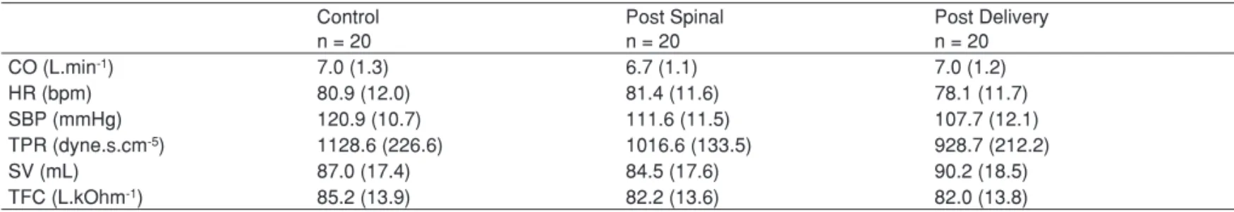
The mean values for each of the hemodynamic variables during the control and each of the study periods are shown in Table I. There were no significant differences in the mean values of hemodynamic variables between the control values and postspinal and postdelivery periods.
The mean values for maximum percentage increase and de-crease for each of the hemodynamic variables assessed, during each study period, are shown in Table II. Systolic blood
pres-sure was maintained within 79.2 ± 14.2 and 105.8 ± 10.0 per-cent of baseline during the postspinal period, and 78.4 ± 11.3 and 100.9 ± 10.7 percent of baseline in the postdelivery period. While the mean values were not significantly different be-tween the control and study periods, the ANOVA analysis revealed significant fluctuations over time in CO, SBP, and HR during the postspinal period (Table III). There were also significant fluctuations in CO and SBP during the postdelivery period. The hemodynamic trends which fluctuated significant-ly over time are shown in Figures 1-5; in contrast, Figure 6 shows the individual HR trend with smoother during the
post-Table I – Mean Values of Hemodynamic Variables in Each Studied Period
Control n = 20
Post Spinal n = 20
Post Delivery n = 20
CO (L.min-1) 7.0 (1.3) 6.7 (1.1) 7.0 (1.2)
HR (bpm) 80.9 (12.0) 81.4 (11.6) 78.1 (11.7)
SBP (mmHg) 120.9 (10.7) 111.6 (11.5) 107.7 (12.1)
TPR (dyne.s.cm-5) 1128.6 (226.6) 1016.6 (133.5) 928.7 (212.2)
SV (mL) 87.0 (17.4) 84.5 (17.6) 90.2 (18.5)
TFC (L.kOhm-1) 85.2 (13.9) 82.2 (13.6) 82.0 (13.8)
Results and mean (SD); CO: cardiac output; HR: heart rate; SBP: systolic blood pressure; TPR: total peripheral resistance; SV: stroke volume: TFC: thoracic fluid content.
Table II - Maximum and Minimum Percent Changes in
Hemodynamic Variables during Postspinal and Postdelivery Period
Post Spinal n = 20
Post Delivery n = 20 CO (L.min-1)
Maximum % 111.0 (11.0) 113.6 (16.5) Minimum % 84.6 (13.0) 87.6 (13.6) HR (bpm)
Maximum % 118.9 (14.7) 103.9 (12.8) Minimum % 76.2 (9.6) 88.6 (11.3) SBP (mmHg)
Maximum % 105.8 (10.0) 100.9 (10.7) Minimum % 79.2 (14.2) 78.4 (11.3) TPR (dyne.s.cm-5)
Maximum % 113.0 (16.2) 100.2 (18.3) Minimum % 77.0 (9.7) 71.2 (11.0) SV (mL)
Maximum % 111.0 (16.1) 115.1 (16.7) Minimum % 84.0 (9.4) 93.4 (10.8) TFC (L.kOhm-1)
Maximum % 99.9 (4.9) 98.5 (4.1) Minimum % 95.2 (3.9) 94.0 (3.8)
Results and mean (SD); CO: cardiac output; HR: heart rate; SBP: systolic blood pressure; TPR: total peripheral resistance; SV: stroke volume: TFC: thoracic fluid content.
Table III - p Values from ANOVA Analysis of Intra-Patient
and Inter-Patient Variability from the Mean with Respect to Time
Variable Control p
Post-Spinal p
Post Delivery p
CO (L.min-1) 0.9846 < 0.0001 * 0.0003 * SBP (mmHg) 0.8075 0.0115 * 0.0095 *
HR (bpm) 0.7062 0.0005 * 0.1154
TPR (dyne.s.cm-5) 0.9393 0.8430 0.1421
SV (mL) 0.6495 0.1941 0.2106
TFC (L.kOhm-1) 0.6916 0.1400 0.9456
CO: cardiac output; HR: heart rate; SBP: systolic blood pressure; TPR: total peripheral resistance; SV: stroke volume: TFC: thoracic fluid content.
Figure 1 - Cardiac Output over Time during the Postspinal Period.
1 2 3 4 5 6 7 8 9 10
1 3
2 4 5 6 7 8 9 10 11 12 12
11
10
9
8
7
6
5
4
3
2
1
Cardiac
Output
(L.min
-1)
delivery period, which did not show significant intra-patient or inter-patient variability on analysis.
The amount of phenylephrine administered during the postspinal and postdelivery periods was 250 ± 131.8 and 205
± 163.8 µg, respectively. The amount of ephedrine adminis-tered during the postspinal period was 1.3 ± 2.2 mg. There was no ephedrine administered during the postdelivery per-iod. Two women received atropine 600 µg secondary to bra-dycardia during the postspinal period. One woman received two boluses of nitroglycerin 100 µg immediately prior to deliv-ery at the request of the attending obstetrician. The block level at the time of delivery, assessed by pin prick, was between T2 and T4 for all patients.
Arterial and venous umbilical blood samples were within normal range. The mean umbilical artery pH was 7.30 ± 0.04,
with a mean arterial CO2 of 55.5 ± 6.26 mmHg, and mean
arterial base excess of -1.56 ± 1.29 mmol.L-1. The mean
umbilical vein pH was 7.33 ± 0.05, with a mean venous CO2
of 49.05 ± 8.46 mmHg, and mean venous base excess of
-1.08 ± 1.32 mmol.L-1 . Figure 2 - Cardiac Output over Time during the Postdelivery Period.
1 2 3 4 5 6 7 8 9 10
1 3 2 4 5 6 7 8 9 10 11 12 12 11 10 9 8 7 6 5 4 3 2 1 Cardiac Output (L.min -1) Time (min)
1 2 3 4 5 6 7 8 9 10
Time (min) 160 160 150 150 140 140 130 130 120 120 110 110 100 100 90 90 80 80 70 70 60 60 50 50 40 40 Systolic Blood Pressure (mmHg )
1 2 3 4 5 6 7 8 9 10
Time (min) 160 160 150 150 140 140 130 130 120 120 110 110 100 100 90 90 80 80 70 70 60 60 50 50 40 40 Systolic Blood Pressure (mmHg ) Hear t R ate (beats/m in)
1 2 3 4 5 6 7 8 9 10
Time (min) 130 130 120 120 110 110 100 100 90 90 80 80 70 70 60 60 50 50 40 40
1 2 3 4 5 6 7 8 9 10
Time (min) 130 130 120 120 110 110 100 100 90 90 80 80 70 70 60 60 50 50 40 40 Heart rate (beats/m in)
Figure 3 - Systolic Blood Pressure over Time during the Postspinal Period.
Figure 4 - Systolic Blood Pressure over Time during the Postdelivery Period.
Figure 5 - Heart Rate over Time during the Postspinal Period.
DISCUSSION
The first important finding of this study is that we were able to demonstrate very diverse hemodynamic profiles in preg-nant women at term through the use of a non-invasive, user-friendly, operator-independent monitor. This may introduce a new era of individualized patient assessment and anesthetic management, without relying on mean values for a cohort which is especially unsuitable in the obstetric population. The baseline cardiac output ranged from 4.4 to 9.4 L.min-1,
making it clear that a mean value for cardiac output is per-haps irrelevant in clinical setting. This widely variable cardiac output in healthy term pregnant women has been previously documented using both Doppler ultrasound and impedance technology 11,12. A high resting heart rate at term was also
seen. The total peripheral resistance showed values that are similar to the non-pregnant state. This has also been noted previously, disproving the usual assumption that low systemic vascular resistance persists throughout pregnancy to term 13.
In fact, our group has recently demonstrated with the same technology that healthy pregnant women at term have a very similar hemodynamic profile when compared to non-pregnant women 14. This should have significant implications for
teach-ing and understandteach-ing clinical practice in the near future. The second important finding of our study is that there are significant fluctuations in cardiac output in postspinal period. The rapid changes in cardiac output in our patients were likely due to fluctuations in heart rate, as stroke volume did not vary significantly and total vascular resistance was maintained. A strong correlation between heart rate and cardiac output has been documented previously 15. The rapid variation in heart rate
was most likely due to the intermittent boluses of phenylephrine given to treat decreases in maternal systolic blood pressure. There was also no significant change in thoracic fluid content, indicating that there was no significant decrease in preload.
Our results differ from those of Langesaeter et al. 9 and Dyer
et al. 15, who have documented a significant increase in
car-diac output in the postspinal period followed by a significant de-crease subsequent to vasoconstrictor administration. The dif-ference between our results and their results may be explained by differences in the vasoconstrictor regimen used, and by the management of hypotension. Particularly, the trigger for vaso-constrictor administration in their studies was a fall in mean ar-terial pressure to 80% of baseline value. Also, Langesaeter et al. 9 used low-dose phenylephrine infusion (0.25 µg.kg-1.min-1),
and Dyer et al. 15 used 80 µg intermittent bolus doses, which
are much lower than the doses used in our study. It is also imp-ortant to note that Langesaeter et al. 9 and Dyer et al. 15 have
used different technologies for hemodynamic monitoring, which may also explain some differences in our findings.
Our findings suggest that we may now be able to review our current practices, and that in general, there is no such a thing as a typical hemodynamic profile during spinal anes-thesia for cesarean delivery. The response of a patient to the anesthetic is unique and dependent on the baseline hemody-namics, the anesthetic technique, and the type and regimen of vasoconstrictor administration. Although one may argue
that comfortable patients and vigorous neonates are good enough outcomes, we may be able to design better plans, and even individualize care; this is especially important in high-risk women and fetuses, situations in which tailored regimens may prove safer and superior.
Current evidence suggests that maintenance of maternal SBP at 100% of baseline results in fewer adverse maternal symptoms and excellent fetal outcomes 7. At our institution
the treatment of any recorded fall in the maternal SBP from baseline is encouraged, and phenylephrine is the first line vaso constrictor used. The ED 95 of intermittent phenylephrine boluses to prevent hypotension is quoted as at least 120 µg
per bolus16, however, in our institution a convenience dose of
100 µg is still routinely used. In this observational study, the attending anesthesiologist was free to manage patient’s SBP as per their usual practice. Despite the goal of maintaining SBP at baseline values the mean SPB for the group during the spinal period was maintained between 78% and 112% of baseline. The early treatment of any fall in maternal SBP using phenylephrine boluses may explain the immediate and rapid fluctuations in cardiac output and heart rate seen in this study. Maternal SBP was maintained at acceptable levels on means assessment, but showed significant fluctuations around the mean. This data suggest that one should either adhere to an all-or-nothing approach to the treatment of any fall in blood pressure, or perhaps consider a phenylephrine infusion regim-en, should a more stable hemodynamic profile be desired. Additionally, if the intermittent bolus dose is the technique of choice, the bolus dose of phenylephrine should be increased, as discussed above.
Another interesting aspect of this study is that it realistically demonstrates the option of stabilizing intraoperative hemody-namics in cesarean deliveries by addressing the exact mech-anisms of decompensation. The fall in cardiac output in our patients was primarily due to the fall in heart rate, due to the reflex bradycardia associated with phenylephrine administra-tion. Thomas et al. studied the effects of phenylephrine and ephedrine on maternal cardiac output using cross-sectional and Doppler echocardiography 17. Bradycardia secondary to
phenylephrine administration was treated with atropine in 11 of 19 patients, and as a result the decreases in cardiac output were comparable to those with ephedrine administration. It is possible that pre-medicating patients with an anti-cholinergic may result in fewer hemodynamic fluctuations. Glycopyrrolate pre-medication could be considered advantageous, as it does not cross the placenta and may avoid adverse fetal effects. Maintaining maternal heart rate may also decrease the dose of phenylephrine required to maintain maternal blood pres-sure. However, this hypothesis requires further study.
Our results also show that the hemodynamic status of our patients was associated with vigorous neonates. Phenyleph-rine is suggested as the vasoconstrictor of choice in obstet-ric anesthesia due to better fetal umbilical gas profiles, as comp ared with ephedrine 6. Ephedrine crosses the placenta,
phenyleph-rine as compared to ephedphenyleph-rine administration, may be due to selective perfusion of the low resistance uteroplacental vas-cular bed.
Finally, our results also showed significant hemodynamic fluctuations in the postdelivery time, but again, unique to our anesthetic management. The hemodynamic effects of oxyto-cin bolus and infusion are well-documented. Oxytooxyto-cin recep-tors are widely present in the cardiovascular system 18. The
activation of oxytocin receptors in the heart releases atrial natriuretic peptide; on the endothelial receptors, it stimulates the release of nitric oxide 17. As a result, vasodilation is
ob-served. Increased cardiac output and heart rate in the post-delivery period usually occur as a response to a fall in vascu-lar resistance due to oxytocin administration. In this study, this response was blunted by the administration of phenylephrine, and a much less-pronounced effect was observed. This effect has previously been noted by Dyer et al. 15.
Hemodynamic assessment during cesarean deliveries in clinical practice is currently by and large limited to blood pressure and heart rate assessment. However, hemodyn-amic changes during cesarean delivery have been assessed through a variety of different methods, including thoracic 15 and
whole body impedance 8, echocardiography and Doppler
ultra-sound 19, all minimally invasive techniques 9, as well as
pulm-onary artery catheterization and thermodilution. All of these methods have limitations, and are unlikely to become routine clinical practice especially as bedside monitoring techniques.
Bioreactance technology is a newer form of non-invasive continuous cardiac output monitoring 10,14, and has recently
been used in the obstetric population for the first time 14. It is
based on an analysis of the relative phase shifts of oscillating currents that occur when the current traverses the thoracic cavity, as opposed to the traditional bioimpedance-based system which relies only on measured changes in signal ampli-tude. Unlike bioimpedance, bioreactance-based non-invasive CO measurement does not use static impedance, and does not depend on the distance between the electrodes for SV and CO calculations, thereby significantly increasing the accuracy of the result. Moreover, its readings were shown to correlate well with results obtained from the pulmonary artery catheter thermodilution-derived measurements of cardiac output 10. In
addition, it has also been shown that the NICOM® system has
acceptable accuracy, precision and responsiveness for CO monitoring in patients experiencing a wide range of circulatory situations 20. Important features of this technology are that it is
completely non-invasive and operator-independent.
REFERÊNCIAS / REFERENCES
01. Clark RB, Thompson DS, Thompson CH – Prevention of spinal hypotension associated with Cesarean section. Anesthesiology, 1976;45:670-674.
02. Macarthur A, Riley ET – Obstetric anesthesia controversies: vaso-pressor choice for postspinal hypotension during cesarean delivery. Int Anesthesiol Clin, 2007;45:115-132.
04. Langesaeter E, Rosseland LA, Stubhaug A – Hemodynamic effects of oxyto-cin during cesarean delivery. Int J Gynaecol Obstet, 2006;95:46-47. 05. Cyna AM, Andrew M, Emmett RS et al. – Techniques for preventing
hypotension during spinal anaesthesia for caesarean section. Co-chrane Database Syst Rev, 2006;(4):CD002251.
06. Ngan Kee WD, Khaw KS, Ng FF – Comparison of phenylephrine infu-sion regimens for maintaining maternal blood pressure during spinal anaesthesia for Caesarean section. Br J Anaesth, 2004;92:469-474. 07. Ngan Kee WD, Khaw KS, Tan PE et al. – Placental transfer and fetal
metabolic effects of phenylephrine and ephedrine during spinal anes-thesia for cesarean delivery. Anesthesiology, 2009;111:506-512.
08. Tihtonen K, Koobi T, Yli-Hankala A et al. – Maternal hemodynamics during cesarean delivery assessed by whole-body impedance cardi-ography. Acta Obstet Gynecol Scand, 2005;84:355-361.
09. Langesaeter E, Rosseland LA, Stubhaug A – Continuous invasive blood pressure and cardiac output monitoring during cesarean deliv-ery: a randomized, double-blind comparison of low-dose versus high-dose spinal anesthesia with intravenous phenylephrine or placebo infusion. Anesthesiology, 2008;109:856-863.
10. Keren H, Burkhoff D, Squara P – Evaluation of a noninvasive con-tinuous cardiac output monitoring system based on thoracic bioreac-tance. Am J Physiol Heart Circ Physiol. 2007;293:H583-H589.
11. van Oppen AC, Stigter RH, Bruinse HW – Cardiac output in normal pregnancy: a critical review. Obstet Gynecol, 1996;87:310-318.
12. Dennis A, Arhanghelschi I, Simmons S et al. – Prospective observa-tional study of serial cardiac output by transthoracic echocardiography in healthy pregnant women undergoing elective caesarean delivery. Int J Obstet Anesth, 2010;19:142-148.
13. Moertl MG, Ulrich D, Pickel KI et al. – Changes in haemodynamic and autonomous nervous system parameters measured non-invasively throughout normal pregnancy. Eur J Obstet Gynecol Reprod Biol. 2009;144(Suppl 1):S179-S183.
14. Ohashi Y, Ibrahim H, Furtado L et al. – Non-invasive hemodynamic assessment of non-pregnant healthy pregnant and preeclamptic women using bioreactance. Rev Bras Anestesiol, 2010;60:603-613. 15. Dyer RA, Reed AR, van Dyk D et al. – Hemodynamic effects of
ephed-rine, phenylephephed-rine, and the coadministration of phenylephrine with oxytocin during spinal anesthesia for elective cesarean delivery. An-esthesiology, 2009;111:753-765.
16. Tanaka M, Balki M, Parkes RK eet al. – ED95 of phenylephrine to pre-vent spinal-induced hypotension and/or nausea at elective cesarean delivery. Int J Obstet Anesth, 2009;18:125-130.
17. Gutkowska J, Jankowski M, Mukaddam-Daher S et al. – Oxytocin is a cardiovascular hormone. Braz J Med Biol Res, 2000;33:625-633.
18. Thibonnier M, Conarty DM, Preston JA et al. – Human vascu-lar endothelial cells express oxytocin receptors. Endocrinology, 1999;140:1301-1309.
19. Thomas DG, Robson SC, Redfern N et al. – Randomized trial of bolus phenylephrine or ephedrine for maintenance of arterial pres-sure during spinal anaesthesia for Caesarean section. Br J Anaesth. 1996;76:61-65.
20. Squara P, Denjean D, Estagnasie P et al. – Noninvasive cardiac output mon-itoring (NICOM): a clinical validation. Intensive Care Med, 2007;33:1191-1194.
Resumen: Doherty A, Ohashi Y, Downey K, Carvalho JCA –
Monito-rización No Invasiva con Base en la Biorreactancia Revela Inestabili-dad Hemodinámica Significativa Durante la Cesárea por ElegibiliInestabili-dad bajo Raquianestesia.
Justificativa y objetivos: La monitorización de la presión arterial
ofrece una comprensión limitada de las consecuencias hemodinámi-cas de la raquianestesia para la cesárea. El objetivo de este estudio fue evaluar, con la ayuda del monitor de débito cardíaco no invasi-vo y con base en la biorreactancia, las alteraciones hemodinámicas durante la cesárea electiva bajo raquianestesia, en la cual bolos in-termitentes de fenilefrina fueron utilizados para prevenir y tratar la hipotensión.
Métodos: Este estudio observacional fue realizado posterior a la
aprobación de la comisión de ética en investigación y de la firma del consentimiento informado. Se evaluaron los pacientes sanos con ce-sárea electiva programada bajo raquianestesia. Bolos intermitentes de fenilefrina fueron administrados para mantener la presión arterial sistólica en los niveles basales, y las pacientes fueron evaluadas con la ayuda del monitor de débito cardíaco no invasivo con base en la biorreactancia. Los datos hemodinámicos se recopilaron con-tinuamente en el momento basal y durante los períodos postraquia-nestesia y después del nacimiento del feto. Los datos se analizaron usando ANOVA para modelos mixtos, y un p < 0,05 fue considerado significativo.
Resultados: La presión arterial sistólica se mantuvo entre 79,2 (14,2)
y 105,9 (10,0) por ciento de los valores basales durante el período postraquianestesia, y 78,4 (11,13) y 100,9 (10,7) por ciento de los va-lores basales en el período postparto promedio ± de. Las fluctuacio-nes significativas se observaron en la presión arterial sistólica, en la frecuencia cardíaca y en el débito cardíaco en el período postparto.
Conclusiones: Un nuevo monitor no invasivo, con base en la
biorre-actancia, reveló fluctuaciones hemodinámicas significativas durante la cesárea bajo la raquianestesia, pese a los intentos de mantener la presión arterial a niveles basales con bolos intermitentes de feni-lefrina.
Descriptores: ANESTESIA, Obstétrica, Regional: raquianestesia;
CIRUGÍA, Cesárea; COMPLICACIONES: Hemodinâmica; TÉCNI-CAS DE MEDICION, débito cardíaco.
Ayuda Financiera: El equipamiento para el estudio fue suministrado