Original article
Evaluation of sucralfate enema in experimental diversion
colitis
*José Aires Pereira
a, Murilo Rocha Rodrigues
a, Daniela Tiemi Sato
a, Paulo Pedroso Silveira
Júnior
a, Alice Moreira Dias
a, Camila Gonçalves da Silva
b, Carlos Augusto Real Martinez
c,* a School of Medicine, Universidade São Francisco (USF), Bragança Paulista, SP, Brazilb Biological Sciences Institute, Universidade Estadual de Campinas (UNICAMP), Campinas, SP, Brazil c Post-graduation Program in Health Sciences, Universidade São Francisco (USF), Bragança Paulista, SP, Brazil
a r t i c l e i n f o
Article history:
Received 20 July 2013 Accepted 23 August 2013
Keywords:
Colon
Experimental colitis Short chain fatty acids Sucralfate
Rats
a b s t r a c t
Diversion colitis (DC) is an inlammatory disease that develops in segments with fecal di-version. Sucralfate (SCF) complex, which consists of sucrose octasulfate and polyaluminum hydroxide, has been demonstrated to be effective in the treatment of different forms of colitis. However, until now, the effects of SCF have not been evaluated in DC.
Objective: to evaluate whether the use of enemas containing SFC improves histological ind-ings in experimental DC.
Methods: Thirty-six rats underwent right colon bypass procedure through the creation of a proximal colostomy and a distal mucous istula. The animals were divided into two groups according to the euthanization procedure to be performed two to four weeks after sur-gery. Each experimental group was divided into three subgroups of six animals, which were submitted to daily application of enemas containing saline solution 0.9% or SCF at con-centrations of 1.0 g/kg/day or 2.0 g/kg/day, respectively. The diagnosis of DC in segments with fecal diversion was established by histopathological study considering the following variables: epithelial loss, formation of crypt abscesses, the population of goblet cells, in-lammatory iniltrate and presence of ibrosis. For statistical analysis, the nonparametric Mann-Whitney and Kruskal-Wallis tests were used, with a signiicance level of 5% (p <0.05).
Results: It was observed that the daily application of SCF enemas decreased epithelial loss, formation of colon crypt abscesses, inlammatory iniltrate and tissue ibrosis (p <0.05), un-related to time of intervention. The intervention with SCF preserves the goblet cell popula-tion. The effects of the substance on the preservation of colonic epithelium; the decrease in the inlammatory process and subsequent abscess formation in the colon crypts are associated with the concentration used, whereas tissue ibrosis decrease is associated with the concentration and time of intervention.
Conclusion: Preventive application of SCF enemas reduces the inlammatory process in the colon with fecal diversion
© 2013 Elsevier Editora Ltda. All rights reserved.
*Study carried out at Post-graduation Program in Health Sciences of Universidade São Francisco (USF), Bragança Paulista, SP, Brazil.
* Corresponding author.
E-mail: [email protected] (C.A.R. Martinez)
Palavras-chave:
Cólon
Colite experimental
Ácidos graxos de cadeia curta Sucralfato
Ratos
r e s u m o
Avaliação dos efeitos da aplicação de enemas com sucralfato em modelo experimental de colite de exclusão
A colite de exclusão (CE) é uma doença inlamatória que se desenvolve em segmentos desprovidos de trânsito fecal. O sucralfato (SCF) complexo formado pelo octossulfato de sacarose e hidróxido de polialumínio vem se demonstrando eicaz para o tratamento de diferentes formas de colite, porém, até a presente data, os efeitos do SCF ainda não foram avaliados na CE.
Objetivo: avaliar se a aplicação de clisteres contendo SFC melhora as alterações histológicas encontradas em modelo experimental de CE.
Métodos: trinta e seis ratos foram submetidos à derivação do trânsito no cólon direito pela confecção de colostomia proximal e fístula mucosa distal. Os animais foram divididos em dois grupos experimentais de acordo com o sacrifício ser realizado após duas ou quatro semanas do procedimento cirúrgico. Cada grupo experimental foi dividido em três subgru-pos de seis animais segundo terem sidos submetidos à aplicação diária com enemas con-tendo solução isiológica a 0,9% ou SCF nas concentrações de 1,0g/kg/dia ou 2,0 g/kg/dia. O diagnóstico de CE nos segmentos sem trânsito foi estabelecido por estudo histopatológico considerando-se as seguintes variáveis: perda epitelial, formação de abscessos nas criptas, população de células caliciformes, iniltrado inlamatório e a presença de ibrose. Para aná-lise estatística adotou-se os testes não paramétricos de Mann-Withney e Kruskal-Wallis estabelecendo-se para ambos, nível de signiicância de 5% (p < 0,05).
Resultados: veriicou-se que a aplicação diária de enemas com SCF diminui a perda
epite-lial, a formação de abscessos nas criptas cólicas, o iniltrado inlamatório e a presença de ibrose tecidual (p < 0,05), não relacionada ao tempo de intervenção. A intervenção com SCF preserva a população de células caliciformes. Os efeitos da substância na preservação do epitélio cólico, na redução do processo inlamatório e consequente formação de abscessos nas criptas cólicas encontram-se relacionado à concentração utilizada, enquanto a redução da ibrose tecidual a concentração e ao tempo de intervenção.
Conclusão: a aplicação preventiva de enemas com SCF reduz o processo inlamatório em
segmentos cólicos desprovidos de transito intestinal.
© 2013 Elsevier Editora Ltda. Todos os direitos reservados.
Introduction
Sucralfate (SCF) is the salt formed by the association of disac-charide sucrose octasulfate and poly-aluminum hydroxide.1
This substance is a cytoprotective complex, which was ini-tially used to prevent or treat several diseases of the upper di-gestive tract, mainly represented by gastroesophageal relux disease (GERD), gastritis, peptic ulcers, stress ulcers and acute gastric mucosal lesions.2 Subsequent studies demonstrated
that SCF had beneicial effects on skin and mucosal wound healing, and being successfully used for the treatment of lesions found in varicose ulcers, aphthous stomatitis, oral and genital ulcers in Behcet’s disease, peristomal dermatitis, burns and post-hemorrhoidectomy or istulectomy surgical wounds.3-9
The therapeutic effects of the topical use of SCF on skin and mucosal lesions are related to the property that the formed complex has to adhere strongly to the raw surface of epithelial ulcers, making it dificult to remove the gelatinous layer that forms on the lesion. The adhesive capacity over the raw surface of the wounds seems to be the main mechanism of action of the drug. However, more recently it was shown that the molecule possesses other properties, such as stimu-lating the formation of mucus and increasing the production of prostaglandins and epithelial growth factor (EGF).10 The
topical use of SCF has also antioxidant activity and is capable of reducing the formation of oxygen-free radicals (reactive oxygen species or ROS) produced by leukocytes present in the inlamed tissue. This antioxidant action prevents the peroxi-dation of cell membrane lipids and, therefore, prevents tissue damage.11
Kochhar et al. were the irst to demonstrate the short-term effectiveness of the application of enemas containing SCF for the control of rectal bleeding due to radiation proctitis.12 The
authors showed that the use of enemas containing a 10% SCF suspension inhibited rectal bleeding in most of the patients with radiation proctitis of moderate or severe intensity. From then on, a sequence of well-conducted studies conirmed the beneicial therapeutic effects of the drug in clinical, endo-scopic and histological improvement in patients with radia-tion proctitis, as well as in other forms of inlammatory bowel disease (IBD) accompanied by the formation of ulcers in the colonic mucosa, such as ulcerative colitis (UC) and solitary rectal ulcer.12-18
Glotzer et al. in 1981, described for the irst time the de-velopment of aphthous ulcers, similar to those found in the UC, in segments of the colonic mucosa with fecal diversion in patients who had no history of IBD.19 The authors called this
sup-ply of intraluminal short-chain fatty acids (SCFA) to epithelial cells of the colonic mucosa, caused by bowel bypass. Recent studies suggest that epithelial injury in DC is caused by the increased production of ROS by the epithelial cells of the co-lonic mucosa itself, along with changes in its metabolism as a result of a deiciency in the supply of its main energy sub-strate represented by the SCFA.20-23
Different therapeutic strategies have been used in the treatment of DC. The restoration of SCFA supply either by reconstitution of the fecal stream or by applying nutritional solutions rich in SCFA is able to ameliorate epithelial lesions. Recent studies have shown that the use of enemas containing substances with antioxidant activity, such as 5-ASA and N-acetylcysteine, as they decrease the levels of oxidative stress in the tissue, they may also improve the histological indings in experimental DC models.21,24 Although DC is accompanied
by the formation of ulcers in the colonic mucosal epithelium , to the best of our knowledge, the effects of SCF have not been evaluated in patients or experimental models of DC. There-fore, the objective of this study is to determine whether the use of SCF enemas is effective in reducing histological altera-tions found in the colonic mucosa of an experimental model of DC.
Method
This study followed the recommendations of the Federal Law N. 11.794 and the guidelines of the Brazilian College of Animal Experimentation (COBEA). The research project was approved by the Ethics Committee on the Use of Animals in Research of Universidade São Francisco.
Experimental animals
A total 36 male Wistar rats, weighing 300 to 350 g, obtained from the Central Animal Facility of Universidade São Fran-cisco were used in this study. The animals were kept in indi-vidual cages in air-conditioned environment with controlled temperature, light, humidity and noise level. On the eve of the surgical intervention, they fasted for 12 hours, except for water which was offered ad libitum. The cages were identiied with the number, experimental group and subgroup to which they belonged and that data was tattooed with India ink on the tail of each animal. The rats were always fed the same chow, suitable for rodents, and weighed weekly.
Surgical technique
The fecal diversion in all animals was performed under gen-eral anesthesia by intramuscular administration of 0.1 mL/100 g of 1:1 (v / v) solution of ketamine (50 mg/mL) and xylazine (20 mg/mL) in the left hind leg. After being anesthetized and ixed to the operating table, the abdominal cavity was opened by making a median incision, 3 cm long. After Peyer’s patch identiication, the distance between the patch and the loca-tion chosen for the left colon secloca-tion was measured with a caliper, 4 cm above the upper edge of the patch.
After ligation of the marginal arcade vessels, the colon was sectioned at the chosen point, externalizing the proximal
seg-ment, as the end colostomy in the left hypochondrium, ixing the colostomy to the skin with interrupted sutures using ab-sorbable 4-0 monoilament thread in the four cardinal points, and between them. After the ixation of the proximal colos-tomy, the caudal segment of the left colon was catheterized and irrigated with 40 mL of 0.9% saline solution (SS) at 37°C until the efluent drained through the rectum showed no fe-cal residues.
After the irrigation, the catheter was removed and the dis-tal colon was exteriorized as colostomy (disdis-tal mucous istula) on the lower left lateral face of the abdominal wall. The distal stoma was ixed with the same technique used in the proxi-mal one. The closure of the abdominal wall was performed in two planes of sutures: peritoneum and aponeurosis with running sutures using 4-0 polyglycolic acid thread and the skin with 4-0 nylon thread.
Experimental groups
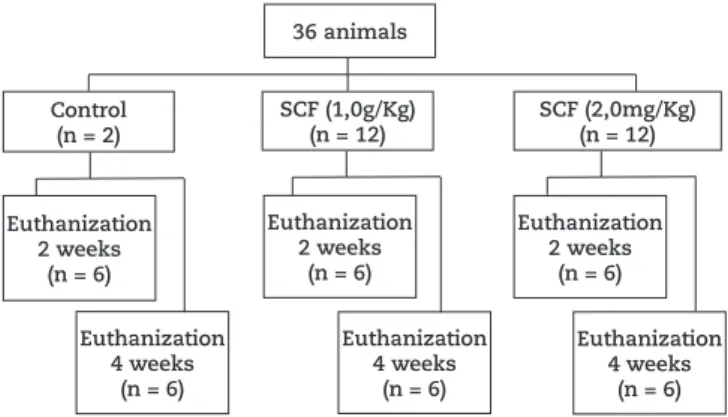
Fig. 1 shows the algorithm for the experimental group formation. The 36 animals were randomly divided into three groups with 12 rats in each. The irst group received daily enemas containing 0.9% saline solution (control group). The second and third groups (experimental groups) received daily enemas containing SCF (EMS do Brasil Ltda., São Paulo, Brazil) at two different concentrations (1.0 g/kg and 2.0 mg/kg, respectively). In each group, six animals were sacriiced two weeks and the other six four weeks after the intervention.
Fig. 1 - Algorithm of the experimental groups formation.
36 animals
Control (n = 2)
SCF (1,0g/Kg) (n = 12)
Euthanization 2 weeks
(n = 6)
Euthanization 2 weeks
(n = 6)
Euthanization 2 weeks
(n = 6)
Euthanization 4 weeks
(n = 6)
Euthanization 4 weeks
(n = 6)
Euthanization 4 weeks
(n = 6) SCF (2,0mg/Kg)
(n = 12)
Sample collection
Histological analysis
The fragments removed for histological analysis were sub-merged in a 10% formaldehyde buffered solution (Sigma, St. Louis, MO, USA) for 24 hours, dehydrated by exposure to increasing concentrations of ethanol and embedded in parafin. From each parafin block, two 5-mm thick histo-logical sections were obtained for slide mounting. Once assembled, they were cleared, hydrated and stained by hematoxylin-eosin (HE) for the diagnosis of colitis and by trichrome Masson (TM) for the assessment of total collagen content (ibrosis).
Slide analysis was performed under a light microscope (Eclipse DS-50, Nikon Inc., Osaka, Japan) by an experienced pathologist in IBD, blinded to the source of the material and the purpose of the study. Histological photographs were taken using a digital videosystem (DS-Fi-50, Nikon Inc., Osaka, Japan) previously attached to the microscope. The specimen analyses were always performed with a 200x final magnification. The analysis of slides in the ir-rigated segments was performed in sites where there were at least three contiguous intact colon crypts.
The following histological parameters were considered for the diagnosis of DC: loss of epithelial surface (epithelial ulcer-ations), colon crypt abscesses, inlammatory iniltrate inten-sity and presence of epithelial ibrosis. The variables epithe-lial loss, presence of colon crypt abscesses and inlammatory iniltrate were stratiied as crosses, according to the degree of each, as follows: a) absent when there were no alterations; b) + when intensity was mild; c) + + moderate d) + + + intense. The intensity of tissue ibrosis was evaluated by the total col-lagen content, quantiied by computerized morphometry and stratiied according to the percentage found per histological ield studied, considering: 0, no ibrosis was identiied; 1 when the content was ≥ 1% and ≤ 5% ; 2 when the content was > 5% and ≤ 10 % and inally 3 when the content was > 10%. For all variables analyzed, the inal value considered for each animal was the mean value after quantiication of three distinct his-tological ields.
The morphometric analysis of collagen content was per-formed using the imaging analysis program NIS-Elements® (Nikon Inc., Japan), release 3.0. The program used RGB (red, green, blue) system color histograms and determined the intensity of the chosen color (in this case blue, the color in which collagen is expressed when the slides are stained with TM) in number of pixels per selected ield, transforming the inal collagen content into percentage per ield (%/ield). The inal value considered for each animal was the mean value obtained after reading three histological ields at the estab-lished magniication (200x).
Statistical analysis
The data was described according to the median with its respective standard error. The comparison between groups was assessed by the median test and analysis of variance with Kruskal-Wallis test. A signiicance level of 5% (p <0.05) was used for the statistical analysis of the results, using the computer program Biostat® release 5.0.
Results
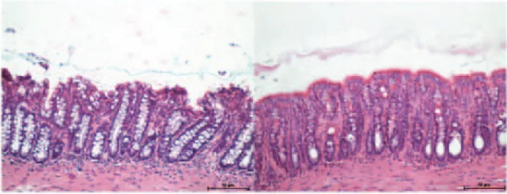
Fig. 2A shows the segment obtained from colon irrigated with 0.9% saline for four weeks, while Fig. 2B shows the colon irrigated with SCF at a concentration of 2.0 g/kg/ day at the same period of time. It can be observed that in animals submitted to intervention with 0.9% SS there is clear epithelial loss, increased goblet cell population, disarray of the architecture and alignment of colic glands. In animals submitted to intervention with SCF 2.0 g/kg/day, the epithelial surface is preserved and the intestinal crypts are aligned, with normal distribution pattern and preservation of the goblet cell population
Fig. 2 – A, Colon segment with fecal diversion submitted to intervention with 0.9% saline solution for four weeks, where we observe the formation of epithelial ulceration, increase in the population of goblet cells and disarray in the architecture of colic glands (HE × 200). B, Colon segment with fecal diversion submitted to therapy with SCF (2.0 g / kg / day) for four weeks, where we can observe the integrity of the colonic mucosal epithelium, maintenance of the goblet cell population and the cytoarchitecture of colic glands (HE 200 ×).
Fig. 3A shows the segment obtained from the colon irrigated with 0.9% saline solution for four weeks, while Fig. 3B shows the colon irrigated with SCF at a concentration of 2.0 g/kg/day. In animals submitted to intervention with 0.9% saline solution, there is mucus accumulation inside the goblet cells, which have replaced part of the absorptive cells of the epithelial surface. One can identify the supericial epithelial ulcers in animals that underwent the intervention with SS. One can verify, through the tissue content of total collagen identiied by TM, that animals submitted to intervention with 0.9% SS have more ibrosis in the mucosal and submucosal layers when compared to animals submitted to intervention with SCF.
tervention in the colon with fecal diversion using a SCF con-centration of 2.0 g/kg/day reduced the formation of abscesses after four weeks of irrigation, when compared to animals ir-rigated with 0.9% SS (p = 0.02) and those irir-rigated with SCF at a concentration of 1.0 g/kg/day (p = 0.02). After four weeks of intervention, the reduction in abscess formation in the intestinal crypts was related to the SCF concentration used. Similarly to what occurred in relation to epithelial loss, the formation of colon crypt abscesses, even though related to the SCF concentration used, did not change with increasing time of intervention regardless of the SCF concentration used.
Fig. 6 shows the scores obtained when analyzing the in-lammatory iniltrate in the mucosal and submucosal layers of the colon irrigated with 0.9% SS, SCF 1.0 g/kg/day and 2.0 g/kg/day for two and four weeks. The results showed that the intervention on the colon with fecal diversion with SCF at a concentration of 2.0 g/kg/day was able to decrease the inlam-matory iniltrate only after four weeks of irrigation, when compared to animals irrigated with 0.9% SS (p = 0.03). There was no worsening in the inlammatory iniltrate in the course of the intervention time, regardless of the SCF concentration used (p = 0.18).
Fig. 7 shows the contents of tissue collagen found in the mucosal and submucosal layers of the colon wall in segments irrigated with 0.9% SS, SCF 1.0 g/kg/day and 2.0 g/kg/day for two and four weeks of intervention. It was observed that ir-rigation of the colon with fecal diversion with SCF at a con-centration of 2.0 g / kg / day for four weeks was able to reduce the total tissue collagen content when compared to animals treated with daily irrigation with SS 0.9% and SCF at a con-centration of 1.0 g/kg (p = 0.009). The presence of ibrosis, al-beit reduced with higher concentrations of the drug, did not change with increasing duration of intervention time, regard-less of the SCF concentration used.
Fig. 3 – A, Colon segment with fecal diversion submitted to therapy with 0.9% saline solution for four weeks, where we observe the intense deposition of collagen in the mucosal and submucosal layers of the colon wall (TM 200 ×). B, Colon segment with fecal diversion submitted to therapy with SCF (2.0 g / kg / day) for four weeks, which shows that there was less collagen deposition when compared to animals irrigated with 0.9% saline solution (TM 200×).
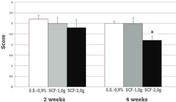
Fig. 4 – Epithelial loss comparing animals submitted to daily irrigation with 0.9% saline solution (SS), sucralfate 1.0 g / kg and 2.0 g / kg after two and four weeks of intervention. Mann-Whitney test. a = p < 0.05 (SCF 2.0 g / kg vs. 0.9% saline solution).
to animals irrigated with 0.9% SS (p = 0.03). The mucosal epithelium of the animals irrigated with SCF was regular and had a smaller population of goblet cells. In animals irrigated with SCF it was observed an increased amount of mucus covering the epithelial surface, when compared to control animals. When assessing the level of epithelial loss with increasing duration of intervention, although it is related to the concentration used, it was observed that it did not worsen with increasing duration of intervention time, regardless of the concentration of SCF used.
Fig. 5 shows the scores obtained by analyzing the presence of crypt abscesses in the segments irrigated with 0.9% SS, SCF 1.0 g/kg/day and 2.0 g/kg/day. The results show that the
in-Fig. 5 – Presence of abscesses in crypts comparing with animals treated with daily irrigation with 0.9% saline solution (SS), 1.0 g SCF / kg and 2.0 g / kg after two and four weeks of intervention. Mann-Whitney Test. a e b = p < 0,05 (a = SCF 2.0 g/kg vs. SS 0.95; b = SCF 2.0 g/kg vs. SCF 1.0 g/kg).
Scor
e
Scor
e
2 weeks
2 weeks 4 weeks
4 weeks
S.S.-0,9% SCF-1,0g SCF-2,0g
S.S.-0,9% SCF-1,0g SCF-2,0g S.S.-0,9% SCF-1,0g SCF-2,0g S.S.-0,9% SCF-1,0g SCF-2,0g
a
Fig. 6 – Inlammatory iniltrate in animals subjected to daily irrigation with 0.9% SS, SCF 1.0 g/kg and 2.0 g/kg after two and four weeks of intervention. Mann-Whitney Test. a = p < 0.05 (a = SCF 2.0 g/kg vs. SS 0.95).
Fig. 7 – Content of tissue collagen in animals submitted to daily irrigation with 0.9% SS, SCF 1.0 g / kg and 2.0 g / kg after two and four weeks of intervention. Mann-Whitney Test. a e b = p < 0.01 (a = SCF 2.0 g/kg vs. SS 0.95;
b = SCF 2.0 g/kg vs. SCF 1.0 g/kg).
Discussion
DC is an inlammatory disease that affects the large intestine segments with fecal diversion.25 Endoscopic examination of
the colon or rectum with fecal diversion shows endoscopic alterations that are characteristic of the disease in all patients after a period of time ranging from three to 36 months the diversion.25 The endoscopic appearance of the intestinal
mu-cosa may vary depending on the intensity and duration of the disease and common indings are: the absence of the vascu-lar pattern of the submucosal layer due to the inlammatory iniltrate, frailty in the excluded mucosa caused by greater vascular congestion, formation of supericial ulcerations that determine spontaneous bleeding or at minimal local trauma and stiffness of the intestinal wall by greater deposition of collagen, leading to ibrosis of the colon wall.26
In more severe cases there may be formation of larger aph-thous ulcers, making it dificult to make the differential di-agnosis with other forms of IBD, particularly URC.27 The
pres-ence of ulcerations on the mucosal surface and the intense inlammatory process are the most signiicant alterations, as they are directly related to the main symptoms of the disease - presence of blood and mucus in stools - reported by most patients. Thus, when establishing therapeutic strategies for the treatment of DC, these aspects should be considered.
The physiopathological bases for the development of DC are not yet fully understood.28 However, most authors believe
that the etiopathogenesis of DC is related to a deiciency in the supply of SCFA to the colonic mucosa.29 This possibility
is supported by the results of studies showing that the dei-ciency in the supply of SCFA to the cells of the colonic mucosa is related to the development of DC, while the restoration of fecal stream or irrigation of the excluded segments with SCFA improves symptoms and reverts endoscopic and histological changes found in the disease.30
Despite the important role played by the maintenance of SCFA supply to prevent the development of DC, the molecu-lar mechanisms that cause epithelial lesions have been un-derstood only recently.20 Experimental studies have shown
that epithelial lesion found in DC models are related to tissue oxidative stress due to an increased production of ROS by the colonic mucosa, a site known to be deicient in antioxidant enzyme systems.31,32 It is well established that ROS such as
superoxide (O2.), hydroxyl (OH), hydrogen peroxide (H2O2) and hypochlorous acid (HClO) are produced in excess by the co-lonic mucosa devoid of fecal stream and are harmful to it.33
ROS are capable of damaging the different defense systems of the colonic mucosa that prevent the migration of antigens and bacteria present in the intestinal lumen to the proximity of the sterile layers of the colon wall.20,22,23,25 The possibility
that deiciencies in the supply of SCFA may trigger the onset of DC gained more support after studies demonstrated that substances that inhibit β-oxidation of SCFA in the intestinal lumen are able to trigger the onset of DC, whereas the use of enemas in the colon without fecal stream with antioxidants, such as 5-ASA and n-acetylcysteine have been successfully used for the treatment of the disease.21,24,34
SCF is a cytoprotective agent that has been used for more than three decades in the treatment of duodenal peptic ul-cers, stress ulcers and GERD. The substance is a sucrose and sulfate-aluminum complex which, when in contact with hy-drochloric acid in the stomach, forms a viscous gel that ad-heres to the gastric mucosa creating a physical barrier that protects the mucosa and prevents the diffusion of hydrochlo-ric acid into the gasthydrochlo-ric wall. The complex formed, in addition to preventing the degradation of the mucus that covers the gastrointestinal epithelium, stimulates the production of bi-carbonate, acting as a buffer with cytoprotective properties. When in contact with the raw surface of epithelial lesions of the digestive tract mucosa, SCF adheres tightly to proteins on the surface of ulcerations, mainly albumin and ibrino-gen, thus forming a stable and insoluble complex, creating a protective layer that covers and protects the ulceration. Re-cent studies have shown that SCF stimulates the production of prostaglandin E2 (PGE2), epithelial growth factor (EGF) and gastric mucus.10,35
It has been previously shown that the PGE-2 is the primary product of arachidonic acid metabolism, playing a critical role in maintaining the integrity of the gastrointestinal
epitheli-Scor
e
Scor
e
2 weeks
2 weeks
4 weeks
4 weeks
S.S.-0,9% SCF-1,0g SCF-2,0g
S.S.-0,9% SCF-1,0g SCF-2,0g
S.S.-0,9% SCF-1,0g SCF-2,0g
S.S.-0,9% SCF-1,0g SCF-2,0g
a
um.36 The increase in PGE-2 production, the main metabolite
of COX-1 and COX-2, can regulate the angiogenesis, motility and survival of epithelial and endothelial cells. A number of studies have shown that SCF increases the production of PGE-2 and prostaglandin-F1 by cells and that this effect is dose-dependent.37
Louw et al. have shown that SCF also signiicantly increas-es the production of TGF-α and that the combination of SCF and TGF-α is able to induce the proliferation of mucosal cells and increase local blood supply, favoring the healing process.38
The inlammatory aggression that develops after tissue damage is often involved in the induction of cell apoptosis, which is considered the main reason for decreased cellularity during the different stages of wound healing. SCF can inhibit cell apoptosis after tissue damage. Matsuu-Matsuyama et al. demonstrated that SCF protects the distal colonic epithelium of rats submitted to radiotherapy, by reducing the level of cell apoptosis by inhibiting the activation of caspase-3 and that this phenomenon may be dependent on p53 protein pathway due to its capacity to decrease, along with p21 protein expres-sion, the Bax/Bcl2 ratio in the colon cells.14
With better knowledge of SCF mechanisms of action, it was observed that in addition to working as a mechanical barrier, it is also able to preserve vascular integrity, increase mucus secretion and EGF production, responsible for stimu-lating epithelial regeneration, angiogenesis and epithelializa-tion, considered the main phase of cutaneous-mucous wound healing. The formation of a stable complex, irmly adhered to ulcers of the digestive tract, maintains EGF production for a long period of time, by exerting a continuous trophic stimu-lation on the gastrointestinal mucosa. This property protects the lining of the digestive tract from new aggressions, as well as stimulates the migration and proliferation of cells from germinal regions of the intestinal crypts.39
Studies have shown that SCF also has a bactericidal effect, effective against Escherichia coli, Pseudomonas aeruginosa and
Staphylococcus aureus. It has been shown that reducing the bacterial population surgical wounds located in the intestinal lumen is an important aspect of the healing process.40
Recently it was shown that the use of high concentrations of SCF decreases the production of ROS by leukocytes or by the xanthine-xanthine oxidase system after cell injury induced by H2O2, showing that the substance has antioxidant activity.10
Fig. 8 shows a summarized version of the known mech-anisms of action of how the SCF molecule acts at different stages of the healing process in surgical wounds.
All these SCF properties combined with the etiopathogenic mechanisms of DC make interesting the assessment of the topical effects of the substance in experimental models of DC. However, as far as we know, this possibility has not yet been studied. The results of the present study conirm the benei-cial effects of the use of enemas with SCF in the prevention of the most frequently found histological alterations in DC. We found that the topical application of SCF in the proposed model reduced epithelial loss, mainly when the substance is applied at a higher concentration and for a longer period of time.
In the animals from the group where intervention was performed with 0.9% SS, the epithelial surface was irregular, with a “brush border” aspect, and in some areas we observed
the formation of small (Fig. 2A) or larger ulcers (Fig. 3A). In all animals treated with SCF, regardless of the concentration used, there was the formation of a gelatinous layer on the colonic mucosa that preserved the epithelial surface, without epithelial ulcerations (Figs. 2B and 3B).
These indings seem to conirm the substance’s cytopro-tective and regenerative properties of the mucosal epitheli-um. Likewise, it was observed that in animals submitted to intervention with SCF, there was a greater amount of mucus covering the epithelial surface and that the goblet cells did not have their cytoplasm illed with mucus as happened in animals submitted to irrigation with 0.9% SS (Fig. 2A, B).
The epithelial surface integrity in animals submitted to intervention with SCF may also be related to the capacity of the substance to increase the local levels of PGE-2, TGF-α, EGF and FGF, all of which, besides exerting a trophic effect on the intestinal epithelium, accelerate the healing process by stimulating cell proliferation and migration.11,36 The
improve-ment in epithelial regeneration in the present study showed to be dose-dependent. One possible explanation for this fact is the greater capacity of mechanical protection when using a higher concentration as well as the increased production of PGE-2, TGF-α, EGF and FGF, which is also dose-dependent.36
The animals submitted to irrigation with higher SCF con-centrations and for a longer period of time had lower rates of abscess formation in crypts and less inlammatory iniltrate (Fig. 4). It is possible that this inding may be related to agrea-ter protection of the epithelial surface provided by the gelati-nous layer that forms on the intestinal mucosa, as well as the increased amount of mucus on the site.
The formation of this additional protective layer can hin-der the migration of antigens and bacteria from the intestinal lumen into the sterile layers of the colon wall, decreasing the local inlammatory response. The lower inlammatory inil-trate, as well as the lower formation of cryptic abscesses may also be related to the bactericidal activities of the substance. It must be also recalled that SCF has an antioxidant activ-ity, neutralizing ROS formed by epithelial cells with energy metabolism altered by a deiciency in the supply of SCFA due to the absence of fecal stream. The neutralization of these ROS can decrease levels of oxidative stress on site, making it dificult for these radicals to attack the epithelial surface cells. At the moment we are measuring in these same ani-mals, the tissue levels of ROS by assessing lipid peroxidation of membranes (tissue levels of malondialdehyde) and of oxida-tive stress to cell DNA (8-OHdG by immunohistochemistry), in order to conirm the importance of the antioxidant activity of SCF in the prevention of oxidative tissue damage found in DC . When analyzing the presence of tissue ibrosis comparing animals preventively irrigated with SCF or 0.9% SS, we found that the use of enemas with SCF at higher concentrations sig-niicantly reduced the content of collagen tissue, suggesting the presence of less local ibrosis. It is possible that this ef-fect is related to a lower local inlammatory process result-ing from mechanical protection given by the substance by reducing bacterial iniltration and inlammation resulting from antioxidant and antibacterial activities. By modulating the production and release of pro-inlammatory cytokines, stimulating the production of PGE-2 and EGF, it is possible that collagen deposition, as well as epithelial replacement, are carried out in a more harmonic way when compared to the animals irrigated with 0.9% SS.
As described by other authors, the protective layer on the epithelium formed by the SCF causes the release of EGF and FGF on site for a longer period of time which improves the healing of epithelial injury.2,11,41 The increased activity of FGF
seems to be a major mechanism of action of SCF.11,42 FGFs are a
class of heparin-binding proteins, mainly represented by basic FGF (bFGF) and acidic FGF (FGF), which stimulate mitogenic, chemotactic and angiogenesis activity in many cell types, including mesenchymal and neural epithelial cells.11 Because
of their myogenic activity on endothelial cells, chondrocytes
and ibroblasts, FGFs play a key role in all stages of wound healing. It is possible that greater stimulation to EGF and FGF production can modulate tissue epithelialization, decreasing collagen deposition as it occurs in animals irrigated with 0.9% SS
The results of this study suggest that the properties of the SCF molecule of improving the mechanical barrier function and accelerating wound healing by stimulating the produc-tion of growth factors, especially FGF, can improve the heal-ing of the colonic mucosa without fecal stream. Moreover, the induction of prostaglandin production, as well as protection against apoptosis, promoting tissue re-epithelialization, can improve the healing process of the colonic mucosa. These biological properties have encouraged the clinical use of the substance as a topical agent for the treatment of different types of colitis caused by inlammation, infection and physi-cal damage, such as in URC and actinic rectitis. According to the clinical evidence reported in the literature, it seems that SCF promotes healing of the intestinal mucosal epithelium in these patients.
The results of this study suggest that for all its properties, topical application of SCF can also be a valid strategy for the prevention and treatment of DC. However, clinical studies in humans, with a signiicant number of cases are still necessary to validate the experimental results found in this study.
Conclusion
Considering the conditions of this study, topical application of enemas containing SCF improves epithelial changes found in experimental DC.
Funding
Fundação de Amparo a Pesquisa do Estado de São Paulo (FAPESP), Process N. 2010/12492-7.
Conlicts of interest
The authors declare no conlicts of interest.
R e f e r e n c e s
1. Volkin DB, Verticelli AM, Maria KE, Burke CJ, Mach H, Middaugh CR. Sucralfate and soluble sucrose octasulfate bind and stabilize acidic ibroblast growth factor. Biochim Biophys Acta.1993;1203:18-26.
2. Szabo S. The mode of action of sucralfate: The 1 x 1 x 1 mechanism of action. Scand J Gastroenterol. 1991;185(Suppl):7-12.
3. Tsakayannis D, Li WW, Razvi S, Spirito N. Sucralfate and chronic venous stasis ulcers. Lance.1994;343:424-5.
4. Altenburg A, Zouboulis CC. Current concepts in the treatment of recurrent aphthous stomatitis. Skin Ther Lett.2008;13:1-4. 5. Alpsoy E, Er H, Durusoy C, Yilmaz E. The use of sucralfate
6. Lyon CC, Stapleton M, Smith AJ, Grifiths CE, Beck MH. Topical sucralfate in the management of peristomal skin disease: An openstudy. Clin Exp Dermatol.2000; 25:584-8.
7. Banati A, Chowdhury SR, Mazumder S. Topical use of sucralfate cream in second and third degree burns. Burns.2001;27:465-9.
8. Gupta PJ, Heda PS, Kalaskar S, Tamaskar VP. Topical sucralfate decreases pain after hemorrhoidectomy and improves healing: A randomized, blinded, controlled study. Dis Colon Rectu. 2008; 51:231-4.
9. Gupta PJ, Heda PS, Shrirao SA, Kalaskar SS. Topical sucralfate treatment of anal istulotomy wounds: a randomized placebo-controlled trial. Dis Colon Rectum. 2011;54(6):699-704.
10. Masuelli L, Tumino G, Turriziani M, Modesti A, Bei R. Topical use of sucralfate in epithelial wound healing: clinical evidence and molecular mechanisms of action. Recent Pat Inlamm Allergy Drug Discov.2010;4:25-36.
11. Wada K, Kamisaki Y, Kitano M, Kishimoto Y, Nakamoto K, Itoh T. Effects of sucralfate on acute gastric mucosal injury and gastric ulcer induced by ischemia-reperfusion in rats. Pharmacology.1997; 54(2):57-63.
12. Kochhar R, Mehta SK, Aggarwal R, Dhar A, Patel F. Sucralfate enema in ulcerative rectosigmoid lesions. Dis Colon Rectum. 1990;33(1): 49-51.
13. Wright JP, Winter TA, Candy S, Marks IS. Sucralfate and methylprednisolone enemas in active ulcerative colitis: a prospective, single-blind study. Dig Dis Sci. 1999;44(9):1899-901.
14. Matsuu-Matsuyama M, Shichijo K, Okaichi K, Ishii K, Wen CY, Fukuda E, Nakayama T, Nakashima M, Okumura Y, Sekine I. Sucralfate protects intestinal epithelial cells from rediation-induced apoptosis in rats. J Radiat Res. 2006;47:1-8.
15. Denton AS, Andreyev HJN, Forbes A, Maher EJ.Systematic review for non-surgical interventions for the management of late radiation proctitis. Br J Cancer. 2002;87:134-43.
16. Henson C. Chronic radiation proctitis: issues surrounding delayed bowel dysfunction post-pelvic radiotherapy and an update on medical treatment. Ther Adv Gastroenterol. 2010; 3(6):359-65.
17. Mendenhall WM, McKibben BT, Hoppe BS, Nichols RC, Henderson RH, Mendenhall NP. Management of radiation proctitis. Am J Clin Oncol. 2013 Mar 11.[Epub ahead of print]. 18. Dehghani SM, Malekpour A, Haghighat M.Solitary rectal
ulcer syndrome in children: a literature review. World J Gastroenterol.2012;18(45):6541-5.
19. Glotzer DJ, Glick ME, Goldman H. Proctitis following diversion of fecal stream. Gastroenterology.1981;80:438-41.
20. Martinez CAR, Ribeiro ML, Gambero A, Miranda DDC, Pereira JA, Nadal SR. The importance of oxygen free radicals in the etiopathogenesis of diversion colitis in rats. Acta Cir Bras. 2010; 25:387-95.
21. Caltabiano C, Máximo FR, Spadari AP, da Conceição Miranda DD, Serra MM, Ribeiro ML, Martinez CA. 5-Aminosalicylic acid (5-asa) can reduce levels of oxidative DNA damage in cells of colonic mucosa with and without fecal stream. Dig Dis Sci.2011;56:1037-46.
22. Martinez CAR, Nonose R, Spadari AP, Máximo FR, Priolli DG, Pereira JA, Margarido NF. Quantiication by computerized morphometry of tissue levels of sulfomucins and sialomucins in diversion colitis in rats. Acta Cir Bras.2010;25:231-40. 23. Nonose R, Spadari APP, Priolli DG, Máximo FR, Pereira JA,
Martinez CAR. Tissue quantiication of neutral and acid mucins in the mucosa of the colon with and without fecal stream: Experimental study in rats. Acta Cir Bras.2009;24:267-75.
24. Martinez CA, Almeida MG, da Silva CMG, Ribeiro ML, Cunha FL, Rodrigues MR, Sato DT, Pereira JA. Enemas with n-acetylcysteine can reduce the level of oxidative damage in cells of the colonic mucosa diverted from the faecal stream. Dig Dis Sci. 2013 Jul 5. [Epub ahead of print]
25. Sousa MV, Priolli DG, Portes AV, Cardinalli IA, Pereira JA, Martinez CA. Evaluation by computerized morphometry of histopathological alterations of the colon wall in segments with and without intestinal transit in rats. Acta Cir Bras.2008;23(5):417-24.
26. Edwards CM, George B, Warren B. Diversion colitis – new light through old windows. Histopathology.1999;34:1-5.
27. Korelitz BI, Cheskin LJ, Sommers SC. The fate of the rectal segment after diversion of the fecal stream in Crohn’s disease: its implication for surgical management. J ClinGastroenterol. 1985;7:37-43.
28. Villanacci V, Talbot IC, Rossi E, et al. Ischaemia: a pathogenetic clue in diversion colitis? Colorectal Dis.2007;9:601-5.
29. Wong JM, de Souza R, Kendall CW, et al. Colonic health: fermentation and short chain fatty acids. J Clin Gastroenterol. 2006;40:235-43.
30. Nassri CGG, Nassri AB, Favero E, et al. Inluência da irrigação de soluções nutricionais no colo excluso de trânsito
intestinal. Estudo experimental em ratos. Rev bras Coloproct. 2008;28:306-14.
31. Liu Q, Shimoyama T, Suzuki K, et al. Effect of sodium butyrate on reactive oxygen species generation by human neutrophils. Scand J Gastroenterol.2001;36:744-50.
32. Longatti TS, Acedo SC, de Oliveira CC,et al.
Inlammatory alterations in excluded colon in rats – a comparison with chemically-induced colitis. Scand J Gastroenterol.2010;45:315-24.
33. Pravda J. Radical induction theory of ulcerative colitis. World J Gastroenterol. 2005;11:2371-84.
34. Keshavarzian A, Morgan G, Sedghi S, Gordon JH, Doria M. Role of reactive oxygen metabolites in experimental colitis. Gut.1990; 31:786-90.
35. Payno A, Lopez-Novoa JM, Rodriguez-Puyol D. Prostanoid production in post-gastrectomy gastritis. Inluence of sucralfate. Am J Med. 1989; 86(6A):17-20.
36. Robert A, Nezamis JE, Lancaster C, Hanchar AJ. Cytoprotection by prostaglandins in rats. Prevention of gastric necrosis produced by alcohol, HCl, NaOH, hypertonic NaCl, and thermal injury. Gastroenterology.1979;77(3): 433-443 37. Slomiany BL, Murty VL, Piotrowski E, Morita M, Piotrowski
J, Slomiany A. Activation of arachidonoyl phospholipase A2 in prostaglandin-mediated action of sucralfate. Gen Pharmacol.1994;25(2): 261-266.
38. Louw JA, Modlin IM, Tang L, Young GO, Lucke W, Marks IN. Changes in mucosal levels of transforming growth factor-alpha from the oxyntic region and ulcer site during duodenal ulcer healing with ranitidine or sucralfate. J Int Med
Res.1998;26(2):82- 86.
39. Nexø E, Poulsen SS. Does epidermal growth factor play a role in the action of sucralfate? Scand J Gastroenterol.1987;127(S uppl):45-49.
40. West AP, Abdul S, Sherratt MJ, Inglis TJ. Antibacterial activity of sucralfate against Escherichia coli, Staphylococcus aureus and Pseudomonas aeruginosa in batch and continuous culture. Eur J Clin Microbiol Infect Dis.1993;12(11):869-871. 41. Braund R, Hook S, Medlicott NJ. The role of topical growth
factors in chronic wounds. Curr Drug Deliv. 2007;4(3):195-204. 42. Yeh BK, Eliseenkova AV, Plotnikov AN, Green D, Pinnell J, Polat