²Study carried out at: Service of Coloproctology of Hospital Heliópolis, São Paulo, SP, Brazil. * Corresponding author.
E-mail: [email protected] / [email protected] (F.B. Formiga) 2237-9363/$ - see front matter. © 2013 Elsevier Editora Ltda. All rights reserved.
http://dx.doi.org/10.1016/j.jcol.2013.09.003
Original article
Predictive factors for temporary defunctioning stoma
permanence in the treatment of rectal adenocarcinoma
²Fernanda Bellotti Formiga
*, Sabrina Miotto, Galdino José Sitônio Formiga, Odilon Victor
Porto Denardin
Service of Coloproctology, Hospital Heliópolis, São Paulo, SP, Brazil
a r t i c l e i n f o
Article history: Received 12 July 2013 Accepted 12 September 2013
Keywords: Rectal neoplasms Surgery
Complications Surgical stomas Surgical anastomosis
a b s t r a c t
Purpose: determine the predictive factors for non-closure of defunctioning stoma in the low anterior resection for the treatment of rectal adenocarcinoma.
Methods: a retrospective cohort study of patients undergoing low anterior resection with defunctioning stoma for a period of nine years was performed. We compared, using uni-variate and multiuni-variate analysis, the group that closed the defunctioning stoma (Group A) with the group that did not (Group B).
Results: eighty-one patients were analyzed; mean age was 61 ± 11 years, with a predomi-nance of women (55.6%). Middle rectal tumors (66.6%), pT3 (59.2%) and pN0 (71.6%) were the most frequent. Stapled anastomosis (65.4%), and loop colostomy (80.2%) were the procedures most frequently performed. Sixty-ive patients (80.2%) underwent stoma clo-sure. The mean time for closure was 8.7 ± 4.4 months. The independent risk factors for non-closure of defunctioning stoma were complications of the anastomosis (p = 0.008) and follow-up complications (p = 0.007).
Conclusion: complications with anastomosis and during the follow-up are factors that may justify a permanent stoma after low anterior resection for treatment of rectal adenocarci-noma.
Palavras-chave: Câncer retal Cirurgia Complicações Estoma cirúrgico Anastomose cirúrgica
r e s u m o
Fatores preditivos da permanência do estoma derivativo temporário no tratamento do adenocarcinoma de reto
Objetivo: determinar quais os fatores preditivos de não fechamento do estoma desfuncio-nalizante na retossigmoidectomia anterior para tratamento do adenocarcinoma de reto. Método: estudo de Coorte retrospectivo dos pacientes submetidos a retossigmoidectomia anterior e estoma derivativo em período de nove anos. Comparou-se o grupo que fechou o estoma derivativo (Grupo A) com o grupo que não fechou (Grupo B) através de análise uni e multivariada.
Resultados: foram estudados 81 pacientes, cuja média de idade foi de 61 ± 11 anos, com pre-domínio de mulheres (55,6%). Tumor de reto médio (66,6%), pT3 (59,2%) e pN0 (71,6%) foram os mais freqüentes. A anastomose mecânica foi a mais realizada (65,4%), assim como trans-versostomia em alça (80,2%). Sessenta e cinco pacientes (80,2%) fecharam o estoma. O tempo médio de fechamento foi 8,7 ± 4,4 meses. Os fatores de risco independentes de não fecha-mento do estoma foram complicações da anastomose (p = 0,008) e do seguifecha-mento (p = 0,007). Conclusão: complicações da anastomose e do seguimento são fatores que podem justiicar a permanência do estoma pós-retossigmoidectomia para tratamento do adenocarcinoma retal.
© 2013 Elsevier Editora Ltda. Todos os direitos reservados.
Introduction
Historically, patients with middle and low rectal tumors were submitted to abdominoperineal resection and consequently to permanent colostomy. Nowadays, with the improvement of the surgical technique by total mesorectal excision, the use of staplers and neoadjuvant chemoradiotherapy, anterior rectal resection with low or ultra-low anastomosis became widely used and well accepted in terms of the oncological radical-ity. This has provided sphincter preservation,1 while adding a
new variable: the derivative or defunctioning stoma.
Low and ultralow anastomosis imply an increased risk of dehiscence.2 Many authors, in the past, indicated a routine
defunctioning stoma for any extra-peritoneal anastomosis: not to prevent dehiscence but to reduce the chance of sys-temic repercussions in case of dehiscence.2-11
However, currently, the selective indication for defunction-ing stoma after low anterior resection predominates.12 The
decision regarding the need for the stoma should consider the following factors: male gender (narrow pelvis),3,5 older
age,6 obesity,2 malnutrition, comorbidities13 or speciically
cardiovascular disease,6 clinical obstruction,5 corticosteroid
use,13 preoperative radiotherapy5,13,14 or transfusion,2
anasto-motic distance less than 5 cm from the anal verge,3 infection5
or pelvic contamination,13 deicient blood supply,2 technical
failure (incomplete rings, positive anastomosis test)1,3 or
dif-iculty (bleeding)1,13 and surgeon’s inexperience.5
The anastomotic dehiscence may have early complica-tions such as localized abscess, intestinal subocclusion or peritonitis, sometimes requiring surgical treatment (reopera-tion) or non-surgical interventions (drainage), which can lead to resolution or sepsis and death. Furthermore, late complica-tions such as anastomotic stenosis, istula, sinus and chronic rectal pain could make a derivative stoma become a perma-nent one.1,2,5
The closing of the stoma should be ideally accomplished in six to 12 weeks.10 However, it is known that this time may
be further extended and, in some cases, the stoma becomes
permanent. Some authors report that the non-closure is re-lated to the patient’s condition (older age, comorbidities) or treatment (chemotherapy, anastomotic dehiscence).13
Given this, it should be considered which patients do not beneit from sphincter-preserving surgery but could be des-ignated for a permanent colostomy, either by Hartmann’s procedure or even abdominoperineal amputation. The impor-tance of determining the factors that lead to a temporary de-functioning stoma to become permanent is, precisely, to tailor the best treatment for each patient and prevent failure.
Thus, the objective of this study is to determine the risk factors for non-closure of defunctioning stoma in low ante-rior resection for treatment of rectal adenocarcinoma, and to determine the predictive factors for closure delay (secondary outcome).
Method
This was a retrospective cohort study carried out through analysis of medical records of patients with the following in-clusion criteria: elective surgery performed by the coloproctol-ogy team of Hospital Heliópolis in São Paulo, between January 2004 and December 2012, submitted to anterior rectal resec-tion with total mesorectal excision and defuncresec-tioning stoma for the treatment of rectal adenocarcinoma, with a minimum follow-up of six months. Poorly documented records regard-ing histological features of the tumor, intraoperative evalua-tions, surgical and follow-up complications and uncertainty regarding the closure, in addition to the patients whose fol-low-up was not carried out by this service were excluded.
presence of metastasis, staging (0-II, III-IV), perineural, lym-phatic and venous invasion. Data related to the treatment was: neoadjuvant chemotherapy and radiotherapy, type of anastomosis (manual and mechanical), stoma type (ileosto-my and transversosto(ileosto-my), radicality of surgery (R0, R1, R2), distal margin (cm), adjuvant chemotherapy and radiotherapy. Data related to treatment complications or follow-up compli-cations included anastomotic and stomal complicompli-cations and those occurring during follow-up.
The rectal tumor was divided into low, middle and upper rectum, considering the measurement in centimeters from the anal verge to the distal end of the tumor through rigid rectosigmoidoscopy. The distal 5 cm were considered as low rectum, from 5 cm to 10 cm, as middle rectum and over 10 cm, upper rectum. The rectosigmoid junction tumor was identi-ied as such when it was found during surgery, completely intraperitoneal at the level of the promontory.
The degree of histological differentiation was divided into well (Grade 1 - G1), moderate (G2) and poorly differentiated (G3), and G0 when the histological differentiation was unde-termined due to absence of lesion in the surgical specimen af-ter neoadjuvant therapy. Histological analysis of the surgical specimen was classiied according to the sixth edition of the American Joint Committee on Cancer (AJCC),15 being pT0 and
stage 0 the absence of tumor in the surgical specimen after neoadjuvant treatment.
The neoadjuvant radiotherapy consisted in 4500 to 5040 cGy irradiation, whereas the neoadjuvant chemotherapy consisted of 5-luorouracil and leucovorin (the irst and last weeks, when combined with radiotheraphy) from six to eight weeks before surgery.
The classiication of surgical radicality regarding tumor resection were R0 = no residual lesion, R1 = microscopic re-sidual lesion and R2 = macroscopic rere-sidual lesion.16
All complications that occurred within 30 days after sur-gery were categorized as postoperative complications. Anas-tomotic complications were considered as late complications (stenosis, sinus, istula, recurrence in the anastomotic line). For this purpose, all patients underwent endoscopic exami-nation of the anastomosis and selected patients were also submitted to radiological evaluation to detect asymptomatic anastomotic complications.
Subsequently, the group that closed the defunctioning sto-ma (Group A) was compared to the group that did not (Group B), using the described variables. The statistical tests used were the chi-square and Fisher’s exact test for qualitative variables and Levene’s test for quantitative variables (age, CEA and margin). Statistical analysis was performed using SPSS (Statistical Package for Social Sciences) software, release 19.0. For variables with p < 0.05 in the univariate analysis, Cox re-gression (multivariate analysis) was performed and indepen-dent predictors for non-closure of the defunctioning stoma in the treatment of rectal carcinoma were identiied.
For the secondary outcome, the same abovementioned pre-dictive factors were analyzed, but patients who did not close the stoma were excluded and the delay in closing was ana-lyzed. Stoma closure delay was deined as interval longer than six months. Patients who closed successfully within six months post-rectosigmoidectomy were compared with those that closed after six months. The statistical analysis
was the same as described for the primary outcome, with 5% statistical signiicance.
Results
Eighty-eight patients underwent low anterior resection with defunctioning stoma for the treatment of rectal adenocarci-noma during nine years at the Coloproctology Service in a tertiary hospital. Seven of them were excluded due to a lack of well-documented information, especially regarding the follow-up and closure veriication.
Eighty-one patients were studied, and the sample charac-teristics are shown in Tables 1 and 2. Sixty-ive of the 81 pa-tients (80.2%) closed the stoma until the end of the study. The mean time for closure was 8.7 months with a standard devia-tion of 4.4 months and a median of eight months.
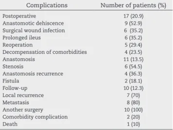
The postoperative, anastomotic and follow-up complica-tions are described in Table 3.
The signiicant variables in the univariate analysis related to the non-closure were: mechanical anastomosis, margin smaller than 2 cm, anastomotic complications and follow-up complications (Tables 4 and 5). At the multivariate analysis, anastomotic and follow-up complications were independent predictors for non-closure (Table 5).
Among the 65 patients who closed the stoma, only 23 of them (35.4%) closed it after six months. The variables that were related to closure delay in the univariate analysis were pT2-3 (p = 0.001), lymph node involvement (pN1-2, p = 0.024) and performance of adjuvant chemotherapy (p = 0.032).
However, none of these factors was signiicant in the mul-tivariate analysis: pT2-3 (p = 0.734), pN1-2 (p = 0.297) and per-formance of adjuvant chemotherapy (p = 0.732).
Discussion
If on the one hand the abdominoperineal amputation of the rectum, created by Miles in 1908, requires a permanent stoma, on the other hand, the rectal anterior resection with low anastomosis and defunctioning stoma allows sphincter preservation. This operation, however, entails a potential risk of permanent stoma4 and morbidity secondary to the stoma,
and especially to its closure.17 It is known that some factors
may affect adversely stoma closure, leading to its delay or even its non-closure.
The rate of non-closure of the defunctioning stoma in our group was 19.8%, similar to that found by other authors who described a rate of 18% to 36%.4,7,13,15,17-20
The mean time of closure was 8.7 months. This number is above the mean value found in international literature, ranging from 4.1 to 5.9 months4,7,13 and coincides with the mean time
found in national studies, of 12 months.20 Some authors report
that 97% of patients closed their stomata within one year after the rectosigmoidectomy,7 a much higher rate than ours of 67.9%.
Some authors have associated older age to non-closure of the stoma,7,11 different from our indings in which age was not
sto-Table 2 – Treatment and complication characteristics of 81 patients submitted to low rectal anterior resection with defunctioning stoma at Hospital Heliópolis between 2004 and 2012. São Paulo, 2013.
Variable Category na (%) / Measure
Neoadjuvant Submitted 23 (28.3)
chemotherapy
Neoadjuvant Submitted 22 (27.1)
radiotherapy
Anastomosis Mechanical 53 (65.4)
Manual 28 (34.5)
Defunctioning stoma Transversostomy 65 (80.2)
(loop) Ileostomy 16 (19.7)
Radicality 0 79 (97.5)
2 2 (2.4)
Distal margin (cm) Mean ± SD 2.3 ± 1.4
Median 2.0
Min – Max 0.2 – 7.0
Adjuvant Submitted 55 (67.9)
chemotherapy
Adjuvant Submitted 28 (34.5)
radiotherapy
Postoperative Present 17 (20.9)
complication
Anastomotic Present 11 (13.5)
complication
Stoma complication Present 38 (46.9)
Follow-up Present 10 (12.3)
complication
a n, number of patients.
Table 1 – Clinical and tumor-related characteristics of the 81 patients submitted to low rectal anterior resection with defunctioning stoma at Hospital Heliópolis between 2004 and 2012. São Paulo, 2013.
Variable Category na (%) / Measure
Gender Male
Female
36 (44.4) 45 (55.6)
Age (years) Mean ± SD
Median Minimum (Min)
-Maximum (Max)
61.05 ± 11.89 62 28 – 84
Comorbidities 43 (53.0)
Smoking status 16 (19.7)
Alcohol consumption 6 (7.4)
Location Lower rectum
Middle rectum Upper rectum Rectosigmoid junction
6 (7.4) 54 (66.6) 18 (22.2) 3 (3.7)
CEA (ng/mL) Mean ± SD
Median Min – Max
17.3 ± 54.2 3.4 0.5 – 393.2 Histological
differentiation b
G0 G1 G2 G3
5 (6.2) 20 (24.7) 50 (61.7) 6 (7.4)
pTc pT0
pT1 pT2 pT3
5 (6.2) 7 (8.6) 21 (25.9) 48 (59.3)
pNc pN0
pN1 pN2
58 (71.6) 16 (19.7) 7 (8.6)
Metastasis 1 (1.2)
Stagingc 0
I II III IV
2 (2.5) 19 (23.5) 32 (39.5) 27 (33.3) 1 (1.2)
Perineural invasion 7 (8.6)
Lymphatic invasion 24 (29.6)
Venous invasion 3 (3.7)
CEA, Carcinoembryonic antigen.
a n, number of patients.
b G0, indeterminate; G1, well-differentiated; G2; moderately
differentiated; G3, little differentiated.
c TNM by AJCC sixth edition.
Table 3 – Complications in 81 patients submitted to low rectal anterior resection with defunctioning stoma at Hospital Heliópolis between 2004 and 2012, São Paulo, 2013.
Complications Number of patients (%)
Postoperative 17 (20.9)
Anastomotic dehiscence 9 (52.9) Surgical wound infection 6 (35.2)
Prolonged ileus 6 (35.2)
Reoperation 5 (29.4)
Decompensation of comorbidities 4 (23.5)
Anastomosis 11 (13.5)
Stenosis 6 (54.5)
Anastomosis recurrence 4 (36.3)
Fistula 2 (18.1)
Follow-up 10 (12.3)
Local recurrence 7 (70)
Metastasis 8 (80)
Another surgery 10 (100)
Comorbidity complication 2 (20)
Death 1 (10)
ma non-closure.11 A possible explanation for those indings
was the increase in the rate of clinical and postoperative com-plications in this group of patients. Their sample, however, did not include only patients with colorectal carcinoma.11 The
present study did not assess obesity, and only 19.7% of pa-tients had a smoking status, thus they were not characterized as predictive factors.
Middle rectal tumors predominated (66.7%) in accordance with the literature data, where the defunctioning stoma is more frequently performed in tumors at that location.7
There are question regarding whether the defunctioning stoma should be avoided in patients with advanced disease, as authors have described that the probability of non-closure in stage IV patients is 30% compared to 3% at stages 0 to III.13
Other authors have described extremely high rates (68.6%) of
non-closure of defunctioning stoma in stage IV.21 In the
pres-ent study, we did not evaluate stage IV alone, as only one pa-tient displayed this stage. Analysis of stages 0 (no residual le-sion after neoadjuvant chemoradiotherapy) to II versus stages III and IV showed no differences between groups A and B, in constrast with a study that demonstrated that stage IV was a predictor of permanent stoma.4
for non-closure. Some authors have demonstrated an associa-tion in the univariate, but not in the multivariate analysis.4,22,23
Our sample shows some association of non-closure with mechanical anastomosis and distal margin smaller than 2 cm in the univariate analysis, but that was not conirmed as an independent factor. There have been no reports of other stud-ies that correlated these variables.
As for the stoma topography, 80.2% were loop transversos-tomy. It is a preference of the service to perform a transver-sostomy rather than an ileostomy. There was no correlation of this variable with the rate of stoma closure. In contrast, other authors have described the colostomy as an indepen-dent variable for permanence of the stoma.4 However, these
authors do not consider the location of the temporary stoma, but the topography of the stoma at the end of treatment.4 In
brief, the recreation of the stoma is very common in patients
who develop pelvic recurrence and this is preferred with the use of colostomy, which generates an interpretation bias of this variable.
Although common sense makes us believe that adjuvant chemotherapy and radiotherapy are factors that inluence closure delay of the defunctioning stoma, these variables were not related to the delay in closure or the permanence of the stoma.
Among the early and late postoperative complications of low anterior resection with defunctioning stoma, the most common are those related to the anastomosis, such as dehis-cence and stenosis.4 These were independent risk factors for
the permanence of the stoma.4,7,8,13,19,22,23 Thus, the
complica-tions of the anastomosis itself are added to complicacomplica-tions 30 days after the surgery. In our sample, anastomotic complica-tions were an independent predictor of failure of closure of
Table 4 – Univariate and multivariate analysis comparing clinical and tumor variables of patients submitted to anterior low rectal anterior resection that closed the defunctioning stoma (Group A) with those who did not close (Group B) at Hospital Heliópolis between 2004 and 2012. São Paulo, 2013.
Variable Category Number of patients (%) Group A Group B
Univariate p Multivariate p Gender Male Female 27 (75) 38 (84.4) 9 (25) 7 (15.6) 0.401 __
Age (years) Mean ± SD
Median Min – Max
62.0 ± 10.5 62.5 48-80
60.8 ± 12.1 62 28-84 0.708 __ Comorbidities Present Absent 32 (74.4) 33 (86.8) 11 (25.6) 5 (13.2) 0.263 __ Smoking Present Absent 11 (68.8) 54 (83.1) 5 (31.3) 11 (16.9) 0.290 __
Alcohol consumption Present Absent 5 (83.3) 60 (80) 1 (16.7) 15 (20) 0.844 __
Location Lower rectum
Middle rectum Upper rectum Rectosigmoid junction 3 (50) 44 (81.5) 15 (83.3) 3 (100) 3 (50) 10 (18.5) 3 (16.7) 0 (0) 0.225 __
CEA (ng/mL) Mean ± SD
Median Min – Max
16.7 ± 56.1 3.7 0.5 – 393.2
19.5 ± 48.5 1.955 0.8 – 184.3
0.863 __ aHistological differentiation G0 G1 G2 G3 3 (60) 19 (95) 38 (76) 5 (83.3) 2 (40) 1 (5) 12 (24) 1 (16.7) 0.200 __
bpT pT0
pT1 pT2 pT3 4 (80) 6 (85.7) 15 (71.4) 40 (83.3) 1 (20) 1 (14.3) 6 (28.6) 8 (16.7) 0.694 __
bpN pN0
pN1 pN2 49 (84.5) 11 (68.8) 5 (71.4) 9 (15.5) 5 (31.3) 2 (28.6) 0.311 __ Metastases Present Absent 1 (100) 64 (80) 0 (0) 16 (20) 0.618 __
b Staging 0-II
III-IV 44 (83) 21 (75) 9 (17) 7 (25) 0.396 __
Perineural invasion Present Absent 4 (57.1) 61 (82.4) 3 (42.9) 13 (17.6) 0.135 __
Lymphatic invasion Present Absent 17 (70.8) 48 (84.2) 7 (29.2) 9 (15.8) 0.222 __
Venous invasion Present Absent 2 (66.7) 63 (80.2) 1 (33.3) 15 (19.8) 0.488 __
CEA, carcinoembryonic antigen.
stoma, but not postoperative complications. Differently from our study, a similar study characterized postoperative compli-cations as a predictive variable.20
We know that complications related to the stoma affect quality of life, especially when they are permanent. Howev-er, there is no correlation between these complications and lower rates of stoma closure. It is noteworthy the fact that if we consider that around 20% of stomata become permanent, these complications should be minimized by good defunc-tioning stoma construction (previous planning, transrectal, adequate size site, good blood supply).7
Regarding the follow-up, the appearance of metastasis and local recurrence can also slow down or prevent closure. Pa-tients with disease progression can have their general status affected and often retain the use of chemotherapeutic agents, thus contributing to the non-closure of the stoma.7
Among all variables analyzed, follow-up complications (local recurrence, synchronous or metachronous tumors, dis-ease progression, surgical indications and clinical complica-tions) are the most obvious predictors of non-closure,4,7,8,13
which corroborated our indings.
Local recurrence is considered the most important factor of stomal non-closure.4,19,22,23 In a critical analysis, one can say
that it is dificult to predict preoperatively if the patient will have a poor evolution, but measures such as adequate stag-ing, radical surgery, effective chemotherapy, radiotherapy if necessary, careful monitoring of patients are the only ways to prevent follow-up complications.
An interesting study reports an overall ive-year survival in patients submitted to low anterior rectal resection without
derivation (81.1%), with temporary stoma (81.5%) and those who progressed to permanent stoma (45.5%).4 This suggests
that it is actually the disease progression that determines non-closure.
Considering the delay in closure, none of the variables was independently related to the closure after more than six months, which suggests that those patients whose stomata become permanent have speciic characteristics, not just be-ing the result of closure delay.
Finally, one should critically consider the limitations of this study: a retrospective design, small sample size, single center and selective decision of defunctioning stoma based on the surgeon’s choice. However, it is undeniable the impor-tance to highlight the indings that point to the need of the construction of a good anastomosis, even with the false safe-ty inherent to the presence of defunctioning stoma, as well as the absence of beneit of this stoma in patients who will potentially progress in the disease.
Conclusion
The rectal adenocarcinoma surgery with defunctioning sto-ma has the potential to become persto-manent. Anastomotic and follow-up complications are predictors of failure.
Conlicts of interest
The authors declare no conlicts of interest.
Table 5 – Univariate and multivariate analyses comparing treatment and complication variables of patients submitted to low rectal anterior resection that closed the defunctioning stoma (Group A) with those who did not close it (Group B) in Hospital Heliopolis between 2004 and 2012, São Paulo, 2013.
Variable Category Number of patients (%) Group A Group B
Univariate p
Multivariate p
Neoadjuvant chemotherapy
Present Absent
15 (65.2) 50 (86.2)
8 (34.8) 8 (13.8)
0.060 __
Neoadjuvant radiotherapy
Present Absent
15 (68.2) 50 (84.7)
7 (31.8) 9 (15.3)
0.120 __
Anastomosis Mechanical
Manual
39 (73.6) 26 (92.9)
14 (26.4) 2 (7.1)
0.044 0.115
Defunctioning stoma
Transversostomy Ileostomy
53 (81.5) 12 (75)
12 (18.5) 4 (25)
0.726 __
Surgical radicality 0 2
0 2
16 (20.3) 0 (0)
0.477 __
Distal margin (cm) Mean ± SD Median Min – Max
2.5 ± 1.4 2 0.6 – 7
1.6 ± 1.3 1.25 0.2 – 5
0.039 0.230
Adjuvant chemotherapy
Present Absent
41 (74.5) 24 (92.3)
41 (74.5) 24 (92.3)
0.077 __
Adjuvant radiotherapy
Present Absent
23 (82.1) 42 (79.2)
5 (17.9) 11 (20.8)
0.755 __
Postoperative complication
Present Absent
11 (64.7) 54 (84.4)
6 (35.3) 10 (15.6)
0.090 __
Anastomosis complication
Present Absent
4 (36.4) 61 (87.1)
7 (63.6) 9 (12.9)
7 (63.6) 9 (12.9)
0.008
Stoma complication Present Absent
30 (78.9) 35 (81.4)
8 (21.1) 8 (18.6)
0.788 __
Follow-up complication
Present Absent
3 (30) 62 (87.3)
7 (70) 9 (12.7)
R e f e r e n c e s
1. Tan WS, Tang CL, Shi L, Eu KW. Meta-analysis of
defunctioning stomas in low anterior resection for rectal cancer. Br J Surg 2009; 96:462-72.
2. Wong NY, Eu KW. A defunctioning ileostomy does not prevent clinical anastomotic leak after a low anterior resection: a prospective, comparative study. Dis Colon Rectum 2005; 48:2076-9.
3. Kumar A, Daga R, Vijayaragavan P, Prakash A, Singh RK, Behari A et al. Anterior resection for rectal carcinoma – risk factors for anastomotic leaks and strictures. World J Gastroenterol 2011; 17(11): 1475-9.
4. Lim SW, Kim HJ, Kim CH, Huh JW, Kim YH, Kim HR. Risk factors for permanent stoma after low anterior resection for rectal cancer. Langenbecks Arch Surg 2013; 398: 259-64. 5. Bax TW, McNevin MS. The value of diverting loop ileostomy
on the high-risk colon and rectal anastomosis. Am J Surg 2007;193(5):585-7.
6. Seo SI, Yu CS, Kim GS, Lee JL, Yoon YS, Kim CW, et al. The role of diverting stoma after an ultra-low anterior resection for rectal cancer. Ann Coloproctol 2013; 29(2): 66-71.
7. den Dulk M, Smit M, Peeters KC, Kranenbarg EM, Rutten HJ, Wiggers T, et al. A multivariate analysis of limiting factors for stoma reversal in patients with rectal cancer entered into the total mesorectal excision (TME) trial: a retrospective study. Lancet Oncol 2007; 8(4):297-303.
8. Bailey CM, Wheeler JM, Birks M, Farouk R. The incidence and causes of permanent stoma after anterior resection. Colorectal Dis 2003; 5(4):331-4.
9. Remzi FH, Fazio VW, Gorgun E, Ooi BS, Hammel J, Preen M, et al. The outcome after restorative proctocolectomy with or without defunctioning ileostomy. Dis Colon Rectum 2006; 49:470-7.
10. Lordan JT, Heywood R, Shirol S, Edwards DP. Following anterior resection for rectal cancer, defunctioning
ileostomy closure may be signiicantly delayed by adjuvant chemotherapy: a retrospective study. Colorectal Dis 2007; 9(5):420-2.
11. Chun LJ, Haigh PI, Tam MS, Abbas MA. Defuncioning loop ileostomy for pelvic anastomoses: predictors of morbidity and non-closure. Dis Colon Rectum 2012; 55: 167-74.
12. Shiomi A, Ito M, Saito N, Hirai T, Ohue M, Kubo Y, et al. The indications for a diverting stoma in low anterior resection for rectal cancer: a prospective multicentre study of 222 patients from Japanese cancer centers. Colorectal Dis 2011;13:1384-9.
13. Gessler B, Haglind E, Angete E. Loop ileostomies in colorectal cancer pacients – morbidity and risk factors for non reversal. J Surg Res 2012; 178: 708-14.
14. Shin US, Kim CW, Yu CS, Kim JC. Delayed anastomotic leakage following sphincter-preserving surgery for rectal cancer. Int J Colorectal Dis 2010;25:843-9.
15. Jessup JM, Gunderson LL, Greene FL, et al. Colon and rectum. In: Greene FL, Page AL, Fleming ID, et al (eds): AJCC Cancer Staging Manual. 6a ed. New York: Springer; 2002. p. 113-24. 16. Edge SB, Byrd DR, Compton CC, Fritz AG, Greene FL, Trotti A.
Colon and rectum. In: AJCC Cancer Staging Manual. 7a ed. New York: Springer; 2010. p.143-64.
17. Mala T, Nesbakken A. Morbidity related to the use of a protective stoma in anterior resection for rectal cancer. Colorectal Dis 2008;10:785-8.
18. David GG, Slavin JP, Willmott S, Corless DJ, Khan AU,
Selvasekar CR. Loop ileostomy following anterior resection: is it really temporary? Colorectal Dis 2010;12:428-32.
19. Junginger T, Gonner U, Trinh TT, Lollert A, Oberholzer K, Berres M. Permanent stoma after low anterior resection for rectal cancer. Dis Colon Rectum 2010;53:1632-9.
20. Formiga FB, Credidio AV, Cruz SHA, Fang CB, Klug WA. Fatores preditivos de não fechamento da estomia desfuncionalizante para anastomose baixa no carcinoma retal [resumo]. Rev bras Coloproct. 2009;29:2-3.
21. Formiga FB. Fatores prognósticos em pacientes portadores de adenocarcinoma colorretal estadio IV. Tese (Mestrado). São Paulo/Brasil: Faculdade de Ciências Médicas da Santa Casa de São Paulo; 2013.
22. Nelson RS, Boland E, Ewing BM, Blatchford GJ, Ternent C, Shashidharan M, et al. Permanent diversion rates after neoadjuvant therapy and coloanal anastomosis for rectal cancer. Am J Surg 2009;198:765-70.