Contents lists available atScienceDirect
Carbohydrate Polymers
journal homepage:www.elsevier.com/locate/carbpol
Review
Cellulose nanocrystals as carriers in medicine and their toxicities: A review
Amedea B. Seabra
a,⁎, Juliana S. Bernardes
b, Wagner J. Fávaro
c,d, Amauri J. Paula
e,
Nelson Durán
a,b,d,f,⁎aCenter of Natural and Human Sciences, Universidade Federal do ABC, Santo André, SP, Brazil
bBrazilian Nanotechnology National Laboratory (LNNano), Brazilian Center for Research in Energy and Materials (CNPEM), 13083-970, Campinas, São Paulo, Brazil cLaboratory of Urogenital Carcinogenesis and Immunotherapy, Department of Structural and Functional Biology, Universidade Estadual de Campinas, Campinas, SP,
Brazil
dNanoBioss, Institute of Chemistry, Universidade Estadual de Campinas, Campinas, SP, Brazil
eSolid-Biological Interface Group (SolBIN), Department of Physics, Universidade Federal do Ceará, Fortaleza, CE, Brazil fInstitute of Chemistry, BiolChemLab., Universidade Estadual de Campinas, Campinas, SP, Brazil
A R T I C L E I N F O
Keywords:
Cellulose nanocrystals Nanomedicine Nanotoxicity
A B S T R A C T
Cellulose nanocrystals (CNCs) are crystalline nanoparticles that present myriad applications. CNCs are produced from a variety of renewable sources, and they can be chemically modified. Although there are promising per-spectives for introducing CNCs into pharmaceutical formulations, prior to achieving commercial products the influence of many parameters such as extraction and toxicity of the resulting products must be revealed. Since there is great physicochemicalflexibility in the steps of obtaining and conjugating CNCs, there are uncountable and complex outcomes from the interactions of those parameters. We present a discussion that helps to unveil the whole panorama on the use of CNCs as drug delivery systems. The methods of producing CNCs are correlated to the resulting nanotoxicity from the cellular to organism level. This review points to relevant concerns that must be overcome to attain safe use of these nanostructures. We also discuss the patents and commercially available products based on CNCs.
1. Introduction
Nanocellulose is a general term for cellulose-based nanostructures, which have been increasingly studied in recent years mainly due to theirflexibility in regard to chemical modifications, physical properties and the possible myriad applications. From a technological point of view, by searching in patent databases with the term“nanocellulose” the versatility and high potential of nanocellulose-based composites in different applications can be revealed (Charreau, Foresti, & Vázquez, 2013;Durán, Lemes, & Seabra, 2012;Trache, Hussin, Haafiz, & Thakur, 2017). These nanostructures were discussed in a previous article (Lin & Dufresne, 2014) that compared three different types of nanocellulose: (i) cellulose nanofibrils (CNFs), (ii) cellulose nanocrystals (CNCs) and (iii) bacterial cellulose (BC) in terms of production, chemical and physical properties and also for their potential applications in medicine. CNCs, or nanowhiskers, represent nanoparticles extracted from lig-nocellulosefibers by hydrolysis with strong acids, while CNFs comprise nanoparticles extracted by the use of strong mechanical (shear) forces (Charreau et al., 2013). In addition, BC represents microbially produced CNFs. The authors emphasized the importance of functional chemical
and physical modification of nanocellulose, since this process will de-termine its toxicity and biomedical properties. On this latter aspect, since CNCs and CNFs are able to bind and release water-soluble mole-cules through ionic interactions, they can be thought of as drug delivery or drug depot systems. Recently, these perspectives were discussed (Plackett, Letchford, Jackson, & Burt, 2014). However, although there is consensus on the recent progress achieved on this topic, including investigation of the biocompatibility and fate of nanocellulosein vivo, Plackett et al. (Plackett et al., 2014) pointed out that most studies were carried out at an academic level, and hence did not focus on preparing specific drugs for practical disease treatment.
Another recent review discussed the chemical structure of CNCs and the available physical and chemical isolation procedures, and also de-scribed optical, mechanical and rheological features of CNCs (George & Sabapathi, 2015). In addition, the review highlighted the novel appli-cations of CNCs in diversefields such as electronics, material sciences, catalysis and biomedical engineering. Furthermore, (Rojas, Bedoya, & Ciro, 2015) presented and discussed recent progress in nanocellulose applications based on the production of face masks, bandages, skin replacements for implants, burns, cuffs for nerve surgery, artificial
https://doi.org/10.1016/j.carbpol.2017.12.014
Received 19 October 2017; Received in revised form 5 December 2017; Accepted 6 December 2017
⁎Corresponding authors at: Center of Natural and Human Sciences, Universidade Federal do ABC, Santo André, SP, Brazil. E-mail addresses:amedea.seabra@ufabc.edu.br(A.B. Seabra),duran@iqm.unicamp.br(N. Durán).
Available online 07 December 2017
0144-8617/ © 2017 Elsevier Ltd. All rights reserved.
blood vessels, cell carriers and support matrices for the immobilization of enzymes. Jorfiand Foster (Jorfi& Foster, 2015) reported advances in the fabrication and design of advanced nanocellulose-based biomater-ials with promising biomedical applications. The authors also discussed the material requirements for each therapeutic application, as well as the challenges that these nanostructures might face in the pharmaco-logical field. More recently, cellulosic nanomaterials were reviewed regarding their production and the influence of the biomass source on the morphological characteristics of the generated nanostructure, as well as the chemical functionalization that can be performed from the hydroxyl radicals on their surface (Mondal, 2017). The nanocellulose physical and chemical properties were correlated with their applica-tions in the contexts of biomedicine, energy, in nanocomposites and especially for environmental issues (Mondal, 2017).
A common concern among researchers is the toxicity of nanocellu-lose. Roman (Roman, 2015) reviewed the literature on the cytotoxicity, oral, pulmonary and dermal toxicity of CNCs. Apparently, no toxicity was demonstrated upon dermal and oral administration of CNCs. However, inconsistent results were reported for pulmonary adminis-tration of CNCs. Moreover, the author highlighted the necessity of further studies focused to evaluate the possible side effects upon in-gestion or skin contact with CNCs. Also, some warnings were raised related to some factors that interfere after processing cellulose (e.g., the presence of endotoxins or toxic chemical impurities). These aspects could be responsible for some adverse health effects through various exposure routes (Roman, 2015). In face of this panorama, a better un-derstanding of the potential human health risk of nanocellulose will come only from the compilation of all possible factors ( Camarero-Espinosa et al., 2016). Firstly, it is important to quantify the toxic dose for human exposure, in particular by skin and inhalation routes. The same applies for environmental applications.
Overall, the aspects that might affect the toxicity of CNCs are (i) size and morphology, (ii) degree of crystallinity, (iii) surface chemistry and (iv) colloidal stability. Another important aspect is that new protocols are necessary for establishing facile and reliable identification of the size and surface chemistry of the nanoparticles, in order to provide a realistic comparison between different studies. This necessity has been previously expressed for other carbon-based nanomaterials, such as oxidized carbon nanotubes (CNTs) and graphene oxide (GO) (Faria et al., 2012; Padovani et al., 2015; Seabra et al., 2014;Seabra, Paula, & Durán, 2013). Rather than having a defined chemical composition, they can have a variety of chemical groups functionalizing the surface, dis-tributed in a random fashion. As a consequence, for these nanos-tructures there is random distribution of surface charges as well as complex stereochemical behavior. Similar behavior occurs for functio-nalized nanocellulose-based nanostructures. Furthermore, CNFs and CNCs intrinsically present a polydisperse size distribution, which also makes it difficult to compare different toxicological studies.
Recently, some authors raised important concerns that justify the study of biointeractions and possible impacts of nanocellulose exposure to humans. This would provide consistent and useful knowledge that can guide the outgrowth of regulatory (Camarero-Espinosa et al., 2016; Endes and Camarero-Espinosa, 2016). The acute and/or chronic toxi-city of nanocellulose during occupational exposure at normal condi-tions or exacerbation of pre-existing disease condicondi-tions must be studied. From the currently available discussions on nanocellulose-based materials in the literature reviews, one can only grasp a general pa-norama involving the wide spectrum of applications as well as the many physical and chemical processing methods to attain products. However, since nanocellulose includes a class of nanostructures with different morphological, physicochemical and biological properties, more de-tailed discussions must be presented for members of this class. From this perspective, CNCs are promising in the biomedical context, espe-cially for drug delivery/depot systems. There are many modification/ functionalization methods for CNCs, which impact their application as drug delivery/depot systems and their toxicological behavior. On this
latter, many concerns were raised on several occasions when these nanocrystals were tested. Consequently, the use of CNCs as drug de-livery/depot systems and the safety aspects involved should be dis-cussed in detail. We have, therefore, compiled a large amount of data on this topic in order to present a suitable discussion on the most re-levant scientific and technological results involving the use of CNCs in biomedical applications. Essentially, we discuss the recent progress in the design of CNCs as drug carrier systems, their nanotoxicity, patents and commercially available CNC products. They are obtained from re-newable sources and CNCs can be extensively modified with the well-established protocols from organic chemistry. This leads to materials with specific mechanical, chemical and biological properties. CNCs can play an important role in future pharmaceutical formulations and medical procedures. However, prior to achieving commercial products based on CNCs, the production–processing–safety relationship re-garding CNCs must be well understood. This review largely contributes towards it.
2. CNCs as drug carriers in aqueous suspensions
This section introduces and summarizes important CNC-based ma-terials that have been explored to date. CNCs have largely been studied when chemically conjugated to other molecules or particles, by cova-lent or non-covacova-lent bonds, in an attempt to provide several function-alities for the system. CNCs were bound to significant amounts of tet-racycline and doxorubicin (DOX), water-soluble and ionizable drugs, which were released during a period of 1 day (Jackson et al., 2011). In order to increase the hydrophobicity of CNCs, cetyl trimethylammo-nium bromide (CTAB) was attached to the CNC surface, which in-creased zeta potential values in a concentration-dependent manner (e.g., from−55 to 0 mV). These functionalized CNC crystallites bound significant quantities of the hydrophobic anticancer drugs docetaxel, paclitaxel and etoposide, which were liberated in a sustained manner in a period of 2 days. In addition, the authors observed uptake of CNC–CTAB complexes by KU-7 bladder cancer cells (Jackson et al., 2011). The surface of torispherical CNCs was also modified by CTAB in order to improve the loading capability of the water-insoluble antic-ancer drugs luteolin and luteoloside (Qing et al., 2016). Although sur-factants, such as CTAB, have been used in conjugation with nanoma-terials, including CNCs, in order to increase the nanoparticle loading with hydrophobic drugs (such as anticancer agents), caution must be taken with this approach (Alkilany & Murphy, 2010). Due to its che-mical property, CTAB might interact with the phospholipid bilayers of the cells, leading to a destabilization of the cell membrane. This de-stabilization might result in cell death. Depending on its concentration, CTAB might create holes in the phospholipid bilayer of the cells (Ulitzur, 1970).
An interesting conjugation was performed from surface-modified CNCs with cationic porphyrin (Por) (Feese, Sadeghifar, Gracz, Argyropoulos, & Ghiladi, 2011). The resulting commonly insoluble crystalline material CNC-Por, although only suspended in an aqueous system, showed high efficacy towards the photodynamic inactivation of Staphylococcus aureusandMycobacterium smegmatis, with only discrete activity againstEscherichia coli. This work describes the synthesis of novel, bioactive and photobactericidal materials that are toxic against several bacteria, with possible utilization in the food and health care industries (Feese et al., 2011). A similar CNC-Por structure showed high efficacy towards the photodynamic inactivation of several micro-organisms (e.g.,Acinetobacter baumannii, methicillin-resistant Staphy-lococcus aureus(MRSA) and multidrug-resistantAcinetobacter baumannii (MDRAB) (Carpenter, Feese, Sadeghifar, Argyropoulos, & Ghiladi, 2012).
Dobberpuhl, Maher, Jaggi, & Chauhan, 2012). Moreover, the anticancer potential of optimized cellulose-CUR formulation was revealed in cell culture models of human prostate cancer cell lines (C4-2, LNCaP and PC-3 cells) by using several techniques, such as cell proliferation, colony formation and apoptosis by 7-amino actinomycin (7-AAD) staining assays. The cellulose-CUR formulation showed increased an-ticancer efficacy in comparison to free CUR. The results demonstrated the availability of the cellulose-CUR formulation and its suitability for application in the treatment of prostate cancer (Yallapu et al., 2012).
A similar approach described the preparation of a nanometric car-rier employing CNCs for aminated biologically active molecules and drugs (Dash & Ragauskas, 2012). CNCs grafted with gamma aminobu-tyric acid (a spacer molecule) were obtained through periodate oxida-tion and a Schiffbase condensation reaction. To control fast delivery of the targeting moiety, a releasable linker such as syringyl alcohol was attached to CNC. The surface morphology of CNCs and their derivatives was studied by transmission electron microscopy (TEM). TEM images showed that the CNCs maintained their characteristic morphology (rod-like) during all steps of the synthetic process. The sizes of CNCs and their derivatives were found to be around 4–8 nm in diameter and 150–300 nm in length (Dash & Ragauskas, 2012).
A three-step covalent binding procedure was performed to convert CNCs intofluorescent labeling nanoparticles conjugated to pyrene (Py-CNC) (Zhang, Li, Zhou, & Zhang, 2012). This nanomaterial showed high selectivity towards Fe3+, as demonstrated by spectroscopic analysis that proved the coordination interaction between Py-CNC and Fe3+as a recognition process. This sensing nanomaterial might be employed as a chemosensor for Fe3+ for many applications in biological, environ-mental and chemical systems (Zhang et al., 2012). In addition, for using CNCs in bioimaging andfluorescence bioassay, the widely used fluor-ophorefluorescein isothiocyanate (FITC) was covalently attached to the surface of CNCs (Dong & Roman, 2007). To this end, initially, the surface of CNCs was decorated with epoxy groups, followed by the introduction of primary amino groups, which reacted with the iso-thiocyanate moiety of FITC, yielding a thiourea. Minimal uptake of untargeted CNCs along with a lack of toxicity was an importantfinding to support the potential of CNCs as carriers in targeted drug delivery applications (Roman, Dong, Hirani, & Lee, 2009).
The preparation of water-soluble photosensitizer–CNCs (PS-CNCs) comprised of CNCs with polyaminated chlorin p6 was reported (Fig. 1) (Drogat et al., 2012). Purpurin-18 (Pp-18) was prepared fromSpirulina
maxima chlorophyll, followed by reaction with different
poly-ethyleneimines (PEIs), yielding a color change from purple to green (Fig. 1A). Hydrolysis of cellulose with sulfuric acid was performed for breaking cellulose chains leading to nanocrystals (Fig. 1B). In a further step, sodium periodate-oxidized glucose units were covalently bound to the nanocrystal surface. Carbonyl moieties reacted with the amine groups of PEI. The cancer cell-targeting potential of PS-CNCs was tested for their antitumor activity against a human immortalized keratinocyte (HaCat) cell line. The half maximal inhibitory concentration (IC50) values were in the nanomolar range, indicating promising biomedical applications of PS-CNCs (Drogat et al., 2012).
Akhlaghi et al. (Akhlaghi, Berry, & Tam, 2013) introduced carboxyl moieties on the surface of CNCs by modification with 2,2,6,6-tetra-methylpiperidin-1-yl)oxyl (TEMPO). In a further step, the poly-saccharide chitosan was reacted with modified CNCs. The obtained nanomaterial was characterized by different techniques. Drug binding and loading efficiencies for procaine hydrochloride on chitosan oligo-saccharide-oxidized CNCs were found to be 21.5% and 14% w/w, re-spectively. At pH 8, the release of procaine demonstrated a large initial burst phase (∼10 min), in whichca.80% of the drug was released. The initial burst was followed by a significantly slower release phase of the drug over the next hour. The authors suggested that the rapid release profile might be appropriate for wound dressing applications, in which high drug release in a short time frame is desirable (Akhlaghi et al., 2013).
In another approach, 5-(4, 6- dichlorotriazinyl) aminofluorescein (DTAF) was grafted onto cotton-derived CNCs (DTAF-CNCs) with dif-ferent surface charge densities (Abitbol, Palermo, Moran-Mirabal, & Cranston, 2013). The labeling efficacy was found to be in the range of nmol/g, and it was influenced by the charge content on the surface of the starting CNCs. Essentially, the amount of DTAF bound to CNCs in-creased with the decrease of surface charge density. The DTAF-CNCs were applied to study the quality of CNC dispersion in electrospun poly (vinyl alcohol) (PVA)fibers. The authors stated that DTAF-CNCs pos-sessed properties that make them adequate as visual markers for dis-persion, cellulase−cellulose interaction and biotoxicity studies (Abitbol et al., 2013).
Dong et al. (Dong, Cho, Lee, & Roman, 2014) reported the pre-paration of folic acid (FA) associated to CNCs and demonstratedin vitro that the folate receptor mediated the uptake by rat brain tumor (C6) and human (DBTRG-05MG, H4) cells (Dong et al., 2014). Initially, CNCs were labeled with fluorescent FITC for cell uptake evaluation. After labeling, in the presence of N-hydroxysulfosuccinimide (Sulfo-NHS) and carbodiimide (EDC),fluorescent CNCs reacted with FA. The obtained material was characterized by several techniques. The folate receptor is overexpressed by several cancer cells and is likely to bind to FA. As expected, in the absence of FA, a lack of binding/uptake was observed. However, considerable binding/uptake of FITC-CNC-FA was reported. The latter was significantly inhibited in the presence of free FA, since it is the biological ligand for the folate receptor. From these data, it was clear that the folate receptor mediates FITC-CNC-FA uptake by cancer cells. To clear the cellular uptake mechanism for FITC-CNC-FA, the cells were pretreated with chlorpromazine or genistin, which are inhibitors of endocytosis through a clathrin-mediated process, or through a caveolae-mediated process, respectively. The results sug-gested that the uptake processes of FA-conjugated CNCs were cell-line sensitive. For instance, the FA-conjugated CNCs were internalized in non-neural cells, such as DBTRG-05MG and C6 cells, mainly via ca-veolae-mediated endocytosis. In neural ganglioma cells, such as H4 cells, the FA-conjugated CNCs were internalized primarily via clathrin-mediated endocytosis (Dong et al., 2014). The data demonstrated that the covalent attachment of FA molecules to the CNC surface was an effective method for targeting CNCs to folate receptors presented in mammalian cells. Moreover, the lack of cytotoxicity of FA-conjugated CNCs to normal cells allied to the special folate receptor-mediated cellular uptake of FA-conjugated CNCs to cancer cells suggests the promising uses of this nanomaterial for the targeted delivery of che-motherapeutic agents to cancer cells (Dong et al., 2014).
A viral inhibitor was associated to CNCs using tyrosine sulfate in order to inhibit infectivity in Vero (B) cells (Zoppe et al., 2014). The surface of cotton-obtained CNCs was derived in two pathways: direct surface conjugation with aqueous media of 4-sulfophenyl iso-thiocyanate (4-SPITC) (cotton-SPTC) or multistep conjugation in or-ganic solvent (DMSA) adding 2,2′-(ethylenedioxy)bis(ethylamine) (EBEA) as a spacer molecule. Vat-EGFP, afluorescent marker expres-sing Semliki Forest virus vector, incubated with CNCs, strongly in-hibited the virus infectivity. Within the concentrations that inin-hibited 100% of the virus, no cytotoxicity to human corneal epithelial (HCE-T) or Vero (B) cells was observed. Following a similar chemistry to poly-anionic inhibitors, these results highlighted the probable application of CNCs inhibiting other viruses (e.g., herpes simplex viruses, human immunodeficiency virus (HIV)) (Zoppe et al., 2014).
cellulose nanocrystals (MA-CNCs) withL-leucine as a spacer molecule (Tang, Huang, Li, Lu, & Chen, 2014). The release behavior of TFLX-A-MA-CNCs in simulated colonicfluid (SCF), gastricfluid (SGF) and in-testinalfluid (SIF) was investigated. The relationship between the ac-cumulative drug release and thefluorescence response was evaluated. The results showed that the drug was efficiently entrapped by the MA-CNC carrier and presented excellent behavior for colon specificity (Tang et al., 2014). This engineered system might be considered as an applicable material for a colon-specific drug delivery system. More studies in this sense are required.
Folate receptor (FR)-positive cancer cells, MDA-MB-468 and KB, were pre-incubated with CNCs conjugated with FA (CNC-FA) as a tar-geting molecule (Colacino et al., 2015). The results revealed an im-portant increase in cytotoxicity induced by irreversible electroporation (IRE) in FR-positive cancer cells, KB and MDA-MB-468. However, CNCs without FA did not potentiate IRE when pre-incubated at the same conditions, as anteriorly shown in protocols for these cell types. How-ever, CNC-FA did not potentiate IRE-induced cytotoxicity in a human adenocarcinoma cell line (A549), which is FR-negative. With un-changed IRE parameters, it was possible to increase the cytotoxic effect on FR-positive cancer cells by using the specific binding of FA to the FR, without damaging FR-negative tissue (Colacino et al., 2015).
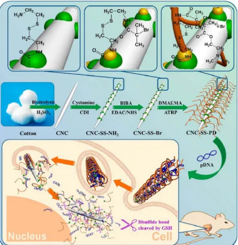
CNCs from cotton wool functionalized with disulfide bond-linked poly(2-dimethylamino)ethyl methacrylate) (PDMAEMA) and especial vectors CNC–graft–PDMAEMA (CNC-SS-PDs) of different molecular weights of PDMAEMA were synthesized (Fig. 2) (Hu et al., 2015). The CNC-SS-PDs presented good transfection efficacy and low cytotoxicity. The antitumor effect of CNC-SS-PDs was assessedin vitroandin vivoby a suicide gene/prodrug system (e.g., cytosine deaminase/5-fluor-ocytosine, CD/5-FC). The authors stated that the modification of CNCs with redox-responsive polycations was an effective method for new gene delivery system development (Hu et al., 2015).
Hyperpigmentation is a skin disease caused mainly due to sun ex-posure or in pregnancy. Some hydroquinone inhibits the generation of melanin and eliminates the skin discoloration. To address this issue, CNCs were complexed to hydroquinone (CNC-hydroquinone) (310 nm), with a binding efficiency of over 80% (Taheri & Mohammadi, 2015). Approximately 80% of hydroquinone was released from CNC-hydro-quinone in 4 h (Taheri & Mohammadi, 2015).
Biocompatible CNCs decorated with poly(2-oxazoline) brushes were recently prepared via UV-induced photopolymerization (Hou et al., 2017). The cationic side chains (amphiphilic and hydrophilic) were used to load indocyanine green (ICG) molecules via electrostatic in-teractions. The authors demonstrated that this complex is an effective platform for photothermal cancer therapy. Taken together, these stu-dies demonstrated the successful complexation of CNCs with different active drugs in cancer and antimicrobial therapies.
3. Selective studies based on the administration of CNCs conjugated with gels
Thein situhydrogels formed with modified CD (CN-CD/F108-2-Dox and CN-CD/F68-2-Dox) presented a sustained release profile with con-trolled release of the drug (DOX) for around one week. In contrast, in the case of neat hydrogels (F108/α-CD and F68/α-CD) an initial burst
of drug release and consequent shortened release periods (2.5 days) were observed. This fact can be explained by an obstruction effect in the components. Furthermore, if the content of CNCs reaches the percola-tion limit, a network structure with linkage among nanoparticles is Fig. 2.Schematic diagram illustrating the preparation of CNC-graft-PDMAEMA (CNC-SS-PD) via atom transfer radical polymerization (ATRP) and the resultant gene delivery process (reproduced fromHu et al., 2015with permission from the American Chemical Society).
induced. This might result in another locking effect, thus resulting in a delay of DOX diffusion. In the case of modified CNCs, sustained drug release of hydrogels in situthrough both effects was observed (Lin & Dufresne, 2013).
CNC aerogel scaffolds produced from red pepper (1–2.5 nm) for releasing beclomethasone dipropionate (BDP) nanoparticles covered with amphiphilic hydrophobin proteins mightfind important pharma-ceutical applications (Valo et al., 2013). Nanostructured particles in-corporated into cellulose aerogels, which are biodegradable, might re-lease BDP in a sustained manner by the change of the matrix compounds. The authors suggested that the morphology and treatment steps of the CNCs and the aerogels are important in controlling the release profile (Valo et al., 2013). Allicin and lysozyme (both anti-microbial agents) were associated to CNC, named allicin-CNC (ACNC), and lysozyme-CNC (LCNC). Both materials were assayed against mi-crobial species by the microdilution method (Jebali et al., 2013). The authors reported that lysozyme and allicin have antifungal and anti-bacterial activity against Aspergillus niger, Candida albicans, Staphylo-coccus aureusandEscherichia colistrains. Interestingly, the same values of minimum inhibitory concentrations MIC50and MIC90were observed for both ACNC and LCNC, even though allicin and lysozyme had dif-ferent MICs against all strains. The authors suggested that these CNCs can be used as antimicrobial agents in several industries, such as foodstuffs, food and in textile materials (Jebali et al., 2013).
Superabsorbent aerogels grafted with CNCs were prepared to pro-mote controlled release of amoxicillin (Anirudhan & Rejeena, 2014). Poly(acrylic acid-co-acrylamide-co-2-acrylamido-2-methyl-1-propane-sulfonic acid) grafted with a nanocellulose/PVA composite loaded with the drug (amoxicillin) showed superior drug release in the intestine in comparison with the gastric region. The authors stated this system would be a promising carrier for the administration of amoxicillin into the gastrointestinal tract (Anirudhan & Rejeena, 2014).
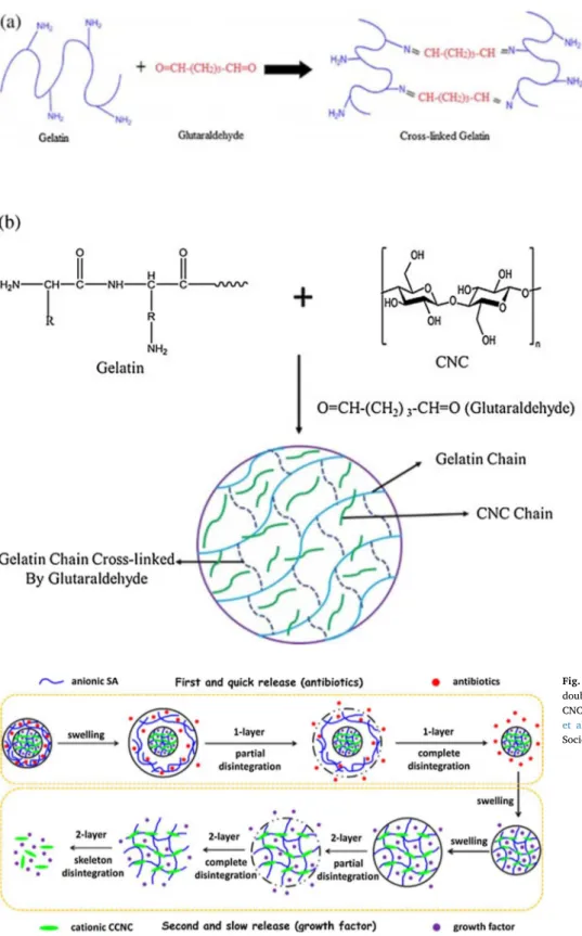
In a different approach, gelatin/CNC hydrogels were synthesized and data from the swelling assays suggest that these hydrogels were pH-and temperature-sensitive (Ooi, Ahmad, & Amin, 2014). Gelatin/CNC hydrogels showed the highest swelling ratio at a pH smaller than the isoelectric point (pI) and the lowest swelling ratio at a pH≈pI. In addition, a greater swelling ratio was observed at higher temperature. These preliminary findings suggest that gelatin/CNC hydrogels might be exploited as drug carriers, as they can be produced as pH- and temperature-responsive systems (Ooi et al., 2014). In this direction, there is a large effort towards the design of external stimulus-responsive materials as a promising feature in biomedical applications. A pro-mising strategy based on micro-hydrogel composites from cellulose nanowhiskers (CNCs) and starch formed from a sonication-assisted emulsion was reported (Mauricio et al., 2015). The incorporation of vinyl bonds into both CNCs and starch led to the creation of the micro-hydrogel composite. In this composite, CNC had an important role as a covalent crosslinker and acted as an emulsifying agent. Vit-B12 release was regulated in response to changes in the CNC amounts. Studies on the release kinetics indicated that the Vit-B12 release was driven by an anomalous mechanism depending on the addition of CNCs (Mauricio et al., 2015). Another approach with a pH-sensitive gelatin hydrogel reinforced with CNCs by using glutaraldehyde as a crosslinker was re-ported (Fig. 4) (Ooi, Ahmad, & Amin, 2016). A swelling test with CNC–gelatin hydrogels showed good pH sensitivity with the highest swelling ratio at pH 3. A model drug, such as theophylline, was used to further evaluate the carrier potentiality of these CNC–gelatin hydrogels. Thefindings suggest that 15% CNC-reinforced gelatin hydrogels were the best candidates suitable for a controlled theophylline delivery system (Ooi et al., 2016).
A smart approach was recently developed to prepare hydrogels with a double membrane (Lin, Geze, Wouessidjewe, Huang, & Dufresne, 2016) to promote co-delivery of two drugs. The shell, composed of al-ginate, promotes rapid drug release (antibiotic drug CH). The anionic alginate/cationic CNC inner membrane provides prolonged drug
release (growth factor EGF) ascribed to the“nano-obstruction effect” (Fig. 5). The double membrane material is biocompatible and can promote this complex simultaneous drug release at different rates (Lin et al., 2016).
A cellulose-based injectable hydrogel with improved mechanical properties was designed to promote sustained release of DOX (You et al., 2016). Quaternized cellulose and cationic CNC were crosslinked byβ-glycerophosphate,β-GP, forming a hydrogel with an increase of temperature. Injection of DOX-encapsulated hydrogels beside the tumor tissues of mice with liver cancer displayed good anticancer therapeutic efficacy (You et al., 2016).
Taken together, these selective publications described recent ad-vances in the preparation and application of CNCs in aqueous suspen-sion and/or incorporated into polymeric matrices and nanocomposites. It should be highlighted that the presence of negative charges derived from sulfate groups on the surface of CNCs prepared by hydrolysis with sulfuric acid represents a site for further conjugation of CNCs with re-levant amounts of drugs, via different techniques. This process allows a control of drug dosage and release. Therefore, due to the unique properties of CNCs, these materials allow versatile surface modification and fabrication of composite materials. These materials have enhanced mechanical and pharmacological properties, suitable for drug delivery applications. However, before biomedical applications, detailed in-vestigation of the toxicity of CNC-based materials is necessary, as dis-cussed in the next section.
4. Nanotoxicity of CNCs–based systems
4.1. In vitro assays
In order to propose the safe use of CNC-based materials for bio-medical applications, it is important to evaluate the toxicity and fate of these nanomaterials. This section presents and discusses in more detail recent progress in the comprehension of the nanotoxicity of these na-nomaterials. Initially, a variety ofin vitrotoxicity assays towards dif-ferent cell lines is described.
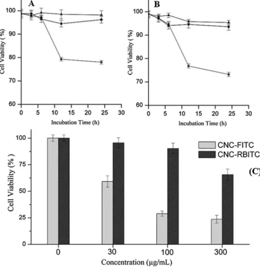
CNCs conjugated with rhodamine B isothiocyanate (RBITC) and with FITC acting on HEK-293 (human embryonic kidney 293) and on Sf9 (Spodoptera frugiperda) cells were evaluated (Mahmoud et al., 2010). No cytotoxicity effect of the conjugate CNC–RBITC was observed (Fig. 6). CNC–RBITC, as a positively charged material, was taken up by both cell lines, causing no damage to cell integrity. In contrast, the negatively charged material CNC–FITC underwent significant cell in-ternalization at pH∼7. The authors suggested that since the surface charge of CNC exhibited an important role in either cellular uptake or cytotoxicity, simple modification of the CNC surface might be tuned for cell penetration without leading to cellular damage. Moreover, surface modification appears to be a suitable strategy for the design of versatile nanomaterials for bioimaging and drug delivery systems (Mahmoud et al., 2010).
In a furtherin vitro study of the nanotoxicity of cellulose-based materials towards mammalian cells, cellulose nanoparticles with spherical morphology (SCNC) (sizes in the range of 80–260 nm) were produced by dialysis initiated from trimethylsilylcellulose (TMSC), with subsequent covalent labeling with FITC. SCNC conjugation with FITC did not change particle size, shape or stability (Fig. 7) (Liebert, Kostag, Wotschadlo, & Heinze, 2011). The incorporation of SCNC–FITC into living humanfibroblasts was followed by using a confocal technique. Confocal micrographs of fibroblasts after 48 h of incubation with SCNC–FITC-labeled cellulose indicated high SCNC loading into cells. In contrast to CNCs, rapid cellular uptake was found for the SCNC without transfection reagents or associated to a receptor molecule. The data presented clearly indicated the influence of morphology on endocytosis, and thus on the biocompatibility of CNCs (Liebert et al., 2011).
cotton led to low cytotoxicity (statistically significant; p < 0.05) and a (pro)inflammatory response (Clift et al., 2011). In other words, CNCs may exhibit in human lung cells some gentle dose-dependent cytotoxic and inflammatory effects. Especially when there is a risk of inhalatory exposure under a high concentration of released CNC powders (Clift et al., 2011). More studies are required on this topic.
The cytotoxicity of CNCs against nine cell lines (RAW 264.7, HBMEC, bEnd.3, MCF-10A, MDA-MB-231, PC-3, MDA-MB-468, KB and C6) was evaluated (Dong, Hirani, Colacino, Lee, & Roman, 2012). CNCs
did not show cytotoxic effects against any of the tested cell lines, at a concentration range of 0–50μg/mL (for 48 h of incubation). Cellular uptake of FITC-labeled CNCs by the cell lines was barely detected. Considering the non-cytotoxicity and the low non-specific cellular up-take, the results indicate that CNCs are promising candidates for na-nomedicine applications (Dong et al., 2012). In a test with a different cell line, dispersions of CNCs (length of 250 nm and diameter of 10 nm) were prepared by hydrolyzing cotton linters. Their cytotoxicity towards L929 cells was investigated by MTT assay. It was observed that Fig. 4.(a) Mechanism of crosslinking between ge-latin chains using glutaraldehyde and (b) proposed mechanism for the formation of semi-inter-penetrating polymer network (IPN) hydrogels (re-produced fromOoi et al., 2016, by permission of Elsevier B.V.).
Fig. 6.(A) Trypan blue Sf9 and (B) HEK-293 cell viability profiles of control (▲) and after addition of CNC-RBITC (□) or CNC-FITC (●) (0.1 mg/mL). Each data point represents the average ± STD of three viability measurements (cell density of 1 × 10⁶ cells/mL). (C) MTT cell viability profile of Sf9 after exposure to different concentrations of CNC-FITC and CNC-RBITC. Each data point represents the average ± SE of three viability measurements (cell density of 1 × 10⁶ cells/mL) (modi
fied from Mahmoud et al., 2010 with permission from the American Chemical Society).
concentrations in the range of 0.01% to 0.2% exhibited low toxicity to L929 cells (Ni et al., 2012).
Still onin vitrotoxicity assays, CNCs derived from different sources, such as hemp,flax and cellulose powder were studied in two different cell lines, Chinese hamster lung fibroblast V79 andSpodoptera frugi-perdaSf9 insect cells (Male, Leung, Montes, Kamen, & Luong, 2012). Cytotoxicity was measured by ECIS (electric cell substrate impedance sensing). Flax exerted the highest inhibition on Sf9 cells compared to cellulose powder and hemp. Interestingly, CNCs obtained from hemp andflax and pretreated with enzymes, such as pectate lyase, were found to cause less cell inhibition compared to CNCs prepared from untreated hemp andflax. Based on the half-inhibition concentration values, CNCs did not show important cytotoxicity in the studied cell lines. The au-thors suggested a correlation of the inhibitory effect with the carboxylic acid content of the CNCs (Male et al., 2012). More studies are neces-sary.
A folate group was bound to carboxymethyl cellulose nanoparticles conjugated with quantum dots in order to obtain specific recognition of cancerous cells (Aswathy et al., 2012). The anticancer agent 5FU was encapsulated in this nanoparticle for targeting cytotoxicity to MCF7 (human breast cancer cells), compared to non-cancerous L929 cells. As expected, the epifluorescent images revealed certain specificity in the internalization of folate-conjugated nanoparticles, since the conjugated nanoparticles were internalized more in the MCF7 cell line due to the presence of FR in the cancer cell line, compared to the L929 cell line (non-cancerous) (Aswathy et al., 2012). HCT116 colon carcinoma and NIH3T3 murine embryofibroblast cells were confronted with CNCs of different sizes by WST-1 assay (Hanif, Ahmed, Shina, Kime, & Um, 2014). The data showed that none of the different sizes (256–1174 nm) of CNCs presented any substantial cytotoxicity at various tested con-centrations (10–250 g/mL) (Hanif et al., 2014).
Recently, thefiber cell interactions of CNCs obtained from cotton (c-CNCs) (average length 237 × 29 nm) and from tunicates (t-(c-CNCs) (average length 2244 × 30 nm) with different aspect ratios were compared (Endes et al., 2015). Mainly, the uptake and translocation of CNCs was investigated through an in vitro lung model (3D), by the above-described fibers. In order to create a typical environment for inhalation exposure experimentation, important doses of rhodamine-labeled CNCs, extrapolated from permissive exposure limits (PEL) for cotton dust, were used (Endes and Müller, 2016429AD;OSHA, 2017) by nebulization with the air–liquid interface cell exposure system (ALICE), as previously described (Endes et al., 2014). This approach allowed elucidation of the influence of CNC dimensions on the cellular interactions with anin vitro lung model. Related to translocation, no rhodamine-functionalized CNCs were found in the lower well
compartment, besides those on monocyte-derived dendritic cells which were settled up at the basal side of the membrane, at all tested con-centrations. This information was confirmed by laser scanning micro-scopy (LSM) and investigated byfluorescence measurement of the su-pernatants at several time points (Fig. 8) (Endes et al., 2015). When lower dose exposure was run the measured values were similar to the limit of quantification (meaning 10-fold the standard deviation of 10 measurements of water (SDH2O)). A control group (rhodamine) was performed by triple-cell co-cultures subjected to a 100μM rhodamine solution via ALICE, conducted for a period of 0–48 h post-exposure periods, andfixation occurred as described in detail in the manuscript. Results also described in the supporting information showed overall diffuse rhodamine staining, different to that of the rhodamine– CNC-exposed co-cultures. The absence of cytotoxicity shown by consistent cell morphology, including multilayer formation after 24 h of post-ex-posure, was observed. However, an increase in cell size, induced by residual ethanol from the rhodamine stock solution, was noted at 1 h and was attenuated after 24 h (Endes et al., 2015). In addition, both the aspect ratio and deposited dose of CNCs exerted a strong influence on thein vitrouptake by a co-culture system (3D) of the human epithelial airway barrier, under experimental conditions due tofiber−fiber in-teractions. The authors stated that these data pointed out the potential implications concerning human exposure to t-CNCs by long inhalation of high aspect ratio nanoparticles (Endes et al., 2015). More studies in this direction are required.
CNCs derived from tunicates and cotton (e.g., differing aspect ratios of∼9 and∼80) were employed as models of high aspect ratio na-nomaterials. Well-dispersed and characterized CNC suspensions were aerosolized by the use of ALICE (Endes and Müller, 2016, 429AD) at a realistic concentration of cells, in the range 0.14–1.57μg/cm2. Oxida-tive stress levels, cytotoxicity and proinflammatory effects, as indicators of the biological impact of any high aspect ratio nanoparticles, were tested using a 3D multi-cellularin vitromodel of ALO (human epithelial airway barrier at the air–liquid interface) 24 h post-exposure. A positive particulate control (crystalline quartz-DQ12) was used as a strategy for validating the ALICE system andfiber amosite asbestos (LFA) to prove the sensitivity of thein vitromodel (Endes et al., 2014).
Regarding the toxicity of cellulose-based hydrogels, a material composed of cellulose nanowhiskers (or CNCs) obtained from linter was dispersed into deionized water using ultrasonic treatment, followed by suspension and centrifugation, thus allowing isolation of the upper aqueous suspension (Zhang et al., 2010). Subsequently,α-cyclodextrin (α-CD) was dissolved in water and, at the same time, the surfactant poly (ethylene glycol)-poly(propylene glycol)-poly(ethylene glycol) (PEO137-b-PPO44-b-PEO137, EPE) was added to an aqueous CNC
suspension and subsequently stirred. An α-CD aqueous solution was poured into the aqueous suspension containing EPE and CNCs. Then, a 2 min ultrasonic treatment was applied and the mixture was condi-tioned at room temperature for the gelation process. The hydrogels were evaluated for cell viability by the MTT assay using the L929 cell line for 48 h of incubation. The results showed that the cell viability was unaffected upon incubation with the nanocomposite, even when the nanocomposite concentration reached 100 mg/mL (Zhang et al., 2010). These results indicate the non-toxicity of CNC-based nano-composites.
CNCs with a high carboxyl content produced a stable colloidal na-nocrystal suspension with no aggregation upon exposure either to high salt concentrations or even high serum-containing media (Hosseinidoust, Alam, Sim, Tufenkjia, & van de Vem, 2015). Interac-tions of all the fracInterac-tions with different tissue cell lines were investigated and demonstrated the uptake of CNCs by specific cell lines without evidence of damage or changes in cell density on the membrane. However, at higher carboxyl contents (over 3.9 mmol/g), a charge-de-pendent decrease in mitochondrial activity was observed. This high surface carboxyl content allowed the conjugation offluorophores to the nanocrystals without affecting the colloidal stability. The cellular up-take offluoresceinamine associated to nanocrystals presented a time– -dose-dependent relationship and underwent a size increase by doubling the surface charge (Hosseinidoust et al., 2015).
Cauda epididymal sperm samples were analyzed for sperm con-centration, motility, morphological abnormalities and DNA damage after continuous pharyngeal aspiration exposure to CNCs (Farcas et al., 2016). CNC exposure altered sperm concentration, motility, cell mor-phology and sperm DNA integrity. High proinflammatory cytokine le-vels and myeloperoxidase (MPO) activity in assays, as well as oxidative stress in both assays and epididymis, were observed. All of these effects damaged the testicular structure. The authors stated that these data demonstrate that exposure of the lung to CNCs induces adverse effects in spermatocytes/spermatozoa, indicating male reproductive toxicity (Farcas et al., 2016).
Burchett et al. (Burchett, 2016) investigated the toxic effects of CNCs on eukaryotic organisms such asSaccharomyces cerevisiaeand on HEK-293 cells, using an autobioluminescent method. TheS. cerevisiae and HEK-293 cells were engineered to expressluxCDABE (a bacterial luciferase operon) for bioluminescent production. This operon allows monitoring of the toxicant exposure to cells. When exposed to CNCs at concentrations ranging from 0.001 to 1 mg/mL, both yeast and human cells reported time- and dose-dependent effects. The authors reported a reduction of bioluminescence in the presence of CNCs (0.001 and 1 mg/ mL) inS. cerevisiaeby 5% and 10%, compared to untreated control cells 8 h post-treatment, respectively. At 12 h, decrease of the signal by 25% and 70% post-treatment was observed, respectively. At the level of 1 mg/mL of CNCs, an important decrease (∼60%) in metabolic activity of HEK-293 after 2 days post-treatment was observed. No significant changes in metabolic activity at a level of 0.001 and 0.01 mg/mL of CNCs throughout the entire period of exposure were observed. The authors stated that these data demonstrated the potential cytotoxic effects of high concentrations of CNCs in many biological systems (Burchett, 2016).
A novel cationic surface-modified CNC derivative was synthesized via a surface-initiated living radical polymerization method. Initial studies indicated that only one of the modified CNCs caused an im-portant decrease (50%) in the viability of J774.A1 cells (mouse monocyte macrophage) at the concentration tested (100μg/mL). It is important to notice that this concentration was higher than that ex-pected for biomedical purposes. However, MCF7 cells were not affected by these cationic CNCs at different tested concentrations (Sunasee, Hemraz, & Ckless, 2016;Sunasee, Hemraz, Ckless, Burdick, & Boluk, 2015).
Overall, thein vitrostudies suggested that CNC-based materials are not toxic. However, a few studies demonstrated that these materials
might cause toxic effects to cells. More studies are necessary to better clarify this issue. The cytotoxicity of CNCs depends on several para-meters such as the source for the synthesis of CNCs, the nanomaterial size and surface chemistry, the cells analyzed and the concentration of CNCs employed.
4.2. In vivo experiments
C57BL/6 mice were exposed to CNCs obtained from wood and formulated as CNCP (powder) (average hydrodynamic size of 304 ± 72 nm) or CNCS (10 wt%, gel/suspension) (average hydro-dynamic size of 88.4 ± 9.8 nm) (Yanamala et al., 2014). Both CNCP and CNCS were compared to asbestos-induced responses for pulmonary outcomes. Pharyngeal aspiration-facilitated innate inflammatory re-sponses were evaluated by an increase in leukocytes and eosinophils retrieved by bronchoalveolar lavage (BAL). Mice exposed to CNCP showed an increase in the number of total inflammatory cells compared to control mice. An acute inflammatory response compared to asbestos was detected by higher overall levels of polymorphonuclear leukocytes (PMNs) and other inflammatory cells upon CNCS or CNCP exposure. Tissue damage was elevated in mice exposed to CNCP. Oxidized protein accumulation of CNCS and up-regulation of inflammatory cytokines as compared to CNCP in the lung was also observed. The data suggested that nanoparticle morphology and nanosize forms of CNCs, in spite of the same source, may be critical factors affecting innate immune in-flammatory responses (Yanamala et al., 2014).
OECD test guidelines 425 and 407 defined acute oral toxicity even as oral toxicity after repeated daily administration of CNCs (O’Connor, Berry, & Goguen, 2014). Acute oral toxicity was estimated by oral ga-vage administration to rats (Crl:CD(SD)BR) of one-time doses of up to 2000 mg/kg of an aqueous suspension form directly into the stomach. The rats’health was monitored for 14 days, followed by necropsy. The median lethal dose was found to be around 2000 mg/mL and no ad-verse effects of CNCs on rats were observed. Crl:(HA)BR guinea pigs were exposed to CNCs by intradermal injection of 1.1 mg/mL of a CNC suspension. After a week, 103 mg/mL of CNC gel was topically applied for 48 h and then repeated 2 weeks later for 24 h. CNCs showed non-sensitizing effects at the tested concentrations. CBA/J mice were treated for three consecutive days with CNC suspension, which was topically applied on the dorsum of each ear, and analyzed by a lymph node assay. The results showed that CNCs were not considered to be contact dermal sensitizers at up to 10.7% CNC concentrations. In ad-dition, this study was carried out according to OECD test guideline 404 (Crl:KBL(NZW)BR albino rabbits). A CNC gel containing 0.5 g of CNCs was topically applied (4 h) with a special semi-occlusive dressing, fol-lowed by 14 days of monitoring signs of erythema and edema. The results showed no corrosive effects and the CNC PII (primary irritation index) score was 0 (non-irritating) (O’Connor et al., 2014).
This section demonstrated that few important studies were elegantly designed to evaluate the possible toxic effects of CNCs in vivo. The preliminary results revealed opposite effects (toxicity and non-toxicity), depending on the parameters of each study. Hence, more studies are necessary to investigate more deeply thein vivo toxicity of CNCs, in addition to evaluating the biodistribution and fate of these nanoma-terials.
4.3. Ecotoxicology of CNC-based systems
CNCs were tested to a broad ecotoxicological panel through toxicity assays including rainbow trout hepatocytes in nine aquatic species,
such as Ceriodaphnia dubia, Daphnia magna, rainbow trout
embryos were used. The most sensitive test was on hepatocytes (EC20 range between 10 and 200 mg/L). However, no genotoxicity was ob-served. In assays with the nine species, CNCs affected the reproduction of the fathead minnow with an IC25value of 0.29 g/L. However, no other effects on survival and growth occurred at concentrations below 1 g/L. The authors stated that on the basis of these data relating to ecotoxicological characterization, CNCs appeared to exhibit low toxi-city potential and probably no environmental risk (Kovacs et al., 2010). For a more complete conclusion, more studies on CNC ecotoxicity are necessary.
Although much progress has been achieved in the evaluation of the toxicity of CNCs, more studies aiming to further investigate thein vitro andin vivocell interactions, biodistribution,in vivofate of CNCs and ecotoxicity are still necessary. Complete understanding of CNC–cell interactions and the materials’fate are currently poorly reported. It should be noted that slight modifications in the preparation and surface chemistry of CNCs can result in distinct toxicity profiles. Due to in-creasing interest in the biomedical applications of CNCs, potential toxicity is an issue of utmost importance that deserves special attention.
5. Patents
Many patents describe inventions using CNCs for several applica-tions. Colloidal suspensions of cellulose crystallite solid films were produced with a new optical property (Revol, Godbout, & Gray, 1997). These solid materials have a helicoidal arrangement of the crystallite content and solid films with iridescent properties. The reflected ir-idescent color renders the materials perfectly suited for optical au-thenticating devices (Revol et al., 1997). Probably, this property could be useful for drug monitoring.
CNC hydrogels and aerogels, with pore diameters around of 100 nm with up to 80% CNCs by weight of cellulose nanoparticles, with or without the presence of one or more metal nanoparticles, might be used in biomedical applications. The applications include drug delivery systems, scaffolds for tissue engineering, contact lenses, biosensors, medical electrodes, breast implants, medical devices, pathogen detec-tion and biocatalysts, as published by Thielemans and Davies (Thielemans & Davies, 2011).
An invention in pharmacology bioassays used in drug discovery, drug screening and toxicity evaluations was reported (Thielemans & Davies, 2011). This invention relates to novel systems and methods used for production and control of the morphology of living tissues and organs produced by mammalian cells using 3D porous scaffolds based on nanocellulose. The resultant nanostructures are also useful tools in highly advanced assays for drugs. In other words, the importance of the present invention was related to methods for evaluating drugs in well plates, in which the well may contain a 3D non-biodegradable, inert, nanocellulose scaffold and cells capable of forming living tissue or or-gans. In addition, the well may contain a drug having biological activity of interest and, optionally, a detector capable of detecting the biological activity in a high throughput format (Gatenholm, 2014).
A patent by Bhattacharya et al. (Bhattacharya, Laukkanen, Lauren, Lou, & Yliperttula, 2012) described a material useful in drug delivery compositions comprised of cellulose nanofibers. According to the au-thors’ description: cellulose nanofibers and/or derivatives thereof or nanofiber bundles in the cellulose nanofibers, less than 1μm, preferably less than 200 nm, more preferably less than 100 nm, and/or derivatives thereof. Examples of drugs that can be added to cellulose nanofibers are hormones, anesthetic, chemotherapeutic, inflammatory, anti-microbial and analgesic drug substances and medicaments, as well as biotechnical and biological drugs, peptides and protein drugs. The in-vention also provides methods for producing these materials and compositions and uses thereof (Bhattacharya et al., 2012).
An interesting invention describes the preparation of a drug delivery system for sustained delivery of bioactive agents, including a matrix comprising CNCs, nanowhisker, nanofibrillated and nanorod cellulose
derived from plant-based material. In this invention, the drug delivery system is composed by at least one bioactive agent (e.g., indomethacin, itraconazole and beclomethasone) and with at least one support se-lected from synthetic polymers, bio compounds and natural polymers (Laukkanen et al., 2013). The drug delivery system can be characterized as a medical device, combination product, implant, transdermal patch or a formulation for oral, sub-lingual, topical, intraocular, intestinal, rectal, subcutaneous, parenteral or mucoadhesive application. The drug delivery system is also an intrauterine delivery system or vaginal de-livery system or subcutaneous implant (Laukkanen et al., 2013).
An invention related toin vivotreatment for cell proliferative dis-eases described methods and devices (Davalos, Rylander, & Arena, 2013). This invention aims to manage solid tumors (e.g., brain tumors) using non-thermal IRE to cause cell elimination in treated tumors in the presence of nanoparticles e.g., CNCs. In the patent, the methods com-prise the use of high aspect ratio nanoparticles with or without mod-ified surface chemistry (Davalos et al., 2013).
Another representative invention describes surface-modified CNCs which were prepared by chemical modification of CNC, as well as their uses in different areas. The CNCs can be used as carriers of particular chemical compounds (e.g., a pharmaceutical with procaine hydro-chloride), in cosmetic or agriculture applications (Parinaz, Tam, & Berry, 2014). The surface of CNC was modified with chitosan oligo-saccharide (CS), by selectively oxidizing the primary alcohol moieties of CNC followed by coupling of the amino groups of CS to the oxidized CNC to provide the desired material (Parinaz et al., 2014).
An invention disclosed the preparation of a colon-targeted prodrug (e.g., tosufloxacin, clinafloxacin, trovafloxacin) with a nanocellulose-based carrier (Tang, Huang, Li, Lu, & Chenxue, 2015). According to the description, nanocellulose acted as a carrier to an amino acid tether, by chemical bonds, connecting the carrier and drug intermediates for colon-targeting prodrug applications.In vitrostudies indicated that the prodrug can effectively achieve colon-specific release, colon-targeted drug carriers meet the requirements. In addition, it can be applied to the development of new therapeutic drugs targeting diseases of the colon (Tang et al., 2015). Finally, another patent described the pro-cesses capable of converting biomass into high-crystallinity nanocellu-lose with low mechanical energy input (Nelson, Retsina, Pylkkanen, & O’Connor, 2015). The process involved fractionating biomass with an acid, a solvent and water to generate cellulose-rich solids, and me-chanical treatment to form nanofibrils and/or nanocrystals. The nano-cellulose material may include nanofibrillated nano-cellulose, nanocrystal-line cellulose or both. Applications of this material are related to pharmaceuticals and drug delivery systems (Nelson et al., 2015).
6. Commercial products
From all of these data, it can be observed that cellulose nanos-tructures have exciting potential in medical applications and also as reinforcement material in nanocomposites. Cellulose-based nanos-tructured materials have surface features that allow a wide variety of chemical modifications. Consequently, these materials can have several applications such as foams, adhesives, specialized barrierfilms, elec-tronic display materials and drug carriers. This latter application has been growing in importance along with the development of nanome-dicine in recent years. It is also important to mention that CNCs asso-ciated to cellulose nanofibers can manifest equal or even better me-chanical properties in regard to the currently available engineering materials (Durán, Lemes, Durán, Freer, & Baeza, 2011). Thus, it is possible to consider that CNC materials can be present in high-end technological applications in the future.
Inc.76many organizations have notified demonstration plants involving CNCs in North America and Europe. The North American companies focus on the production of CNCs (Bio Vision (Canada), CelluForce (Canada) and US Forest Service Forest Products Laboratory (USA)). On the other hand, European companies interested are CNF (Centre Technique du Papier (France)), Innventia AB (Sweden), Stora Enso (Finland), UPM fibril cellulose (Finland) and Borregaard ChemCell (Norway). According to the report of Future Markets Inc. the global market for nanocellulose to 2017 (published in October 2012) would be about $97 billion (estimated) related just to life sciences and medical markets impacted by nanocellulose.
In 2012, a large step was made by CelluForce Inc. in regard to CNC production and characterization. Consequently,NCC™–the trade name for CNCs produced by CelluForceInc.–was granted approval for un-restricted manufacture and implementation in Canada. CelluForce NCC™has been shown to have a good toxicological profile, which al-lows it to be a good performance base for new drug delivery products. It uses an organic growth platform that is compatible with tissues and bone. The surface of CelluForce NCC™is also reactive, allowing the addition of different functionalities and development of a wealth of new applications (http://www.celluforce.com/en/applications).
Nanocellulose represents the development of a wide range of pro-ducts from scaffolds for biomedical applications to biodegradable food packaging. New applications in polymer reinforcement and anti-microbial films will probably be important in the market soon. FP Innovations estimated that the market would be around $250 million in North America by 2010 (http://www.fpinnovations.ca). Future Markets estimated the market to 2024 (https://www.marketresearch.com/ Future-Markets-Inc-v3760/Global-Nanocellulose-8553279/).
The industry reportCellulose Fiber Market Size By Application (Spun Yarn, Clothing, Fabrics), Industry Analysis Report, Regional Outlook (Australia, Brazil, Canada, China, France, Germany, India, Indonesia, Italy, Japan, Malaysia, Mexico, Spain, South Africa, UK, US.), Growth Potential, Price Trends, Competitive Market Share & Forecast, 2016–2024by Global Market Insights Inc. stated that the cellulosefiber market will exceed $39 billion by 2024.76The companies involved in all of these processes are: Innventia AB, CelluForce Inc., Nippon Paper Group Inc., Kruger Inc., Borregaard ASA, FP Innovations and others (http://industryarc. com/Report/11681/nanocellulose-market-nalysis.html). Therefore, cellulose-based nanomaterials have been significantly impacting the industry with positive perspectives.
7. Conclusions and perspectives
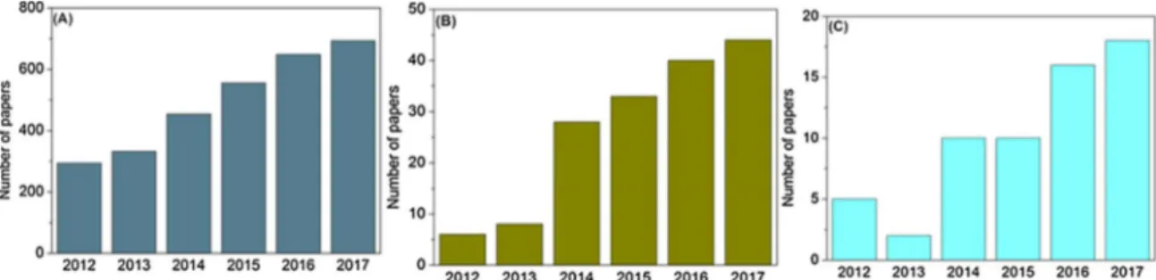
Recently, CNCs have attracted the attention of the industrial and scientific communities due to their interesting characteristics such as low cost, extraction from renewable sources, low toxicity, high me-chanical properties. In addition, CNCs have the ability to be modified by a wide range of chemical modifications, thus allowing conjugation with therapeutic drugs for several applications,(Eyley & Thielemans, 2014; Mondal, 2017). Fig. 9 shows the increase in the number of
publications relating cellulose nanocrystals according to the Web of Science®, in the last 5 years. It should be noted that the uses of cellulose nanocrystals in medicine is still in a nascent stage.
As discussed in this article, CNCs have been successfully conjugated with other nanomaterials and/or drugs for drug delivery applications, yielding new nanocomposites. In this sense, there are several patents, innovations and commercial products based on CNCs. Although some progress has been achieved in the preparation, characterization and application of CNCs from the scientific and technological point of views, there are still some questions to be answered.
Firstly, as discussed in this article, CNCs allow surface modification and conjugation with therapeutic molecules, making CNCs suitable nanocarriers. It should be noted though that it is highly desirable to develop controllable methods for chemical or physical modification of the CNC surface, preferably by using mild or green conditions. In ad-dition, the development of methods for deep characterization of mod-ified CNCs is necessary, thus allowing investigation of the chemical interactions at all length scales, from the molecular to the organism levels.
Regarding the toxicity of CNC-based materials, several papers de-scribedin vitrostudies and, even more importantly, alsoin vivostudies of CNCs. Overall, CNCs have very low toxicity in several models used. However, some studies reported that CNCs could cause some important toxic outcomes. Further investigation of the toxicity of CNCs is thus necessary, since these nanomaterials have great potential for several biomedical applications. In this sense, nanotoxicity is an important topic that should be considered as a priority in the field of nano-technology. More studies based on in vitro and in vivofate, biodis-tribution, cell interactions and ecotoxicity of CNCs are still required. Since the size, morphology and surface chemistry of nano-based ma-terials dictate their toxicity, CNC-based mama-terials produced and pro-cessed differently will lead to different toxic effects. Therefore, special attention to the complete investigation of CNC-based materials is ne-cessary.
Finally, there are several patents, innovations and commercial products based on CNCs highlighting the scientific and technological impacts of this versatile nanomaterial. We hope that this comprehen-sive review article will help to increase the interest in further research on the preparation of CNCs, their characterization and nanotox-icological evaluation, especially considering the biomedical context.
Acknowledgements
We would like to thank Proof Reading Service (Proof-Reding-Service.com) for revising the manuscript. We have appreciated the support from CNPq, FAPESP, INCT-INOMAT, NanoBioss-SisNANO, MCTI) and the Brazilian Network on Nanotoxicology −Cigenanotox (CNPq/MCTI).