Iranian Journal of Basic Medical Sciences
ijbms.mums.ac.ir
The effects of
Eucheuma cottonii
on alveolar macrophages
and malondialdehyde levels in bronchoalveolar lavage fluid
in chronically particulate matter 10 coal dust-exposed rats
Romadhiyana Kisno Saputri
1, Bambang Setiawan
2, Dian Nugrahenny
3, Nia Kania
4, Endang
Sri Wahyuni
5, M Aris Widodo
31 Faculty of Medicine, Brawijaya University, Malang, East Java, Indonesia
2 Department of Medical Chemistry and Biochemistry, Research Center for Toxicology, Cancer and Degenerative Disease, Medical Faculty,
Lambung Mangkurat University, Banjarmasin, South Kalimantan, Indonesia
3 Laboratory of Pharmacology, Faculty of Medicine, Brawijaya University, Malang, East Java, Indonesia
4 Department of Pathology, Research Center for Toxicology, Cancer and Degenerative Disease, Ulin General Hospital, Medical Faculty,
Lambung Mangkurat University, Banjarmasin, South Kalimantan, Indonesia
5 Laboratory of Physiology; Faculty of Medicine, Brawijaya University, Malang, East Java, Indonesia
A R T I C L E I N F O A B S T R A C T
Article type:
Short communication Objective(s)
:To investigate the effect of Eucheuma cottonii on alveolar macrophages (AM) and
malondialdehyde (MDA) levels in bronchoalveolar lavage fluids (BALF) in particulate matter 10 (PM10) coal dust-exposed rats.
Materials andMethods: Ten groups, including a non exposed group and groups exposed to coal
dust at doses of 6.25 (CD6.25), 12.5 (CD12.5), or 25 mg/m3 (CD25) an hour daily for 6 months with or
without supplementation of ethanolic extract of E. cottonii at doses of 150 (EC150) or 300 mg/kg
BW (EC300). The number of macrophages was determined using a light microscope. MDA levels
were measured by TBARS assay.
Results: EC150 insignificantly (P > 0.05) reduces the AM in CD groups compared to non treatment
groups. EC150 and EC300 significantly (P < 0.05) decreased MDA levels in CD12.5 and CD25 groups
relative to non treatment groups.
Conclusion:E. cottonii attenuated oxidative stress in chronic exposure of PM10 coal dust.
Article history: Received: Nov 5, 2013 Accepted: Apr 30, 2014
Keywords:
Alveolar macrophage Coal dust
Eucheuma cottonii
Malondialdehyde
►
Please cite this paper as:Kisno Saputri R, Setiawan B, Nugrahenny D, Kania N, Sri Wahyuni E, Widodo MA.The effects of Eucheuma cottonii on alveolar macrophages and malondialdehyde levels in bronchoalveolar lavage fluid in chronically particulate matter 10 coal dust-exposed rats. Iran J Basic Med Sci 2014; 17:541-545.
Introduction
Coal dust exposure can induce an acute alveolar and interstitial inflammation leading to chronic pulmonary diseases. The essential mechanism of coal dust-induced lung damage is likely to be mediated by macrophage activation and the recruitment of
polymorphonuclear cells (1). The increase in lung
macrophages has been reported to be the primary lung response to air pollutants in animal and human. Alveolar macrophages are cells first encountering inhaled pollutants in the lower respiratory tract. Their phagocytic capacity provides an efficient nonspecific defense against inhaled particles. Macrophages and other phagocytic cells produce reactive oxygen species (ROS) during phagocytosis or stimulation with a wide variety of agents (2). ROS can randomly react with lipids, proteins, and nucleic
acids. When ROS target lipids, they can initiate the lipid peroxidation, a chain reaction that produces
multiple breakdown molecules such as
malondialdehyde (MDA) (3).
The cultivated edible red seaweed, Eucheuma
cottonii (Kappaphycus alvarezii), contains vitamin C,
-tocopherol, -carotene, zinc, selenium, phenols,
and sterol. These compounds have antioxidant and anti-inflammatory properties, and thus directly or indirectly contribute to the inhibition or suppression of free radical generation (4). Previous studies also showed that the seaweed suppresses thiobarbituric
acid reactive substances (TBARS) formation of H2O2-
induced lipid peroxidation in red blood cells (5). Moreover, consumption of the seaweed is already proved not exert any damage to the liver, heart, kidney, brain, spleen, and eyes in normal rats (6).
*Corresponding author:Bambang Setiawan. Department of Medical Chemistry and Biochemistry, Research Center for Toxicology, Cancer and Degenerative
The present study aimed to investigate the effect
of E. cottonii on the absolute number of
bronchoalveolar lavage fluids (BALF) alveolar macrophages and BALF MDA levels in rats
chronically exposed to particulate matter (PM) 10
coal dust.
Materials and Methods
Preparation and extraction of E. cottonii
E. cottonii was harvested from the coastal areas of Tamiang, Kotabaru (South Kalimantan, Indonesia). The preparation and extraction of the seaweed were
performed according to the method of Fard et al (7).
ABTS reducing activity in vitro
2, 2’-Azinobis-(3-ethylbenzothiazoline-6-sulfonic
acid) (ABTS•+)-reducing activity was determined
according to the previous method (8).
Animal
A total of 40 male Wistar rats were randomly divided into ten groups. One group was a negative control group (without any treatment). Three groups were positive control groups exposed to coal dust at
doses of 6.25 (CD6.25), 12.5 (CD12.5), or 25 mg/m3
(CD25) one hr per day for 6 months. Six groups were
treatment groups exposed to coal dust with
supplementation of ethanolic extract of E. cottonii at
doses of 150 (EC150) or 300 mg/kg BW (EC300).
E. cottonii administration
The ethanolic extract of E. cottonii was given
orally using a gavage (gastric tube), and started
together with coal dust exposure. Doses of E. cottonii
and coal dust were based on previous study (7).
Coal dust exposure
Coal dust exposure was performed according our
previous study (9). The PM10 coal dust exposure was
conducted using equipment (0.5 x 0.5 x 0.5 m) that was designed by and available in Laboratory of Pharmacology, Faculty of Medicine, Brawijaya University, Malang. This equipment provided an ambient environment containing coal dust for inhalation by the animal. The airstream of the apparatus was set at 1.5-2 l/min mimicking an environmental airstream. To prevent hypoxia and discomfort, we also provided oxygen supply in the chamber. The non-exposure group was only exposed to filtered air in laboratory. In order to provid coal dust aerosolization, we put weighed coal dust in bottom hole of equipment; then the coal dust will circulate and enter the chamber again via upper hole. This aerosol will inhaled by rats in plastic chamber.
Ethics
Animal care and experimental procedures were approved by the Institutional Research Ethics
Committee of Faculty of Medicine, Lambung Mangkurat University, Banjarmasin, Indonesia.
Isolation of bronchoalveolar lavage fluids
The rats were euthanized by anesthetizing with ether inhalation and exsanguinated by cardiac puncture. Briefly, the thorax was opened by a median incision, the left bronchus was clamped with forceps, and a small incision was made in the the right bronchus and a plastic catheter attached to a 5-mL syringe was inserted. Bronchoalveolar lavage was performed a total of two times by repeated flushing with 5 ml ice-cold PBS, with gentle massage of the lungs. The bronchoalveolar lavage fluids were pooled and stored on ice until further use (10).
Analysis of the number and viability of BALF alveolar macrophages
BALF were centrifuged at 1000 rpm for 10 min at
4°C to obtain the cells and supernatants. The total
number of alveolar macrophages was determined using a light microscope by counting the number of cells in at least five squares of a hemocytometer after excluding dead cells using Trypan blue staining (10).
Analysis of BALF malondialdehyde levels
The supernatants obtained after centrifugation of BALFs were measured for the MDA levels (11). The principle of the method is the spectrophotometric measurement of the color generated by the reaction of thiobarbituric acid (TBA) with MDA. For this purpose, 250 µl of trichloroacetic acid solution was added to 100 µl of supernatant in a test tube. Next, 100 µl of TBA solution, 200 µl of HCl, and 0.5 ml of aquabidest were respectively added to the mixture. The tube was placed in a boiling water-bath for 15 min and then cooled in tap water. The absorbance of the colored product was read at 532 nm using a spectrophotometer. The values obtained were compared with a series of MDA tetrabutylammonium salt (Sigma-Aldrich, St Louis, MO, USA) standard solutions.
Statistical analysis
Data are presented as mean ± SD. The collected data were analyzed using Kruskal-Wallis test.
Probability value of P < 0.05 indicated a significant
difference between groups and later subjected to
Mann-Whitney Post hoc test. All statistical analyses
were performed using SPSS software 17.0 for Windows.
Results
ABTS reducing activity
In vitro, the ethanolic extract of E. cottonii at
concentration of 5 μg/ml was a weak scavenger of the
ABTS radical (8.43%) compared to quercetin at the
same concentration (94.30%). This finding means E.
Analysis of BALF alveolar macrophages
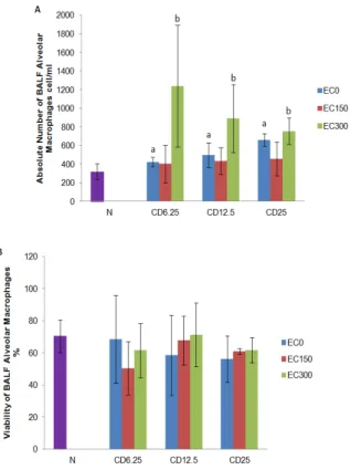
Figure 1 (A) showed that coal dust exposure at all
doses significantly (P < 0.05) increased the absolute
number of BALF alveolar macrophages compared to the negative control group. The supplementation of
E. cottonii had fluctuative effect on the absolute number of BALF alveolar macrophages. The
administration of EC150 insignificantly (P > 0.05)
decreased the number of BALF alveolar
macrophages, whereas EC300 significantly (P < 0.05)
increased the number of BALF alveolar macrophages in coal dust-exposed groups compared to the positive control groups.
Figure 1 (B) showed that coal dust exposure at all
doses insignificantly (P > 0.05) increased the viability of
BALF alveolar macrophages compared to the negative
control group. The administration of EC150 and EC300
insignificantly (P > 0.05) increased the viability of BALF
alveolar macrophages in CD12.5 and CD25 groups
compared to the positive control groups.
Analysis of BALF malondialdehyde levels
Figure 2 showed that coal dust exposure at all
doses significantly(P< 0.05)increasedtheBALF MDA
Figure 1. (A)The number of bronchoalveolar lavage fluids alveolar macrophages. (B) The viability of broncho alveolar lavage fluid alveolar macrophages. Data are presented as mean ± SD (n = 4 each group). aP < 0.05 in comparison with negative control group, bP < 0.05
in comparison with its positive control group. Negative control group (N); group exposed to coal dust at dose of 6.25 (CD6.25), 12.5 (CD12.5), or
25 mg/m3 (CD25); group supplemented with the ethanolic extract of
Eucheuma cottonii at dose of 150 (EC150) or 300 mg/kg BW (EC300);
non-supplemented group (EC0)
Figure 2. The bronchoalveolar lavage fluids malondialdehyde levels. Data are presented as mean ± SD (n = 4 each group). aP <
0.05 in comparison with negative control group, bP < 0.05 in
comparison with its positive control group. Negative control group (N); group exposed to coal dust at dose of 6.25 (CD6.25), 12.5
(CD12.5), or 25 mg/m3 (CD25); group supplemented with the
ethanolic extract of Eucheuma cottonii at dose of 150 (EC150) or
300 mg/kg BW (EC300); non-supplemented group (EC0)
Analysis of BALF malondialdehyde levels
Figure 2 showed that coal dust exposure at all doses
significantly (P < 0.05) increased the BALF MDA
levels compared to the negative control group. The
administration of EC150 and EC300 significantly (P <
0.05) decreased the BALF MDA levels in CD12.5 and
CD25 groups compared to the positive control groups.
Discussion
In this study, we found that the number of BALF alveolar macrophages significantly increased after
chronic PM10 coal dust exposure. This is consistent
and extended with previous study that alveolar macrophages increased after acute coal dust exposre
by intratracheal instillation (1). The increasing may
be due to the accumulation of coal dust particles in the alveoli leading to increased number of alveolar macrophages to engulf the particles and activation of alveolar macrophages and neutrophils. Coal dust exposure causes acute inflammation in lung and recruitment of macrophages and neutrophils to the site of the injury. This study also showed that the viability of alveolar macrophages decreased after coal dust exposure. This is consistent with previous study that coal dust significantly decreased the viability of alveolar macrophages due to apoptosis compared to untreated controls (9, 12, 13).
The administration of EC150 insignificantly
decreased number of BALF alveolar macrophages,
otherwise EC300 significantly increased number of BALF
alveolar macrophages in coal dust-exposed groups compared to positive control group. The increased number of BALF alveolar macrophages in coal
dust-exposed groups due to the administration of EC300 may
neutrophils and monocytes (14). Otherwise, the increased alveolar macrophages in this study may be also due to the method of anesthesia conducted using ether.
Coal dust exposure significantly decreased the viability of BALF alveolar macrophages compared to the negative control group. The reduction in cellular viability may be as a consequence of the coal dust-induced apoptosis of macrophages. Previous study found that coal dust increases pulmonary apoptosis and increases the expression of the pro-apoptotic
mediator Bax (15). The administration of E. cottonii
tended to increase the viability of BALF alveolar macrophages but not reach significant level in coal dust-exposed groups compared to the positive
control groups. This finding indicated that E. cottonii
inhibit apoptosis pathway of macrophage.
This study showed that coal dust exposure significantly increased the BALF MDA levels, an index of lipid peroxidation of the lung tissue. Previous study also showed the same result that coal dust exposure for 60 days caused an increase in lipid peroxidation in rats. Inorganic components of coal dust such as iron, which can stimulate hydroxyl radical formation, and also chromium, nickel, manganese, and vanadium, have oxidative properties able to induce lipid peroxidation. The higher concentration of these active components in coal
dust may result in higher oxidative stress (1, 9). This
study also indicated that E. cottonii was able to
significantly reduce BALF MDA levels in coal-dust exposed rats. This is consistent with previous study
that E. cottonii significantly reduces plasma lipid
peroxidation in rats with high-cholesterol diet (15).
According to the result of ABTS reducing activity in
vitro, E. cottonii was a weak scavenger of the ABTS radical compared to quercetin. However, the decreased BALF MDA levels in the present study may be still due to inhibition of lipid peroxidation via free radicals scavenging mechanism or metals chelating
mechanisms. The antioxidant contents of E. cottonii
such as -tocopherol, -carotene, ascorbic acid, and
polyphenols are able to scavenge the hydroxyl radical and superoxide anions. Phenolic compounds also have a metal chelating potency depending upon their unique phenolic structure as well as the number and location of the hydroxyl groups.
Conclusion
The present study showed that chronic coal dust exposure increases oxidative stress and the absolute number of alveolar macrophages in rat BALFs. The
ethanolic extract of E. cottonii is able to significantly
decrease oxidative stress but not the inflammatory cells.
Acknowledgment
The authors gratefully acknowledge to the Ministry of Research and Technology, Indonesia, for
the SINas research grant of 2012 (Grant ID: RT-2012-1350). We thank all technicians in Laboratory of Pharmacology, Faculty of Medicine, Brawijaya University, for valuable technical assistances, especially for Mrs. Ferrida dn Mr Mochamad Abuhari.
References
1. Pinho RA, Bonatto F, Andrades M, Frota ML Jr, Ritter C, Klamt F, et al. Lung oxidative response after acute coal dust exposure. Environ Res 2004; 96:290-297.
2. Forman HJ, Torres M. Reactive oxygen species and cell signaling: respiratory burst in macrophage signaling. Am J Respir Crit Care Med 2002; 166:S4-8. 3. Lodovici M, Bigagli E. Oxidative stress and air pollution exposure. J Toxicol 2011; 2011. Article ID 487074, 9 pages
4. Nagarani N, Kamaguru AK. Evaluation of anti-inflammatory, antidiabetic, cytotoxic activity of Kappaphycus alvarezii. Int J Pharm Bio Sci 2013; 4:921-929.
5. Devi KP, Suganthy N, Kesika P, Pandian SK. Bioprotective properties of seaweeds: In vitro evaluation of antioxidant activity and antimicrobial activity against food borne bacteria in relation to polyphenolic content. BMC Complement Altern Med 2008; 8:38.
6. Mohamed S. Healing power of Malaysian
seaweeds: cover story. Synth 2011; 32-33:4-5. 7. Ghasemi Fard S, Tash Shamsabadi F, Emadi M, Meng GY, Muhammad K, Mohamed S. Ethanolic extract of Eucheuma cottonii promotes in vivo hair growth and wound healing. J Anim Vet Adv 2011; 10:601-605.
8. Adedapo AA, Jimoh FO, Afolayan AJ, Masika PJ. Antioxidant activities and phenolic contents of the methanol extracts of the stems of Acokanthera
oppositifolia and Adenia gummifera. BMC
Complement Altern Med 2008; 8:54.
9. Kania N, Setiawan B, Widjajanto E, Nurdiana N, Widodo MA, Kusuma HMSC. Peroxidative index as novel marker of hydrogen peroxide involvement in lipid peroxidation from coal dust exposure. Oxid Antioxid Med Sci 2012; 1:209-215.
10.Curry-McCoy TV, Venado A, Guidot DM, Joshi PC. Alcohol ingestion disrupts alveolar epithelial barrier function by activation of macrophage-derived transforming growth factor beta1. Respir Res 2013; 14:39.
11.Draper HH, Hadley M. Malondialdehyde
determination as index of lipid peroxidation. Methods Enzymol 1990; 186:421-431.
12. Ghanem MM, Battelli LA, Mercer RR, Scabilloni JF, Kashon ML, Ma JY, et al. Apoptosis and Bax expression are increased by coal dust in the polycyclic aromatic hydrocarbon-exposed lung. Environ Health Perspect 2006; 114:1367-1373. 13. Huang YC, Soukup J, Harder S, Becker S. Mitochondrial oxidant production by a pollutant dust and NO-mediated apoptosis in human alveolar macrophage. Am J Physiol Cell Physiol, 2003; 284:24-32.
non-specific immunity in rats. Anim Sci Papers Rep 2009; 27:149-157.
15.Matanjun P, Mohamed S, Muhammad K,
Mustapha NM. Comparison of cardiovascular