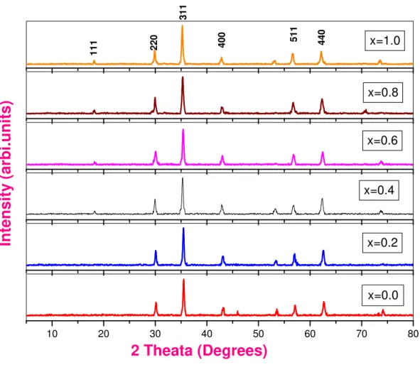
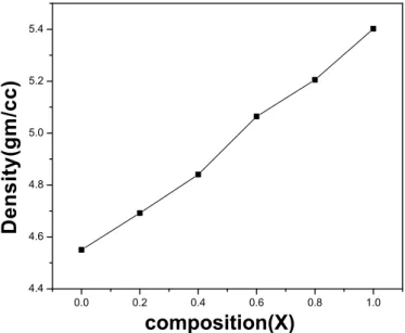
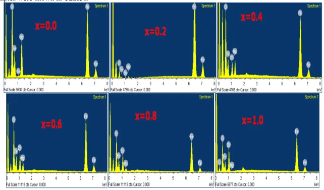
Structure, Morphology and Chemical Synthesis of Mg
1-xZn
xFe
2O
4Nano-Ferrites Prepared by Citrate-Gel Auto Combustion Method
D.Ravikumar
1,B.Kiran Kumar
1, D.Ravinder
2*, Ch.Abraham Lincoln
1*.
1.Department of Chemistry, Osmania University, Hyderabad 500007,AndhrapradeshIndia
2.Department of Physics, Nizam college, Basheerbagh, Osmania University Hyderabad 500001,Andhra Pradesh,India.
ABSTRACT
Mg-Zn Nano ferrites having chemical formula Mg1-xZnxFe2O4(where x=0.0, 0.2, 0.4, 0.6, 0.8, 1.0) were
synthesized by the citrate-gel auto combustion method. Synthesized powders were sintered at 500oC for four hours in air and characterized by XRD, SEM and EDS.XRD analysis shows that cubic spinal structure of the ferrites and the crystalline sizes (D) were found in the range 25-35 nm. The values of the lattice parameter (a) increases and X-ray density (dx) increases with doping of Zn content. Scanning Electron Microscopy (SEM)
studies revealed Nano nature of the samples. An elemental composition of the samples was studied by using Energy Dispersive Spectroscopy (EDS). The observed results can be explained on the basis of composition and crystallite size.
Keywords
:
Mg-Zn Nano- Ferrites; Citrate Gel Auto Combustion Method; X-Ray Diffraction; ScanningElectron Microscope; Energy Dispersive Spectroscopy.
I.
Introduction:
Research on nanoparticles has opened as avenue for various potential applications due to their novel properties. More specifically materials with nano scale dimensions 1-100 nm show extraordinary physical and chemical properties. The reduction in the size of the particles leads to increase in relative surface area which results the increase in the surface to volume ratio. The quantum size effect and the large surface area of nanoparticles dramatically change some of the magnetic properties and exhibit super paramagnetic phenomena and quantum tunnelling of magnetization, because each particle behaves as single magnetic domain [1]. Nanomaterials therefore play a very prominent role in physical, chemical and biomedical applications due to their high surface energies. Among such materials Ferrites are useful magnetic materials because of their versatility, low cost and high electromagnetic performance over a wide frequency range [2]. Spinel type ferrites are commonly used in many electronic and magnetic devices due to their high magnetic permeability and low magnetic losses [3]. Usually ferrite materials have low conductivity i.e. high resistivity which greatly influences the dielectric and
behaviour and the associated applications, magnetic data storage, etc [6].
Magnesium ferrite belongs to a class of compounds having the general formula M+2Fe+32O4
(and also AB2O4) crystallizing with Spinel structure
(where M represents tetrahedral site and Fe represents the octahedral site)[7]. The interesting properties of Spinel ferrites arise from their ability to distribute the cations among the Tetrahedral (A) and Octahedral (B) sites [8].For obtaining the specific properties ferrites can be fabricated by substituting various magnetic and non-magnetic ions which greatly affect the magnetic moments, lattice parameters and exchange interactions [9,10]. Magnesium nano ferrites are the potential materials for various applications due to their high electrical resistivity, low magnetic and dielectric losses. [11, 12]. Doping of MgFe2O4 with one or several metals is
the best method to alter its properties. Several researchers have reported the synthesis of Mg-Cr ferrites using different techniques such as Double Sintering technique [13], Solid state ceramic sintering technique [14], Micro emulsion method [15], Co precipitation technique [16]. To my knowledge a little information is available about the nano sized Mg-Zn ferrites formed by Citrate-Gel Auto
also produces homogeneous and stochiometricoxides[17].
In this work we prepared Nano-ferrites of the composition
Mg1-xZnxFe2O4 (where x=0.0, 0.2, 0.4, 0.6,
0.8, and 1.0) by Citrate –Gel auto combustion method with a very low particles size with the assumption that the magnetic, electrical and dielectric properties would be improved by substitution of Zn with Mg ions. As per the authors knowledge it is the first observation of Mg-Zn nano ferrites obtained with low particles size by citrate-gel auto combustion method. In this paper, we present the Synthesis, XRD, SEM
II. Experimental:
2.1 Materials:
Mg-Zn Nano ferrites having the chemical formula Mg1-xZnxFe2O4 (where x=0.0, 0.2, 0.4, 0.6,
0.8 and 1.0) were synthesized by Citrate-Gel auto combustion technique using magnesium nitrate (Mg(NO3)26H2O, 99%, SD Fine Chem. Ltd. Mumbai,
India), ferrite nitrate (Fe(NO3)29H2O, GR grade, Otto
Chemie Pvt. Ltd. Mumbai, India), Zinc nitrate (Zn(NO3)26H2O, GR grade, Otto Chemie Pvt. Ltd.
Mumbai, India) and Ammonia (NH3, AR grade, SD
Fine Chem. Ltd. Mumbai, India).
Chemical Reaction:
Mg(NO3)26H2O + 2Fe(NO3)29H2O +C6H8O7H2O MgFe2O4 + 29H2O + 4N2 + 6CO2 + 5.5O2
2.2 Synthesis:
Calculated quantities of above nitrate salts were dissolved in double distilled water and required amount of aqueous citric acid solution was added as chelating agent. The mixture was thoroughly stirred to get a homogeneous solution. Ammonia solution was added to this nitrate-citrate mixture to adjust the pH to 7. The mixed solution was heated at about 100oC with uniform stirring and evaporated to obtain a highly viscous gel denoted as precursor. The resultant gel was further heated on a hot plate maintained at a temperature of 180oC to 200oC. When finally all water molecules were removed from the mixture, the viscous gel began frothing. The gel gave a fast flameless auto combustion reaction with the evolution of large amounts of gaseous products (shown in the above chemical reaction). It started in the hottest zones of the beaker and propagated from the bottom to the top like eruption of a Volcano. The reaction was completed in a minute giving rise to dark grey voluminous product with a structure similar to Branched Tree. Finally the burnt powder was ground and was calcined in air at temperature 500oC for four hours to obtain a spinal phase.
2.3 Characterisation:
The structural characterization of the synthesized samples was carried out by Rigaco X ray diffractometer using Cu Kα radiation (λ=1.5405A0) at room temperature by continuous scanning in the range of 50 to 800at the scanning rate of 0.020 per sec to investigate the phase and crystallite size. The Morphology of the samples was studied by Scanning Electron Microscope (SEM). Elemental analysis was carried out by using Energy Dispersive Spectroscopy (EDS).
III. Results and Discussions
:
3.1 XRD Analysis:
10 20 30 40 50 60 70 80
In
ten
sit
y
(
arbi.
u
n
it
s)
2 Theata (Degrees)
x=0.0
x=0.2
x=0.4
x=0.6
x=0.8
440
511
400
111 220
311
x=1.0
Figure 1.XRD patterns of Mg1-xZnFe2O4 nano ferrites
The crystallite size (D) was calculated for all the composition using maximum intensity peak fromScherrer’s formula [18].
Crystallite size = D = 0.91λ/βCosθ
Where λ = wavelength of X-Ray; β = Full width half maximum (radians); θ = Bragg’s angle at the peak position
The crystallite size was in the range of 25 nm to 35 nm (shown in the Table 1) for different composition Conventional methods need a very high temperature and prolonged heating time [13, 14]. Advantages of Citrate gel method over the conventional methods are (i) simple and economic method (ii) use of relatively simple equipment (iii) formation of high purity products (iv) good stoichiometric control (v) production of ultra-fine particles with a narrow size distribution (vi) short processing time (vii) very low processing temperature (viii) low sintering temperature.
Table1. Mg1-xZnxFe2O4 Nano Ferrites Particle size &Latice parameter
Name of the composition Particle size(D) nm Latice parameter(A) nm
MgFe2O4 34.55 8.356
Where, a = lattice parameters; d = interplanar spacing and h k l are the miller indices
A plot of the lattice parameters Vs compositions is shown in Fig.2, which indicates the variation of the lattice parameter with composition. The lattice parameters are found to decrease linearly with increase of ions in the Mg-Zn nano-ferrites system indicating that the system obeys Vegard’sLaw [19].
0.0 0.2 0.4 0.6 0.8 1.0
8.35 8.36 8.37 8.38 8.39 8.40 8.41 8.42
L
a
tt
ic
e
P
a
ra
m
e
te
r(
A
o
)
composition
Figure2.Variation of latice parameter with composition for Mg-Zn nano-ferrites The X-Ray density (dx) is calculated using the following formula [20] and were tabulated in Table2
X-Ray density (dx) = 8M / Na3 (gm/cc)
Where, M = molecular weight of the sample N = Avogadro’s number
a = lattice parameter
Table2. Density and unitcell volume of Mg1-xZnxFe2O4 nano ferrites with composition
Name of the composition Densitygm/cm3 Unit cell Volume (Ao)3
MgFe2O4 4.52 583.438
Mg0.8Zn0.2Fe2O4 4.692 589.323 Mg0.6Zn0.4Fe2O4 4.84 591.434 Mg0.4Zn0.6Fe2O4 5.064 591.857 Mg0.2Zn0.8Fe2O4 5.205 592.704
ZnFe2O4 5.4021 594.187
A plot of the x-ray density (dx) Vs compositions is shown in Fig3. The X-Ray density depends on the
0.0 0.2 0.4 0.6 0.8 1.0 4.4
4.6 4.8 5.0 5.2 5.4
D
e
n
s
it
y
(g
m
/c
c
)
composition(X)
Figure 3.Variation of x-ray density with composition
Volume of the unit cell is calculated as V=a3
The calculated values are tabulated in Table 2. It is observed that volume of this unit cell increases with increase in Zn content. It is because Volume of the unit cell depends on the lattice parameter which increases with increase in Zn content. The X-Ray density (dx) and volume of the unit cell (V) of Magnesium Ferrite were
in good agreement with the reported values 4.518 gm/cc and 588.06(A0)3 from ICDD data.
0.0 0.2 0.4 0.6 0.8 1.0
582 584 586 588 590 592 594 596
V
o
lu
m
e
o
f
u
n
it
c
e
ll
(
A
o
)
composition(X)
Figure4. Variation of Volume of the unit Cell with composition
composition
da dB
MgFe2O4
3.618 2.954 Mg0.8Zn0.2Fe2O4
3.63 2.964 Mg0.6Zn0.4Fe2O4 3.634 2.967
Mg0.4Zn0.6Fe2O4
3.635 2.968 Mg0.2Zn0.8Fe2O4
3.637 2.969 ZnFe2O4
3.64 2.972
The relation between hopping length for Octahedral and Tetrahedral sites as a function ofZn content (x) was shown in Fig.4. It is observed that the distance between the magnetic ions increases as the Zn content increases. It may be explained due to the fact that Mg ion has smaller radius (0.714A0) than Zn ion i.e. (0.828A0) which makes the magnetic ions become closer to each other and hopping length decreases.
0.0 0.2 0.4 0.6 0.8 1.0
3.615 3.620 3.625 3.630 3.635 3.640
H
o
p
p
in
g
l
e
n
g
th
(A
o
)
Composition
0.0 0.2 0.4 0.6 0.8 1.0 2.952
2.954 2.956 2.958 2.960 2.962 2.964 2.966 2.968 2.970 2.972 2.974
H
o
p
p
in
g
l
e
n
g
th
(
A
0
)
composition(X)
Figure5. Variation of hoping length dB with composition
3.2Morphology by SEM:
The morphological analysis was performed using SEM (Scanning Electron Microscope). The secondary electron images were taken at different magnifications to study the morphology by SEM.Fig.6 shows SEM representative micrographs of the sample with different amounts of substitution.
It can be seen from SEM micrographs of various compositions that the morphology of the particles is similar. They reveal largely agglomerated, well defined nano particles of the sample powder with inhomogeneous broader grain size distribution. Such broader size distribution is characterized of mechanically activated nano sized particles. The agglomeration of particles is also because they experience a permanent magnetic moment proportional to their volume [22].
Heating results in the well-faced grains to form solid bodies. It is a porous network of sintered bodies’ exhibiting foam like structure.
samples were shown in Table4.
Figure6. Mg1-xZnxFe2O4 EDS with composition
Table 4.Elements of each sample composition analysed by % of weight and atomic % obtained by EDS
Element
FerriteCompositi on
O Fe Mg Zn
Weight % Atomic % Weight %
Atomic % Weight %
Atomic %
Weight %
Atomic %
MgFe2o4 36.37 61.56 51.56 25.00 2.06 3.44 0.00 0.00
Mg0.8Zn0.2Fe2O4 5.47 16.81 84.78 74.61 0.98 1.98 8.77 6.59
Mg0.6Zn0.4Fe2O4 34.85 62.99 47.00 24.34 6.21 7.38 11.94 5.28
Mg0.4Zn0.6Fe2O4 30.49 59.46 47.30 26.42 4.36 5.60 17.85 8.52
Mg0.2Zn0.8Fe2O4 32.98 63.26 43.13 23.70 2.31 2.91 21.58 10.13
ZnFe2O4 16.08 41.05 60.55 44.29 0.00 0.00 23.32 14.57
Conclusions
Citrate-Gel Auto Combustion Technique is a convenient way for obtaining a homogeneous nano sized mixed Mg-Zn ferrites.
X-Ray Diffraction pattern confirms the formation of cubic Spinel structure in single phase without any impurity peak. It is in good agreement with the standard data from ICD.
The size of various Mg-Zn Nano- ferrites was in the range of 20-35nm.
The lattice parameters are increases with the increase of Zn substitution in Mg-Znferrites which indicates that the mixed Mg-Zn nano-ferrite system obeys Vegard’s Law.
SEM micrographs of various compositions indicate the morphology of the particles is similar. They reveal largely agglomerated, well defined nano particles of the sample powder with inhomogeneous broader grain size distribution.
EDS data gives the elemental % and atomic % in the mixed Mg-Zn ferrites and it shows the presence of Mg, Zn, Fe and O without sprecipitating cations.
IV. Acknowledgements:
The authors are very thankful to Prof.V.Uma Head Dept. Of Chemistry, University College of Science, Osmania University,Hyderabadand Prof.M.Vithal Dept. Of Chemistry,University College of Science, Osmania University, Hyderabad and Prof. T.L.N. Swamy, Principal, Nizam College for their support and encouragement to carry out this research work.
References:
[1.] Surender Kumar, Tukaram J. Shinde, Pramod N. Vasambekar. Adv. Mat. Lett. 2013, 4(5), 373-377Doi: 10.5185/amlett.2012.10429 [2.] Dixit, Gagan.; Singh, J.P.; Srinivasa, R.C.;
Agarwal, H.M.; Chaudhury, R.J. Adv. Mat. Lett.2012, 3(1),21-28Doi: 10.5185/amlett.2011.6280
[3.] T. Giannakopoulou; L. Kompotiatis; A. Kontogeorgakos; G. Kordas. J.magn. Magn. Magn. Mater.246,360.Doi: 10.1016/S0304-8853(02)00106-3
[4.] M.A. Ahmed, Phy.Status solidi A.1989,111,567.Doi:
10.1002/pssa.2211110222
[5.] E.Olsen;J.Thoustad,J.Appl.Electrochem.1999, 29,293.Doi:10.1023/A:1003464304448 [6.] Kaur, Balwinder.;Arora, Manju.; Shankar,
Ajay.; Srinivasa, Avanish Kumar.; Pant, Rajendra Prasad. Adv. Matt. Lett. 2012, 3(5), 399-405.Doi:10.5185/amlett.2012.7288
[7.] E.W. Gorter,
Magn.Mater.2003,265,311.Doi:10.1016/S030 -8853(03)00280-4
[9.] A.K.M. Zakaria ; M.A. Abgar; S.G. Erikson; F.U. Ahmed; S.M. Yunus; R. Delaplane; V.Stanliu;P.Svedlindle,Mater.Res.Bull.2004,3 9,1141.Doi:10.1016/j.materresbull.2004.02.0 15
[10.] L.B.Kong;Z.W,Li;G.Q.Lin;Y.B.Gan,J.Am.Ce ram.Soc. 2007,90,2104.Doi:10.1111/j.1551-2916.2007.01691.x
[11.] Y.Konseogolu;H.Kavas;B.Aktas,PhysStatusS olidi 36,1595.Doi:10.1002/pssa.20053104 [12.] V.B Kawade; G.K. Bichile; K.M. Jadhav;
Mater.Lett. 2000,42,33.Doi:10.1016/S0167-577X(99)00155-X
[13.] FaizunaNesa; A.K.M. Zakaria; M.A. Saeed Khan; S.M. Yunus; A.K. Das; S.G. Eriksson; M.N.J. Khan; D.K.Saha;M.A. Hakim,World J. Condens. MaterrPhy.@012, 2, 27.Doi:10.4236/wjcmp.2012.21005
[14.] MuhammedJavedIqbal; ZahoorAhmad; TurqutMeydan; YevgenMellikov, J.Appl. Phy.2012,111,033906.
Doi:10.1063/1.4704197
[15.] N.M Patil; S.D. Jadhav; U.B. Sankpal; M.R. Kadam; B.K. Chougule; N.S. Gajbhiye,
Mater.Chem.andPhy. 2009,
113,233.Doi:10.1016/j.matchemphys.208.07. 066
[16.] K. Rama Krishna; D. Ravinder; K. Vijaya Kumar; Ch. Abraham Lincoln, World J.
Condens. Mater
Phy.2012,2,153.Doi:10.4236/wjcmp.2012.23 025
[17.] CullityB.D.,Elementspf XRD, Addison Wesley publishing Company, Inc., Reading, Machusetts,1,1959,132.
[18.] M.J. Iqbal; m.N. Ashiq; P. Hernandez-Gomezb; J.M. Munoz, J. Magn. Magn.
Mater.2008, 320,
881.Doi:10.1016/j.jmmm.2007.09.005 [19.] R.C. Kumbale; P.A. Sheikh; S.S. Kamble;
Y.D. Kolekar, J. Magn. Magn. Mater.2009,478,599.Doi:10.1016/j.jmmm.20 05.03.007
[20.] B.Viswanatham; V.R.K. Murthy; Ferrie Materials: Science and Technology, Narosa Publishing House, 1990,4.