UNIVERSIDADE DE TRÁS-OS-MONTES E ALTO DOURO
Histopathological comparison between two methods
of hepatic biopsies obtained by Tru-cut needle and
Laparoscopic cup Forceps in dogs
Dissertação de Mestrado em Medicina Veterinária
Cláudia Isabel Oliveira Santos
Orientador:
Professora Doutora Anabela Gouveia Antunes Alves, UTAD
Coorientador:
Professor Doutor Carlos Alberto Antunes Viegas, UTAD
II
UNIVERSIDADE DE TRÁS-OS-MONTES E ALTO DOURO
Histopathological comparison between two methods
of hepatic biopsies obtained by Tru-cut needle and
Laparoscopic cup Forceps in dogs
Dissertação de Mestrado em Medicina Veterinária
Cláudia Isabel Oliveira Santos
Orientador:
Professora Doutora Anabela Gouveia Antunes Alves, UTAD
Coorientador:
Professor Doutor Carlos Alberto Antunes Viegas, UTAD
Composição do Júri:
Professora Doutora Anabela Gouveia Antunes Alves
Professora Doutora Cristina Maria Teixeira Saraiva
Professora Doutora Isabel Cristina Ribeiro Pires
III
DECLARAÇÃO
Nome:
Cláudia Isabel Oliveira Santos
C.C.:13755959
Correio eletrónico: Clauosantos27@gmail.com
Título da Dissertação de Mestrado em Medicina Veterinária: Histopathological
comparison between two methods of hepatic biopsies obtained by Tru-cut needle
and Laparoscopic cup Forceps in dogs
Orientador: Professora Doutora Anabela Gouveia Antunes Alves
Coorientador: Professor Doutor Carlos Alberto Antunes Viegas
DECLARO QUE ESTA DISSERTAÇÃO DE MESTRADO É RESULTADO DA MINHA
PESQUISA, TRABALHO PESSOAL E INDICAÇÕES DOS MEUS ORIENTADORES.
TODAS AS FONTES CONSULTADAS ESTÃO DEVIDAMENTE REFERENCIADAS
NO TEXTO E BIBLIOGRAFIA FINAL. ESTE ESTUDO NÃO FOI APRESENTADO
EM NENHUMA OUTRA INSTITUIÇÃO PARA OBTENÇÃO DE QUALQUER GRAU
ACADÉMICO.
Vila Real, 28 de Janeiro de 2015
IV
“O saber não ocupa espaço, e sim o vazio de nada saber”
Eça de Queiroz
Para o Meu Querido Avô, eterna fonte de inspiração e força.
Sem os seus ensinamentos o meu gosto pelo saber e conhecimento nunca seriam os mesmos.
V RESUMO
Existem numerosas técnicas para a obtenção de biopsias hepáticas, 2 das quais foram avaliadas no presente estudo.
O uso dos 5 mm oval cup Forceps, permite a obtenção de amostras em número e tamanho significativo de um modo minimamente invasivo, além de fornecer visualização direta do órgão (Tams, 2003; Twedt & Monnet, 2013). No entanto alguns autores afirmam que estas amostras não revelam as lesões mais profundas e exacerbam outros componentes morfológicos normais. (Rothuizen & Twedt 2009)
A Tru-Cut needle, uma das técnicas mais populares, económica e fácil de realizar necessitando apenas de sedação profunda rapidamente tem sido ultimamente alvo de duras críticas quanto à sua capacidade de diagnóstico e respetiva validade. (Kirberger & Stander, 2011; Radlinsky, 2013)
Existem opiniões divergentes sobre estas técnicas e poucos estudos onde a sua morfologia histopatológica seja descrita, inclusive não encontramos nenhum estudo referenciado nas bases de dados disponíveis, onde ambas as técnicas sejam estritamente comparadas. Daí a realização do presente estudo, efetuado em 16 cães com afeções hepáticas, dos quais se obtiveram biopsias hepáticas pelos 2 métodos, e que consistiu numa comparação histopalógica com o principal objetivo de clarificar as diferenças encontradas, entre os 2 métodos nos seguintes parâmetros: fragmentação; número de tríades portais; alterações dos ductos biliares, da veia Porta, da artéria Hepática, da veia Centrolobular; presença de hemorragia, congestão, vacuolização, pigmento, colestase, fibrose, necrose e inflamação; localização da vacuolização, da fibrose, da necrose e da inflamação.
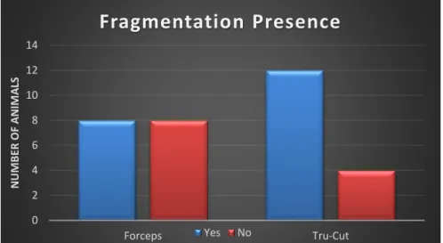
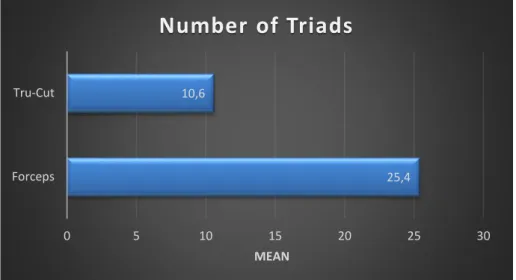
Verificou-se uma elevada fragmentação em ambas as técnicas, o que não era esperado nas amostras obtidas pelos Forceps. Quanto ao número de tríades portais, uma das grandes desvantagens apontadas à técnica por Tru-Cut, o número mínimo de 6 tríades só não foi atingido em 3 animais. A congestão apresentou o dobro da prevalência nos Forceps comparativamente às amostras de Tru-Cut, podendo-se colocar a hipótese deste resultado ter origem na compressão provocada pelo respetivo instrumento. As alterações nos ductos biliares, na veia Porta, na veia Centrolobular; a presença de hemorragia, fibrose, necrose e inflamação foram facilmente identificadas em ambas as técnicas durante a avaliação, apresentando normalmente uma prevalência ligeiramente superior nas amostras dos Forceps. Apenas a presença de vacuolização, pigmento, colestase e as alterações da A. Hepática apresentaram uma concordância estatisticamente significativa na utilização dos 2 métodos. A falta de concordância estatística encontrada nos outros parâmetros, facilmente identificados, deve-se ao baixo número de amostras utilizadas neste estudo.
VI A maior dificuldade encontrada nas amostras obtidas por Tru-Cut foi a localização das lesões, sendo que a necrose e a vacuolização foram os parâmetros onde a dificuldade foi notoriamente maior, apresentando prevalências significativamente inferiores às obtidas por Forceps. Com base na bibliografia esta dificuldade deve-se ao baixo número de tríades portais obtidas, onde são necessárias um mínimo de 15 tríades para localizar lesões e apenas 4 superaram ou igualaram este valor.
A incapacidade de visualizar a totalidade do lobulo hepático impossibilitou o diagnóstico em algumas amostras obtidas por Tru-Cut, no entanto foi possível atingi-lo noutros casos. Quanto aos Forceps, tirando a provável congestão artefactual e a elevada fragmentação não foram sentidas outras complicações para obtenção de um diagnóstico.
Palavras-chave: Biópsias hepáticas, Tru-Cut needle, 5 mm oval cup forceps, doenças hepáticas, avaliação histopalógica, tríades portais
VII ABSTRACT
Innumerous techniques exist for hepatic biopsy obtention, 2 were evaluated in the present study.
The use of 5 mm oval cup Forceps ensures the acquisition of significative numerous and size samples in a minimal invasive way, besides ensures a direct visualization of the organ. (Tams, 2003; Twedt & Monnet, 2013). However some authors affirm that this samples don’t reveal deeper lesions and exacerbate other normal morphological components. (Rothuizen & Twedt 2009)
The Tru-Cut needle is one of the most popular, economical and easy techniques, using only heavy sedation, has been lately target of tough critics due to its diagnosis capacity and credibility. (Kirberger & Stander, 2011; Radlinsky, 2013)
Divergent opinions exists over this techniques and few studies where their morphologic histopathology is described, inclusively we don’t found any study in the data base available, where both are strictly compared. That’s why the present study was carried in 16 dogs with hepatic affections, where hepatic biopsies were obtained by the 2 methods, which consisted in a histopathological comparison with the principal objective of clarify the differences found between the 2 methods in the next features: fragmentation, number of portal triads; ductal, portal vein, hepatic artery and centrolobular vein changes; hemorrhage, congestion, vacuolization, pigment, cholestasis, fibrosis, necrosis and inflammation presence; vacuolization, fibrosis, necrosis and inflammation localization.
A high fragmentation prevalence was found in both techniques, but wasn’t expected in the samples obtained by Forceps. Concerning the number of portal triads, a great disadvantage pointed to Tru-Cut technique, the minimum number of 6 triads didn’t get it only in 3 animals. Congestion presented the double of the prevalence on Forceps when compared with Tru-Cut samples, which take us to consider the hypothesis of this result have origin in compression provoked by this instrument. The ductal, portal vein, centrolobular vein changes; the hemorrhage, fibrosis, necrosis and inflammation presence were easily detected in both techniques during evaluation, presented normally a slight superior prevalence in Forceps samples. Just the presence of vacuolization, pigment, cholestasis and hepatic artery changes presented a significant statistical agreement in the 2 methods. The lack of statistical agreement found in the other features, easily identified, is due to the small number of samples used in this study.
The major difficulty found in samples obtained by Tru-Cut was the localization of lesions, necrosis and vacuolization were the features where the greatest difficulty was found, presenting prevalence’s significantly inferiors to Forceps. Based on bibliography this difficult is
VIII due to the small number of portal triads obtained, where a minimum of 15 are necessary to localize the lesions and just only 4 presented this or superior value.
The incapacity of assess to an entirely hepatic lobule, make it impossible to some Tru-Cut samples arise into a diagnosis, however it was possible to get it in another cases.
In spite of the probable artefactual congestion and high fragmentation it wasn’t sensed other complications to diagnosis obtention in Forceps technique.
Key words: Hepatic biopsies, Tru-Cut needle, 5 mm oval cup Forceps, liver diseases, histopathological evaluation, portal triads
IX ACKNOWLEDGEMENTS
A long journey has been experienced to elaborate this dissertation, final mark in 6 years essentially of learning, dedication and hard work. Unfortunately it´s impossible to express my gratitude to all that have accompanied this passage, that’s why my public thanks are only directed to the people that helped me in the elaboration of this dissertation or supported me in uncertainly and difficult moments lived, while the material that gave origin to the present study had not arrived to my hands.
My gratitude to Professor Anabela Alves by her indispensable assistance and cooperation in interpretation of biopsy slides, for all the kindness, dedication, availability and advices given during the realization of this investigation work.
My gratitude to Professor Viegas since always a reference as professor with his visionary and captivating personality of becoming the Veterinary Medicine in Portugal an excellence. Thank you for your dedication, initial orientation, knowledge and advices transmission.
My gratitude to Dr. Lecoindre by supplying the samples and proposing me the collaboration in this magnificent study. I’m also really thankful for all the knowledge transmitted, availability and patience during my fabulous 3 months of stage at the Clinique Vétérinaire des Cerisioz.
My gratitude to Professor Colaço by his precious help in statistical analysis, availability and patience with my doubts.
Thank you to my dear childhood friend Rafaela by her psychological support and linguistic revision in this work.
My gratitude to my friends and thesis partners Rafa, João and Ricardo that have accompanied from near my write and laboratorial part in Vila Real. Thank you very much by your companionship, support, laugh and despairing moments eased with chocolate or oily food.
Thank you to Sandra, Lita, Dora, André, Peres, Mariana, Carolina, Joh, Lipa, Leo, Ália, Diana, Isabel and Cecília among others friends and partners that give me strength and motivation to continue in the difficult moments.
An especial thank you to my mother Isabel by her dedication, education, sense of work and responsibility implemented in me since I was young. To her and my sister Inês my deepest gratitude for all the help provided in good and bad times.
X LIST OF CONTENTS
CHAPTER 1-LITERATURE REVIEW ... 1
1. Liver biopsy: Initial considerations ... 1
1.1. Indications to perform a hepatic biopsy ... 2
1.2. Pre-biopsy considerations: Patient preparation ... 4
1.2.1. Fasting ... 4
1.2.2. Blood coagulation tests ... 4
1.2.3. Ultrasound examination ... 6
1.2.4. Anesthesia ... 6
2. Hepatic biopsy techniques ... 7
2.1. Laparoscopy or Celioscopy ... 7
2.1.1. Material necessary for the procedure ... 8
2.1.3. Post-operative care ... 13
2.1.4. Advantages of laparoscopy ... 13
2.1.5. Disadvantages ... 15
2.2. Needle biopsies devices ... 16
2.2.1. Cutting or core needle biopsy ... 16
2.2.1.1. Available instrumentation and advantages: ... 17
2.2.1.2. Procedures ... 19 2.2.1.3. Post-operative considerations ... 21 2.2.1.4. Advantages ... 21 2.2.1.5. Disadvantages ... 22 2.2.2. Menghini needle ... 24 2.2.2.1. Procedure ... 24 2.2.2.2. Advantages ... 25 2.2.2.3. Disadvantages ... 25
XI 2.2.3.1. Procedures ... 26 2.2.3.2. Advantages ... 26 2.2.3.3. Disadvantages ... 27 2.3. Laparotomy ... 28 2.3.1. Procedures ... 29 2.3.2. Advantages ... 30 2.3.3. Disadvantages ... 31 3. Sample Management ... 33
CHAPTER 2. Histological comparison between two methods of hepatic biopsies obtained by Tru-cut needle and Laparoscopic cup forceps in dogs ... 34
1. Aim ... 34
2. Material and Methods ... 34
2.1. Sample collect ... 34 2.2. Sample Analysis ... 36 2.3 Data analysis ... 36 3. Results ... 38 3.1. Histopathological evaluation ... 38 3.2. Analytical Statistics ... 48 4. Discussion... 49 5. Conclusion ... 58 CHAPTER 3- REFERENCES ... 60
XII LIST OF FIGURES
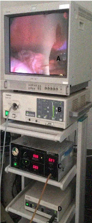
Figure 1. Videolaparoscopy tower………..………...……….8
Figure 2. 5 mm trocar………..…..………….…………..………8
Figure 3. 5-mm endoscopic cup biopsy forceps ……….……..………8
Figure 4. Ventral approach by Hasson Technique ………..….………...…..11
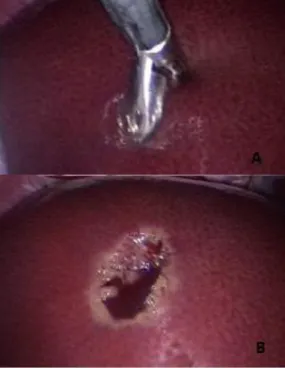
Figure 5. Liver Biopsy ……….….………..………12
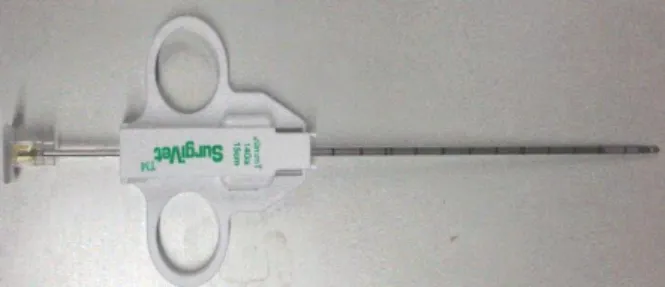
Figure 6. Manual Tru-Cut needle ………..…………...………17
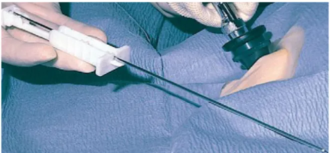
Figure 7. 14G Semiautomated Tru-cut needle ………...…………18
Figure 8. Automated Tru-Cut needle ……….………..………18
Figure 9. The Menghini needle ……….………..……….24
Figure 10. Material necessary to perform the Fine Needle Aspiration Technique……….25
Figure 11. Skin Punch biopsy technique………..………29
Figure 12. Guillotine technique ………..………..………30
Figure 13. Graphic representation of Fragmentation’s presence……….38
Figure 14. Mean of triads number achieved in samples ………39
Figure 15. Intense glycogen accumulation in centrolobular zone. PAS stain ……….….40
Figure 16. Biliary pigment in a Forceps sample with Fouchet stain……….………41
Figure 17. Porto-portal or bridging necrosis. Reticulin stain...42
Figure 18. Carcinoma of undetermined origin with comedo necrosis in Portal zone………...45
XIII LIST OF TABLES
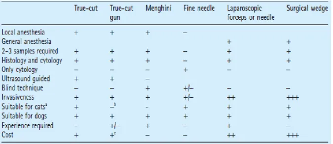
Table 1. Summary of advantages, disadvantages and requirements for liver biopsy…………32 Table 2. Patient relevant information: Personal and Clinic……...……….….33 Table 3. Prevalence of different Vacuolization locations in Tru-Cut and Forceps technique...41 Table 4. Different Fibrosis Grades and Prevalence found in Tru-Cut and Forceps technique..42 Table 5. Prevalence of different Fibrosis locations in Tru-Cut and Forceps technique………43 Table 6. Prevalence of different Necrosis locations found in Tru-Cut and Forceps technique..43 Table 7. Prevalence of different Inflammation locations in Tru-Cut and Forceps technique…44 Table 8. Prevalence of diagnosis found in Tru-Cut and Forceps technique………..47 Table 9. Prevalence of main features found in Tru-Cut and Forceps technique…..………….47 Table 10. Summary of statistical agreement between Tru-Cut and Forceps technique……..48
XIV ABBREVIATIONS, SYMBOLS AND ACRONYMS
US- Ultrasound
WSAVA- World Small Animal Veterinary Association ALT- Alanine transaminase
AST- Aspartate transaminase ALP- Alkaline phosphatase
GGT- Gamma glutamyl transpeptidase PU/PD- Polyuria/Polydipsia
PSSs- Portosystemic shunts PTT- Prothrombin time
APTT- Activated thromboplastin time
PIVKA- Proteins Induced by Vitamin K Absence UAS- Ultrasonographic Harmonic Scapel
FNA- Fine needle aspiration G- Gauges
1 CHAPTER 1-LITERATURE REVIEW
1. Liver biopsy: Initial considerations
Liver biopsy ideally consists in a representative sample of liver tissue, which can be obtained by numerous different techniques. (Wang et al. 2004) There is no absolute recommendation for the use of one specific method. Regardless of being obtained percutaneously by needle biopsies devices blind or ultrasound (US guided), surgically by laparoscopy or laparotomy it’s important to realize the risks and restrictions of each method. Diverse authors have divergent opinions about different techniques but all agree that the method of choice should always be based on each particular case, clinician’s experience and material available. (Burger et al. 2006; Rothuizen et al. 2006; Richter 2013)
Usually is indicated on suspicion and evaluation of hepatobiliary disease or systemic disease that affects the liver. (Wang et al. 2004; Webster 2010) For the majority of authors, the histological analysis is referred as the golden standard to accurately achieving into a diagnosis. (Richter 2003; Richter 2012)
Biochemical tests, radiographs and ultrasonography can determinate if hepatic disease exists and identify the chronicity or acuteness of the process. Different diseases produce similar alterations in hepatic function or laboratory tests. Consequently none of these alterations are able to accurately identify the cause, appropriate treatment or predicts prognosis. (Bexfield 2012; Lorenzi 2010)
Liver illnesses are classified mainly on histopathologic appearance of biopsy specimens obtained. This samples only represent a small percentage of the liver, therefore the examination of an adequate amount of tissue, able to represent the entire liver is required, essentially to distinguish not only changes in the hepatobiliary and vascular structures but also identify the location and typical distribution patterns of any kind of lesions. (Barnes et al. 2006; Rothuizen et al. 2006) Thus it is also important to classify the stage, reversibility, severity, progression and the origin of hepatic disease. (Richter 2003)
A great difference, even disagreement in the histopathological evaluation made by different pathologists revealed unclear and non-uniform classifications of hepatic diseases. Due the great need showed of standard patterns to classify hepatic diseases in small animals equally around the world the International Liver Standardization Group of World Small Animal Veterinary Association (WSAVA) was created and a book with guidelines was published in 2006. According to the information on it, the final diagnosis of a hepatic disease doesn’t depend only from histopathology, should always be based on the interactions between the clinician, the pathologist and the ultrasonographer. (Rothuizen 2006; Rothuizen & Twedt 2009)
2 With the continuous improvements in biopsy methods and in noninvasive imaging of the liver, hepatic biopsy starts to be finally a routine and better recognized as an essential tool and not only on diagnosis but inclusively in the management of patients with hepatic disease. The clinicians can accompany the disease evolution and make a treatment more specific instead of supportive care only or even in the worst cases counter-productive or dangerous (Richter 2003; Watson 2005).
The liver biopsies considerations discussed in this dissertation are far away from a simple histopathological comparison between two techniques. This work is also a help in a resumed and simple way to standard and consolidate a confident approach at liver diseases in dogs, by identify properly the indications, when is the right time to perform a liver biopsy, to choose the most indicated technique in spite of their limitations, management and importance of size sample, the value of histopathology and standards but also sensitize the veterinary class to the importance of cooperation between the clinician and the pathologist to have a successful representation of the liver’s patient.(Cullen 2007; Rothuizen 2006)
1.1. Indications to perform a hepatic biopsy
After a clinical history revision, careful clinical examination, laboratory testing and diagnostic imaging, obtaining samples of liver tissue is the next step on the investigation of liver disease. There are several indications to perform it and all the clinicians should be aware of them.
The most common indication is detection of abnormal hepatic function. (Bexfield 2012) An increasement of serum biochemical hepatic and cholestatic enzymes activity (alanine transaminase (ALT), aspartate transaminase (AST), alkaline phosphatase (ALP) and gamma glutamyl transpeptidase (GGT) of unknown origin is an example (Watson 2014; Webster 2010).A wisely evaluation, where combinations of all the complementary exams in patients with elevation of serum hepatic and cholestatic enzymes activity should be made first, to rule out other pathological causes as hyperadrenocorticism, diabetes mellitus and congestive heart failure. In an asymptomatic patient, for example, exposed to a preanesthetic analysis or in routine biochemical profile, when an increase of hepatic enzyme activities is detected, the majority of the authors recommend the assessment of hepatic function with serum bile acids measurement. If the results are not significantly elevated, the biochemistry profile is repeated in 4 to 6 weeks. If the activity of hepatic enzymes persists elevated, a hepatic biopsy is then indicated(Richter 2003; Twedt 2005).According to the guidelines, an indication to perform a hepatic biopsy also exits in asymptomatic patients that have moderate to severe elevations of serum hepatic enzymes persistent for at least 3 months. Or if there are mild to moderate
3 elevations that persists for at least 6 months for no reason. However in any of these situations, where clinical signs suitable with hepatic disease develop, the biopsy should not be delayed. The same is applied in concurrent elevations of serum bile acids. (Richter 2013)
Abnormal hepatic function tests (pre and post-prandial bile acids, bilirubin and blood ammonia measurements) obtained in patients that manifests clinical signs compatible with hepatic disease (polyuria/polydipsia, vomit, anorexia and jaundice) is another example of indication. The evaluation of serum albumin, glucose, urea nitrogen and clotting factors, as
another functional tests, are indicated to access the hepatic function in cases where the liver impairment is so markedly abnormal despite the maintenance of hepatocellular membrane. Therefore serum activities of hepatic enzymes are normal in spite of the pronounced liver dysfunction. (Richter 2003; Tams 2005) The portosystemic shunts (PSSs), terminal cirrhosis, and metastatic hepatic neoplasia are some of the examples. (Richter 2013)
Diagnostic imaging can reveal specific anomalies (PSSs and extrahepatic bile duct obstruction) but unfortunately, not every imaging results indicate a specific diagnosis. Studies confirm that focal hepatic masses or diffuse echotextural changes (hypoechoic or hyperechoic, uniform or mottled) aren’t exclusive from one pathology to be considered pathognomonic, so a biopsy may be warranted, depending on the other laboratory tests results and clinical signs. (Richter 2012; Richter 2013)
Abnormal hepatic size of unknown cause (microhepatia or hepatomegaly) is also a common indication. As for elevated enzymes, in cases of hepatomegaly is important rule out the same nonhepatic causes. Other indication to perform a biopsy is suspicion of diffuse or focal neoplasia by hepatic enlargement and as complementary to US exam. (Burger et al. 2006; Richter 2003)
In animals, for example, that have been through chemotherapy, hepatic biopsy it’s an important process to evaluate the progression of the disease as the adjustment of treatment that sometimes isn’t based on histological liver condition and consequently became nonspecific. Serial biopsies are often required to confirm remission of hepatic malignancy, lymphoma is one of this examples, and response to the treatment in hepatobiliary diseases. (Richter 2003; Webster 2010) The chronic hepatitis cases, also examples of biopsy indication, because it is difficult to determinate if there is ongoing inflammation and resolution or progression of fibrosis during long-term therapy. Especially in patients that receive glucocorticoid therapy, the serum hepatic enzymes activity will always be high independent of the underlying disease. (Bexfield 2012; Richter 2013)
Some multisystemic disorders are interesting, from the clinical view, therefore the hepatic involvement assess can be evaluated through a simple biopsy. (Richter 2003; Rothuizen 2008)
4 Even with the existence of a significant amount of literature with several indications to perform a hepatic biopsy, the right time to pursue is often a judgment call. (Richter 2013) Every case is a particular case and most of the times there is not only one right approach and timing. The owners will, economic availability as well as the clinician’s knowledge, experience and material available are also factors that can weigh in this decision-making. (Casaús & Pérez 2008)
1.2. Pre-biopsy considerations: Patient preparation
To choose the right technique to perform a liver biopsy, some considerations about the patient and procedures must be arranged for every particular case. (Rothuizen & Twedt 2009)
1.2.1. Fasting
The animal that is going to be submitted to a hepatic biopsy should be fasted at least for 12 hours. It’s a very important instruction, since the visceral surface of the liver is covered by the stomach, and with a full stomach this process may be harder and more dangerous. (Rothuizen et al. 2006; Rothuizen 2008) In case of sedation or an anesthesia needed, the fact that the animal is fasted is also helpful besides the obviously need for the US performance. (Rothuizen & Twedt 2009) When the evaluation of glycogen deposits in hepatocytes is required, it is easier to compare the samples in a fast liver since the glycogen stores fluctuate according to carbohydrate ingestion. Many patients are already fasted since some of the common symptoms observed in hepatic disease are nausea and decrease of appetite. (Rothuizen et al. 2006)
1.2.2. Blood coagulation tests
One of the majors concerns for the clinic, in a potential candidate to a hepatic biopsy, is the possibility of uncontrolled bleeding.(Richter 2006; Rothuizen et al. 2006)The average blood loss from a liver biopsy is reported to be around 2 ml in normal dogs. (Rothuizen & Twedt 2009) The extent of bleeding has always been associated with a prolonged time action of coagulation factors. This abnormal extension can be due to hepatobiliary diseases that reduce the production of clotting factors, inadequate intestinal resorption of vitamin K or disseminated intravascular coagulation. Different coagulation factors can be evaluated, based on the most
5 recent literature. The advised factors to measure are prothrombin time (PTT), activated thromboplastin time (APTT), and platelet count included on complete blood count. A buccal mucosal bleeding time is also recommended specially in breeds associated with Von Willebrand disease (Rothuizen & Twedt 2009; Rothuizen et al. 2006). In this cases desmopressin acetate should be given 30 to 60 minutes before the procedure. (Bunch 2002; Rothuizen 2008).
The evaluation of this factors should be performed shortly before the biopsy (no more than 24 hours) because in cases of hepatic diseases they may change quickly. (Rothuizen et al. 2006). However, the reserve capacity of the liver in producing clotting factors is so huge
that, for majority of diseases, the production is rarely decreased to the point of becoming a limited factor. (Rothuizen & Twedt 2009) In cases of severe clotting factors deficiency, the literature recommend avoid liver biopsy realization, despite of none indicate which is the limit to consider that biopsies are unsafe. Therefore the most common complications cannot be predicted with prolonged PTT, APTT or thrombocytopenia. According some authors experience at least the doubling of the PTT can be tolerated. (Bunch 2002; Rothuizen et al. 2006)
Fibrinogen is the most critical indicator for bleeding predisposition. Especially when reductions below 50% of the reference level (1g/l) occurs. (Favier 2009; Rothuizen et al. 2006)
Inclusively in a study made on 1000 dogs subject to hepatic biopsy, where animals with fibrinogen less than 6% were excluded, in the others 94% only three cases have significant hemorrhage. (Rothuizen et al. 2006)
Some authors refer thatVitamin K administration may be helpful in certain situations. In one report vitamin K administration improved proteins induced by vitamin K absence (PIVKA) times in 10 of 23 dogs with hepatic disease. According to other authors experience the bleeding following hepatic biopsy does not correlate with coagulation tests, including the PIVKA test. (Richter 2003; Monnet 2010)
In the most common cases where significant hemorrhage occurs, technical errors such as damaging large vessels were committed. (Richter & Arnell 2010; Richter 2012; Richter 2013) As prevention, when the clinic have doubts about animals status, fresh frozen plasma can be given 2 hours before the procedure (Rothuizen et al. 2006; Rothuizen & Twedt 2009).
6 1.2.3. Ultrasound examination
Independently the clinician suspects of a vascular, biliary or parenchymal hepatopathy, an US examination of the abdominal cavity, focusing on the liver, biliary tract and portal vein, is an essential preparation before liver sampling (Rothuizen et al. 2006).
Systematic evaluation is required to identify carefully the size, presence of local (discrete nodules, cysts, masses, or focal areas of heterogeneous mottling) or diffuse changes (increased or decreased echogenicity, diffuse mottling) in the architecture of liver (less sensitive for the last one). (Mayhew & Weisse 2012; Richter 2013) Diameter and wall thickness of extrahepatic, intrahepatic bile ducts and gall bladder; vascular changes or the presence of free abdominal fluid (Barnes et al. 2006).Vascular diseases in spite of usually leading to a typical and uniform histological pattern, US colorflow doppler is the majority of times an essential tool to diagnose several changes as congenital PSSs or arteriovenous fistulas. (Rothuizen & Twedt 2009; Webster 2010)
Local changes should be sampled selectively with US guidance needle or by direct view as laparoscopy and laparotomy. While diffuse changes can be unsystematically sampled with one of the many techniques existents.
These US evaluation provides important information for the clinician and the pathologist. It is very important, for example, to rule out severe congestion in an enlarged liver, which is a contraindication for tissue sampling. Besides US complements the histopathologic diagnostic and helps the clinic to choose which is the best biopsy approach for every particular case (Rothuizen et al. 2006; Rothuizen & Twedt 2009).
1.2.4. Anesthesia
The majority of drugs normally administered during anesthesia undergo hepatic metabolism. In patients with moderate to severe dysfunction, a large group should be avoided and some used in reduced doses. In hypoalbuminemic animals it’s important have in mind that the drugs have a superior duration in the organism. (Bennett 2007; Radlinsky 2013)
A liver biopsy can be taken under local anesthesia and sedation when a fast sampling method is chosen, if the animal is cooperative or isn’t stable for general anesthesia (Rothuizen et al. 2006). Local anesthesia is commonly performed with lidocaine which is enough because the liver is completely painless if the larger bile ducts are avoided. When the clinic opt by general anesthesia premedication can be done with midazolam, the preferred benzodiazepine in alert patients. The induction agents of choice are etomidate and propofol, especially this last
7 one if there is the risk of occurring seizures. A constant rate infusion of fentanyl is the opioid of choice during the procedure. Both isoflurane and sevoflurane inhalant anesthetics can be used to maintenance (Radlinsky 2013).
All the clinicians should have in mind, that considerations in respect of the animal (fasting, coagulation profile, US examination and anesthesia) are not the only factors associated with complications in a liver biopsy. Potential complications came associated within patient preparation, material available and the clinician’s experience. With an experienced operator that knows well the procedure and equipment in good conditions most of the techniques are safe and have a low complication rate (Rothuizen & Twedt 2009; Watson 2014).
2. Hepatic biopsy techniques
When the decision on biopsy the liver’s patient is taken the next step is to choose one of the existent techniques. Consultation of the pathologist prior to biopsy can be useful since different diagnosis techniques require different handling and amounts of tissue. Hepatic biopsies can be obtained by laparotomy, percutaneously with core needles or aspiration needles, with or without US imaging guidance and by laparoscopy. ( Bexfield 2012; Cullen 2013; Richter 2003; Vasanjee et al. 2006) Each one has advantages and disadvantages, inclusively in veterinary literature it is controversial the superiority of different techniques between different authors. Though, no restricted recommendation exits for the use of one specific method. It is important to understand the possibilities and limitations under the given circumstances. Independent of that, all biopsies should be performed under sterile conditions (hair clipping, washing and disinfection). Therefore the technique selected depends on clinician’s preference, animal stability, available equipment and biopsy size needed to diagnostic tests. (Casaús & Pérez 2008; Rothuizen et al. 2006; Vasanjee et al. 2006) A detailed description about material, methods, advantages and disadvantages which includes sample size, fast cost, bleeding of each technique is described in this chapter. (Brovida & Rothuizen 2010)
2.1. Laparoscopy or Celioscopy
Laparoscopic technique is a minimal invasive way to assess the internal organs. It’s achieved by distention of the abdominal cavity with gas and using a rigid telescope that allows the examination of all peritoneum (Monnet & Twedt 2003; Monnet 2010). At least six types of
8 tissue samples can be collected by laparoscopy: Fine needle aspiration, Tru-cut needle, excisional biopsy (with grasping forceps, ultrasonographic harmonic scalpel (UAS), endoloop and snare suture) peritoneal washing, aspiration of fluid (from gallbladder or hepatic cysts) and brush cytology of visceral surface. (Freeman et al. 1999)
2.1.1. Material necessary for the procedure
• Videolaparoscopy tower with monitor, light source, insufflator and camera. (Figure 1) • 5-mm laparoscope
• A Veress needle
• 5-mm trocars (Figure 2)
• 5-mm endoscopic cup biopsy forceps (Figure 3) • 5-mm endoscopic blunt probe
• Gelfoam absorbable gelatin sponge (optional) (Case & Alvarez 2014; Monnet & Twedt 2003)
Figure 1. Videolaparoscopy tower.
(A) Monitor; (B) Light source; (C) Insufflator; (D) Camera;
Figure 2. 5-mm trocar:
Formed by obturator and cannula.
9 2.1.2. Procedure
The primary step is to determine the best entry location for the initial port, according to the lesion’s place and surgeon’s preference. The two most common are ventral midline and the right-side midabdominal approach. (Monnet et al. 2008; Monnet 2010)
A left-side midabdominal entry site is rarely used since gallbladder is not satisfactorily visualized and the entry lies over the spleen location, turning the procedure more difficult. (Twedt & Monnet 2005; Twedt 2011) However, it can be used in cases of left lateral or medial hepatic lobe affection. (Tams 2003)
The first trocar for pneumoperitoneum establishment and laparoscope placement can be inserted by two classic methods, the Veress needle and the Hasson technique. (Kolata & Freeman 1999; Twedt & Monnet 2005) An advantage of the first technique over the second is that can be used on the side or midline, in contrast with this last one that is routinely done in the ventral midline. (Twedt 2011)
The right-side approach is normally used for evaluation of the liver (85% of the hepatic surface can be examined), the extrahepatic biliary system can be followed easily to its entry into the duodenum and the majority of the abdominal viscera can also be visualized. Yet with this approach the left lateral liver lobe is more difficult of examine. (Freeman 2009; Twedt 2008; Twedt 2011)
The patient is placed in left dorsal oblique decubitus at a 45° angle and the punctures sites are surgically prepared. The Veress needle, which consists in a sharp outer trocar and a blunt inner stylet, which is inserted through the skin just on the right side, laterally to the umbilicus and perpendicularly to the abdominal wall. (Kolata & Freeman 1999; Monnet 2010; Richter 2010) By the hanging drop technique the clinician can ensure that the tip of the needle is the peritoneum: Attach a syringe partially filled with sterile saline to the Veress needle, aspirate and check for any body fluid. If none is detected, inject a small amount of saline. Detach the syringe and lift the abdominal wall. The saline in the hub of the needle will drop, if the tip of the needle is in the abdominal cavity. (Kolata & Freeman 1999; Monnet & Twedt 2003; Richter 2012)
The abdomen is then insufflated with gas (generally CO2) to a suitable pressure (10 to
12 mm Hg), determined by a pressure gauge, an automatic insufflator or when the abdominal wall is tympanic to touch. Over distension should be avoided, because decrease of venous return or deterioration of ventilation status can occur. (Monnet 2010; Richter 2010) Once the desired degree of pneumoperitoneum is reached, a 0.5- to 1.0-cm skin incision is made between the last rib and the flank on the right lateral abdomen. (Case & Alvarez 2014; Richter 2010) An incision larger than the cannula will cause gas leak and lead to removal or fall during
10 de portal insertion so careful must be taken. (Freeman 2009; Moore & Ragni 2012) In patients with a small liver, or in large dogs, the entry site should be positioned more cranially. In the opposite cases a more caudal entry site allows an increased working space. (Moore & Ragni 2012; Richter 2010) The trocar and cannula together are then inserted into the abdominal cavity a twisting motion. The surgeon’s forefinger can be extended down with the shaft or grasp the cannula 3 cm from the tip with the free hand to prevent excessive insertion into the abdomen. (Monnet & Twedt 2003; Twedt & Monnet 2005)
Once the trocar is removed the valve inside the cannula closes avoiding insufflation loss. The remote light sourceand the camera are connected to the laparoscope with a fiber optic cable and when it is inserted through the cannula the valve opens automatically. All the viscera should be inspected carefully to ensure that hemorrhage or iatrogenic trauma didn’t occurred. The insufflation line is then exchanged, from the Veress needle to this cannula. The needle is removed and the incision is prolonged to 0.5 cm at minimum to introduce by direct visualization a second cannula. (Kolata & Freeman 1999 Moore & Ragni 2012; Richter 2010) The main disadvantages of Veress needle technique, beside of the inadvertent abdominal viscera damage, are the insufflation of omentum, falciform ligament or even the space between the abdominal musculature and air embolism. (Twedt 2011)
When multiple organ examination and biopsy is required, the animal can be placed in dorsal recumbency. If the patient is positioned in a head-up position, the convex surface of the liver is easily visualized, except in obese patients that can be obscured by the falciform ligament.To access to the concave surface, the animal has to be in a Trendelenburg position (with the head down). Angling the head to the side increases visualization to the contralateral liver lobes. (Kolata & Freeman 1999; Monnet et al. 2008; Moore & Ragni 2012) In this decubitus the pneumoperitoneum can be established by the open or Hansson technique. The classic technique uses an especial component, a cone known as an olive, which fits over the cannula of a 10 mm trocar, with flanges for attaching sutures. This initial port is placed just caudal to the umbilicus, avoiding the fat pad. Make a 5- to 7-mm incision through the skin and fascia place two stay sutures in the last one. (Kolata & Freeman 1999; Moore & Ragni 2012; Twedt & Monnet 2005)A blunt obturator-cannula is then inserted, also with a twisting motion. A gas-tight seal is achieved by tying the stay sutures to olive or by placing a purse-string suture around it. The pneumoperitoneum is established with the same cautions described earlier
11 The main advantages of this technique when compared with the Veress needle, are reduction of visceral trauma and insurance that insufflation is achieving intraperitoneally. As disadvantages the placement can be only on ventral midline and the initial skin incision needs to be larger, raising the risk of subcutaneous emphysema. The introduction of cannulas that do not require a trocar has diminished even more the risk of this technique. (Moore & Ragni 2012; Twedt 2011)
Secondary ports are inserted in locations that provide optimal access to the liver and other organs in which biopsy is required. The ports should be separated enough (30º to 60º) to allow manipulation of instruments and should not be placed too close or too far from the place to biopsy. (Kolata & Freeman 1999) When only one secondary portal is used, it is usually placed in a paramedian position either right or left cranial quadrant of the abdomen, normally on the side of the telescope operator’s dominant hand. (Figure 4) The placement of the second cannula, under laparoscopic visualization, should be made with the same cares mentioned earlier, especially not to put it cranial to the last rib. If a second secondary port is needed is usually placed on the contralateral side. In cases of focal lesions should be placed ipsilateral. (Moore & Ragni 2012; Twedt 2011)
The liver should be evaluated cautiously for size, texture, colour, margins and presence of adhesions, mass lesions or nodules. A blunt probe, introduced carefully by one of the secondary ports can be useful for move omentum away, lift and inspect the liver lobes individually. (Case & Alvarez 2014; Monnet 2010; Tams 2003) The normal liver should be uniform, smooth, dark red, with distinct margins, and should not tear or bleed when manipulated. The gall bladder and biliary system can also be examined by retracting the scope slightly, elevating the right lateral and right middle lobes. The presence of anomalies as swelling discoloration areas and obstruction should be discarded. A normal gallbladder should be thin walled and easily compressible. (Monnet et al. 2008; Moore & Ragni 2012; Richter 2010) Grasping forceps should not be used to manipulate liver lobes, because often too friable and the risk of traumatic injury and bleeding is big. (Freeman et al. 1999; Monnet 2010; Twedt & Monnet 2013)
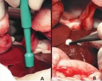
The majority of authors recommend the use of a 5-mm oval cup biopsy Forceps to perform liver biopsies. Some are presented with two small sharp spikes in the tip for grasping Figure 4. Ventral approach by Hasson Technique:
Prepared for a person right-handed with the secondary port on the right side. (Twedt 2011)
12 the tissue. In small dogs (<10 kg) pediatric laparoscopic equipment is preferred, this 3-mm oval cup biopsy Forceps generally provide enough tissue for analysis. When the 5-mm Forceps are used in this patients, only half a cup size should be used to avoid sampling too deep and damage larger vessels causing consequently excessive bleeding. When a deep hepatic lesion is identified by US or suspected but not evident on the liver's surface, a needle core biopsy can be directed to the lesion under direct visualization. (Mayhew 2009; Twedt 2008; Twedt 2011)
In cases of large focal liver masses extreme care should be taken because these lesions are often very vascular, some authors recommend the use of an UAS. When hemorrhage is a concern a prettied loop ligature can be applied before taking samples. (Mayhew 2009; Monnet 2010; Twedt & Monnet 2013)
The next step is identify the areas to biopsy, generally 3 to 4 samples should be taken
from both “normal” and “abnormal” areas, as from the edge and surface of the liver. However, the number of biopsies obtained is conditioned by the risk of bleeding and the tissue amount needed. (Freeman 2009; Monnet & Twedt 2003; Twedt & Monnet 2005).
For obtaining an edge biopsy, insert the closed instrument under the liver lobe next to the region to be sampled. This ensures that no other tissue is in the biopsy cups.The forceps are opened and slowly pull out to the lobe drops into
it, then are closed and held like that for nearly 30 seconds or longer to promote coagulation. Then gently twist and pull firmly to retrieve the tissue sample. To sample the liver surface open and direct the cups about 90-degree angle to the surface, advance 5 to 10 mm into the liver and then close and proceed as for the edge sample (Figure 5). (Case & Alvarez 2014; Twedt 2007)
The sample dimension depend on the clinician’s technique and depth of lobe penetration. The forceps should be retracted closed to prevent any type of damage. (Freeman et al. 1999; Twedt 2011)
The biopsy sites especially but all the abdominal cavity should be examined for active bleeding. It is important have in mind that the telescope can magnify it and, 1 or 2 mL of blood may appear excessive. If there is concern about
Figure 5. Liver biopsy
(A) 5 mm cup forceps taking a biopsy from the liver surface;
(B) Post biopsy from the liver’s surface. (Moore & Ragni 2012)
13
active bleeding, several measures can be taken. The blunt probe, an electrocautery scalpel, hemostatic clips or hemostatic material (Gel-Foam) can be efficiently used on the bleeding biopsy site. (Richter & Arnell 2010; Rothuizen & Twedt 2009; Twedt 2007)
To conclude the procedure the instruments and telescope are removed. The pneumoperitoneum is decompressed, by opening one of the cannula valves. The trocars are
removed and for every incision site the fascia and skin should be sutured. (Case & Alvarez
2014; Richter 2012)
2.1.3. Post-operative care
Post-operative care passes by provide local analgesia as bupivacaine or lidocaine, at the cannula sites, for 1 to 2 days and tramadol for 48 to 72 hours after the procedure. The patient is kept quiet and monitored closely (blood pression, US exam) during 5 hours after procedure, time necessary to decreased of hematocrit as evidence of bleeding. In cases of severe bleeding, fresh frozen plasma, fresh whole blood should be used. In last case entry in surgery to identify and stop the cause of bleeding (Freeman 2010; Twedt & Monnet 2005; Twedt 2011).
2.1.4. Advantages of laparoscopy
In a quick, effectively and minimal invasive way, all the abdominal cavity can be examined. An excellent vision of the liver, biliary tree is allowed regardless of the hepatic size or patient’s conformation (Rothuizen & Twedt 2009). If any other organ involvement exists or in cases of unsuspected disease, a detailed inspection and biopsy not exclusively from the hepatobiliary system can occur (Vasanjee et al. 2006). Small masses (0.5 cm or smaller) and metastatic lesions that may be missed with US imaging, can be individually sampled, as other desired areas, avoiding other structures. Acquisition of multiple larger pieces of tissue, approximately 45 mg from each sample, are sufficient for histopathology, mineral analysis and culture; Inclusively provide identical information to laparotomy samples and risk of sampling artifact is also low and useful especially in cases of heterogeneous liver affections. (Radlinsky 2013;Richter 2006; Richter & Arnell 2010)
A study made by Vasanjee et al. 2006 with 12 dogs, confirms that the 5-mm cup Forceps results in consistent diagnostic samples, with a mean of 16 to 18 portal triads per central and peripheral biopsy specimen. In another study made by Petre et al. 2012 in 80 dogs submitted to laparoscopic liver biopsy, a credible diagnosis was also obtained with a mean of
14 8 to 13 portal triads. However histologic disagreement in the diagnosis was found in 7 of 49 (14%) dogs who multiple slides were made from different biopsy sites. This numbers enforces why multiple samples from different lobes should always be obtained, as the importance of associate it with macroscopic appearance of the hepatic parenchyma and clinical data, despite of considering that samples present a sufficient number of portal triads to reach into a histologic diagnosis. (Richter 2012).
Hemorrhage normally occurs at low quantity, when compared with needle core biopsies, in spite of the laparoscope magnify it. (Radlinsky 2013; Richter 2006) The majority of times the procedures described early are enough to control the unexpected (Twedt 2008). The possibility of deal directly with it by visual confirmation of hemostasis, in a minimal invasive way, is definitely one of the great advantages of laparoscopy (Moore & Ragni 2012). In Petre et al. 2012 study only 3 (4%) of the dogs required blood transfusion after biopsy, and all of them were anemic prior to the procedure. In the Vasanjee et al. 2006 study, that evaluate the hemorrhage grade by different liver biopsies techniques, the UAS by laparoscopy has the lesser hemorrhagic grade, inclusively lesser than the 5-mm oval cup biopsy Forceps. Besides it provides a good sample for histologic analysis. Another study from Barnes et al. 2006 also supports that UAS has the lesser hemorrhagic effect in liver biopsies but compared with 10-mm oval cup forceps.Twedt & Monnet 2013 reinforce and complement this opinion in affirming that the amount of collateral tissue damage induced with an UAS is similar to the damage induced by the oval cup biopsy Forceps.
Generally is performed under general anesthesia but in some rare situations as a debilitated patient, can be executed under sedation with IV propofol and local anesthesia, in the cannula areas, with bupivacaine for example. Takes less time to obtain multiple hepatic biopsies, usually 10 to 15 minutes, when compared with laparotomy so less anesthetic is needled. (Moore & Ragni 2012; Twedt & Monnet 2013; Vasanjee et al. 2006) Routinely the patient is able to be dismissed within hours after the procedure, this fast recovery is attributed to the small surgical sites used. (Radlinsky 2013; Richter 2012) In Petre et al. 2012 study, 76 of dogs (95%) survived to hospital discharge with 1 day mean of hospitalization, after undergoing laparoscopic liver biopsy, only 3 (4%) required conversion to laparotomy (to extraction of 2 big hepatic masses and 1 inadvertent laceration of spleen). Besides the lower postoperative morbidity, decreased infection rate and slight postoperative pain are other advantages of laparoscopy over the laparotomy. (Richter 2012; Rothuizen & Twedt 2009)
15 2.1.5. Disadvantages
All the procedures have potential complications, in laparoscopy all the risks related with a general anesthesia should be also considered. Involuntary organ damage through instrument introduction and manipulation, uncontrollable hemorrhage, overdistension of the abdomen with gas, air embolism and pneumothorax by inadvertent puncture of the diaphragm are the most related problems. With a meticulous and toughly attention to the technique procedures and by being performed for an experienced operator the probability of complications and limitations are minimal. (Moore & Ragni 2012; Richter 2010)
On a review of 250 animals, 217 dogs, the complication level was less than 2% and the severe complications that include anesthetic or cardiovascular-related death or fatal bleeding were very rare. Controlled hemorrhage, organ trauma from the Veress needle, trocar, or instruments were the minor complications more commonly detected. (Rothuizen & Twedt 2009; Twedt 2007;Twedt 2011)
The truly disadvantages pointed by some authors to laparoscopy, when compared with the other techniques, is that sampling the edge of the liver cannot always reflect profounder lesions. This critic supports that the histopathology at the subcapsular level, show more fibrous tissue with nonspecific inflammatory changes and that the probability of compression artifacts by biopsy forceps is big. Consequently this facts can give a misleading and non-representative sample of the entire liver. Other authors have a divergent opinion by affirming that the samples collected by cup Forceps are so large that this circumstances should not be considered a major concern. Inclusively some authors affirm that the truth limitations of this technique are the expense of the equipment, more execution time, the general anesthesia required and higher cost to the client when compared with needle biopsy techniques. (Rothuizen & Twedt 2009; Twedt 2006; Rothuizen et al. 2006)
When a unique, large resectable mass is founded but cannot be removed with security, the procedure should be altered to laparotomy to remove it appropriately. (Moore & Ragni 2012; Twedt 2011) In spite of all advantages pointed to UAS in Barnes et al. 2006 and Vasanjee et al. 2006 studies, the samples obtained with this technique presented significantly more coagulation necrosis, cavitational fragmentation and fibrosis then cup forceps.
Prolonged clotting times, and ascites are relative contraindications for laparoscopy, instead should be considered as an advantageous approach in this situations. (Monnet 2010) When abdominal distension from ascites is present the first cannula can inclusively be placed directly, if it’s possible view through the liquid. To not intensify hypoalbuminemia, examine and sampling, only the necessary fluid should be removed and replaced by CO2. (Twedt 2011;
16 2.2. Needle biopsies devices
Three different types of needles can be used to obtain a liver biopsy:
1. A cutting or core needle which entangles the tissue after cutting it off. The original one used was the Vim-Silvermann needle, which later was replaced by the Tru-Cut type.
2. The Menghini needle which is attached to a syringe for tissue obtention by aspiration (with saline solution) was first reported by Menghini and is still referred as the Menghini technique.
3. A fine needle attached to an empty syringe obtains by aspiration an amount of isolated cells for cytological examination. (Rothuizen et al. 2006)
The abdominal US is several times associated to fine needle aspirates or tissue-core biopsies which became popular due to the ability of visualizing lesions and needle path during biopsy. Many clinicians have assumed that the complications would be reduced and the efficiency optimized. Several studies have examined the utility of different biopsy needles by percutaneous approaches, which were considered safety procedures, in a minimal invasive way, therefore they continue to be required for cytology or histopathologic diagnosis. (Fife 2010; Cole et al. 2002; Richter & Arnell 2010)
2.2.1. Cutting or core needle biopsy
Consists in an inner needle with 2-cm long indentation, which is advanced into the hepatic parenchyma and the tissue is then pushed into the indentation. Thereafter, the outer sleeve with its cutting edge is advanced over the inner needle and the tissue is cut off, after which the entire instrument is withdrawn. They have a sharp tip, which can easily penetrate other structures, so they should only be used under US guidance or direct visualization. (Rothuizen et al. 2006; Rothuizen 2008; Rothuizen & Twedt 2009)
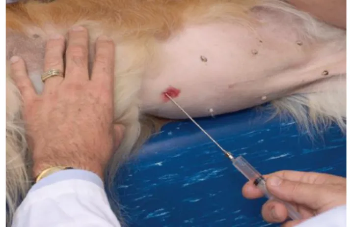
The US guided Tru-cut needle biopsy is the technique most widely used in veterinary medicine for liver biopsy due its minimal invasive approach, compared with laparotomy or laparoscopy. Can be performed under local anesthesia and minimal sedation, heavy sedation or in some rare cases under general anesthesia. (Richter 2006; Richter 2012; Watson 2005) Very small lesions or in close proximity to large vascular structures and a nervous patient are examples where general anesthesia is preferred (Rudloff 2010). Drugs that cause splenic enlargement or panting (acepromazine and thiopental for example) should be avoided as pre anesthetics or inducers. (Fife 2010; O’Brien et al. 2004) Three sizes 18, 16 and 14 gauges (G)
17 are referred in literature for hepatic biopsy in dogs. (Kirberger & Stander 2011) The smaller, 18 G needles collect approximately 5 mg of tissue, so only 14 G needles, which collect 15 to 20 mg, are recommended to use in medium and large sized dogs, as 16 G needles in small ones. (Radlinsky 2013; Richter 2006; Rothuizen et al. 2006) Ideally each individual needle biopsy sample should have a minimal core length of 1 cm, but 2 cm is always preferable. (Rothuizen et al. 2006) The general idea that samples obtained are often too small and nonrepresentative can be minimized and the accuracy of reaching to a reliable diagnosis raised by multiple biopsy samples collection (2 to 4 specimens in animals over 10 kg), that should be taken with the largest Tru-cut needle indicated. (Richter & Arnell 2010; Richter 2013 Watson 2005) However the number of biopsy specimens obtained will also depend of the patient coagulation status, type of diagnostic tests planned, and acceptability of tissue retrieved. (Richter 2003)
2.2.1.1. Available instrumentation and advantages:
Tru-cut needles can be manual, semi-automatic, or for use with an automatic biopsy-gun. ( Rothuizen et al. 2006; Rothuizen 2008; Rothuizen & Twedt 2009) The choice of which tissue-core biopsy is going to be used in each particular situation is usually based on the size, type of lesion, the patient’s safety and personal preference of clinician. (Fife 2010; Richter 2012; Richter 2013; Rudloff 2010)
A manual Tru-Cut needle (Figure 6) needs two hands to operate, so two people are crucial for the procedure. One to place the transducer and another to operate the Tru-cut needle. This makes the devices more difficult to control and their handling takes more time but, in contrast, with practice the operator can control the depth of the needle and the
length of tissue to be sampled. (Fife 2010) With this technique the probability of induced rupture of the liver lobe during respiratory excursions raises. (Rothuizen et al. 2006) Manual devices are cheap but their use is not advised other than during surgery under direct visual control. (Rothuizen & Twedt 2009)
18 Semiautomated needles (Figure 7)
requires manual placement of the internal obturator, which is advanced to the desired depth into the liver, followed by an automatic thrusting of the outer sheath by a spring-loaded mechanism. These needles have the additional advantage of control over it the
final position, since the tip of the needle can be precisely localized before the outer cutting sheath is deployed. (Richter 2012; Richter 2013; Rudloff 2010) Semiautomatic biopsy needles are the most expensive but are very easy to use inclusively are available in various sizes. (Fife 2010; Rothuizen et al. 2006; Rothuizen & Twedt 2009)
After the needle loading, out of abdominal cavity, automatic biopsy guns (Figure 8) advance automatically the cutting needle and external shaft when triggered into the liver in a fraction of a second. The inner cutting shaft advances a specific distance
(1.5 to 2 cm) which can be precisely adjusted, to control the length of tissue sampled, beyond the external shaft when it is triggered. (Fife 2010; Richter 2003; Richter 2006) Which is particularly useful in small dogs and in the case of small lesions. Though a careful handling should be applied when it is used. This guns are reusable and can be sterilized, most of them accept disposable needles of varying sizes. (Fife 2010) The biopsy gun is expensive, but single-use gun needles are cheap. This technique is generally recommended for centers where biopsies are collected frequently. (Rothuizen et al. 2006; Rothuizen 2008) There is minimal displacement of the liver, a shorter intraparenchymal phase with rapid cutting action, and a more reliable yield of tissue which tends to be less fragmented. (Richter & Arnell 2010; Richter 2013)
Actually automated needles are preferred and should be either completely automated or semiautomated. The needles can be operated with one hand, while the other hand operates the US transducer allowing the precise placement of biopsy instrument. (Richter 2006; Richter 2012)
Figure 7. 14G Semiautomated Tru-cut needle.
Figure 8. Automated Tru-Cut needle. (Kirberger & Stander 2011)
19 2.2.1.2. Procedures
The patient is typically placed in dorsal recumbency for the majority of procedures. A rubber trough or V tray can be useful for positioning the animal. (Richter 2003) In patients with particularly small livers, it may be difficult to satisfactorily visualize the needle without gas interfering of the stomach. Frequently helps to place the patient in a 45-degree right ventral oblique position. (Richter 2006; Richter & Arnell 2010; Richter 2013) When the animal is under volatile anesthesia, the assistant can compress the breathing bag to hold the patient in deep inspiration, which improve visualization by consequently move the diaphragm and liver caudally. If a lesion cannot be seen due to overlying gas or bone, changing patient or transducer position usually allows adequate visualization. (Richter 2003; Richter 2012; Richter 2013)
In spite of core biopsies taken generally from only one liver lobe, (normally from the left one to minimize the chance of lacerating the bile ducts or gallbladder which are on the right side), especially in cases that diffuse disease is suspected (Radlinsky 2013; Richter 2013) a careful US examination, normally with linear and convex probes, allows a detailed planning of the procedure based on the type of echo pattern, size of the lesion, proximity to other organs, to blood vessels, determination of cystic or solid tissue, and needle path. If color flow doppler is available it should be used to check the vascularity of the lesion and to avoid major vessels, as arteries which are usually not visible ultrasonographically. For diffuse lesions the transducer is usually placed just caudal and to the left of the xiphoid and aimed at the left medial or lateral lobes. (Kirberger & Stander 2011; Richter 2003; Richter 2006) For focal affections the location of the lesion will determine the position of the animal and needle path (it’s important to have in consideration that 2 cm are required for needle advancement). (Proot & Rothuizen 2006; Radlinsky 2013; Richter 2012)
The area is surgically prepared, a sterile covering should be applied to the transducer (a sterile surgical glove is commonly used), coupling gel is put on it and small stab incision is made on the skin for the needle insertion site. (Richter & Arnell 2010; Richter 2013; Tams 2003)
Three different US guided techniques, normally performed with linear and convex transducers, can be associated with any of the three core needle types, early mentioned, for liver tissue obtention. (Kirberger & Stander 2011)
The first technique is called the indirect guidance method, where the US probe is only used to find the structure of interest, determine the needle depth and then is removed. Generally this technique is not recommended and should only be used when the target is
20 enormously large and when direct visualization of the needle is not required while it is passed blindly into the structure. (Kirberger & Stander 2011; Radlinsky 2013)
Under no circumstances the location of the needle should be determined by moving the needle side to side into the liver, unnecessary trauma can be caused. (Fife 2010; Richter 2013).
The other two techniques allow direct visualization of the needle, while it is passed into the liver. In both techniques, the needle needs to be angled obliquely to be seen on the image. Needles can be scarified or have an echo-enhancing coating to improve visibility. In rare instances when the needle cannot be visualized, the gentle movement of transducer or needle movement in and out, applied gently, should resolve. Ideally this movement should be minimal. Indirect evidence of puncture can also be identified by the organ movement or by motion at the border. Care must be taken to prevent going too deep or completely through the liver with the needle and lacerate other structures under the hepatic lobe. (Radlinsky 2013; Richter 2003; Richter & Arnell 2010; Richter 2012)
The second technique is the freehand technique. It is normally easy to perform with linear transducer. (Kirberger & Stander 2011) The majority of sonographers prefer this technique because allows to the operator handling the transducer with one hand, usually the non-dominant hand, while the other advances the needle into the organ, under direct visualization. This method confers a great flexibility in spite of requiring good hand-eye coordination and taking practice to master it.
The last one is the needle guide technique. Some US machines have a manufactured needle guide that fits into the transducer. This equipment will guide the needle at a pre-set angle and will maintain the orientation to the transducer. The machine software projects the needle path in the screen, allowing accurate placement of it. After the guide attachment, the needle is finally passed through the guide that should also be sterilized before used. One problem with this approach is that the needle guide is often huge and thus the choice of approaches may be limited, particularly in small animals. (Fife 2010; Radlinsky 2013; Richter 2003)
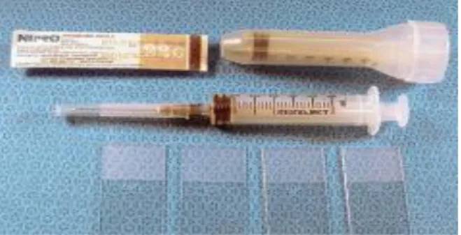
Once the biopsy specimen is obtained, the needle is removed. When multiple samples of different organs are required a separate needle should be used for each organ. (Fife 2010) The core should be resting within the specimen notch of the needle. A 25G needle can be carefully used to transfer the tissue or can be flushed with saline into a 10% formalin vessel. (Cullen 2007;Richter 2003)
21 2.2.1.3. Post-operative considerations
External digital pressure or abdominal compression wrap may be used to control hemorrhage. The patient can also be placed at a position that the weight of the body compresses the biopsy region. If the procedure has occurred without any complications, an US examination with colour flow doppler, normally 1 minute after the compression cessation, is performed to check if excessive hemorrhage occurs, and then 30 minutes after the procedure. Color and mucosal repletion time as packed cell volume/total proteins should also be checked for up to 6 hours after the procedure. Perforation of glisson’s capsule can turn the procedure painful so analgesia post-operatory should be provided.(Bunch 2002; Kirberger & Stander 2011; Richter & Arnell 2010; Watson 2014)
A great diversity of opinions exists about the different techniques and use of different types of needle biopsies. A compilation of the main advantages e disadvantages are presented in this subchapter.
2.2.1.4. Advantages
Percutaneous core biopsies have a low to moderate cost, are considered rapid and easy techniques, especially when the liver is normal or large in size. This is a relatively noninvasive procedure that rarely requires general anesthesia. Inclusively is viewed as the best approach for clinicians with less experience in liver biopsy techniques. (Kirberger & Stander 2011; Radlinsky 2013; Richter 2003)
US guided biopsy when compared with blind percutaneous biopsy offers the advantage of providing more guidance by real-time monitoring nearby vessels as other structures that should be avoided during the needle placement and trajectory following. It’s useful for intraparenchymal lesions that can be visualized, selectively biopsied, enough sensitive and specific for focal lesions (tumors such as carcinomas and lymphosarcoma are an example). Re-check the patient immediately after procedure and search for evidence of complications, as hemorrhage, with this technique is more practical. (Richter & Arnell 2010; Rothuizen et al. 2006; Rothuizen 2008)
A great advantage of needle biopsies is that they collect tissue from as deep as 3 cm, and when associated with US guidance, from any depth. Sampling in depth improves the representation and decreases artefactual sampling of superficial liver tissue. In majority of