Universidade de Aveiro 2010
Departamento de Biologia
Beatriz Lázaro
Pinto
Plasmídeos com Gama Alargada de Hospedeiros em
Ambientes Estuarinos
Broad Host Range Plasmids in Estuarine
Environments
Universidade de Aveiro 2010
Departamento de Biologia
Beatriz Lázaro
Pinto
Plasmídeos com Gama Alargada de Hospedeiros em
Ambientes Estuarinos
Broad Host Range Plasmids in Estuarine
Environments
dissertação apresentada à Universidade de Aveiro para cumprimento dos requisitos necessários à obtenção do grau de Mestre em Biotecnologia, realizada sob a orientação científica da Doutora Cláudia Oliveira, Investigadora em Pós-Doutoramento do CESAM e do Professor Doutor António Correia, Professor Catedrático do Departamento de Biologia da Universidade de Aveiro
o júri
presidente Prof. Doutora Maria da Conceição Lopes Vieira dos Santos
professora associada com agregação do Departamento de Biologia da Universidade de Aveiro
Prof. Doutora Paula Maria Lima e Castro
professora auxiliar da Escola Superior de Biotecnologia da Universidade Católica Portuguesa
Doutora Cláudia Sofia Soares de Oliveira Investigadora em Pós-Doutoramento do CESAM
Prof. Doutor António Carlos Matias Correia
agradecimentos Em primeiro lugar quero agradecer à minha orientadora, Doutora Cláudia Oliveira, pela orientação, pelo grande apoio prestado e por todos os sacrifícios que fez para ajudar para a concretização deste trabalho. Sinceramente muito obrigada por tudo!
Não poderia deixar de agradecer ao Professor António Correia pela oportunidade de desenvolver este trabalho no seu laboratório e pelo seu contributo e apoio ao longo deste tempo.
Agradeço a todos os companheiros de laboratório, e especialmente à Doutora Isabel Henriques e à Juliana Nina de Azevedo pela ajuda e disponibilidade prestada.
Finalmente devo agradecer à Universidade de León (Espanha) por tornar possível a minha estadia em Aveiro.
palavras-chave Camada superficial do mar, Trasferência horizontal de genes, Análise filogenética, Plasmídeos IncP-1, Gene trfA
resumo A transferência horizontal de genes permite a adaptação microbiana a nichos
especiais, dos quais dois bons exemplos são a camada superficial do mar e a coluna de água. Os plasmídeos com gama alargada de hospedeiros (BHR), responsáveis pelo fluxo de material genético entre cromossomas bacterianos (inclusivamente entre microorganismos muito afastados filogeneticamente) têm um papel essencial na evolução das comunidades microbianas. Entre eles, os plasmídeos pertencendo ao grupo de incompatibilidade IncP-1 têm um
interesse especial por causa da sua extraordinária flexibilidade na iniciação da replicação e da estabilidade na sua manutenção numa ampla gama de
hospedeiros. Estes elementos genéticos móveis, além de constituírem ferramentas úteis na engenharia genética, representam uma grande fonte de genes que codificam para características tão significativas como a resistência a antibióticos e a degradação de xenobióticos. No entanto, a diversidade nesta família de plasmídeos BHR tem sido subestimada até agora: actualmente sabe-se da existência de cinco subgrupos divergentes, mas embora alguns plamídeos modelo tenham sido estudados ao detalhe, ainda há muito para investigar.
Em trabalhos anteriores na Ria de Aveiro (costa NW de Portugal), foi feita a captura exógena de plasmídeos bem como o isolamento de bactérias potencialmente hospedeiras de plasmídeos endógenos. Neste trabalho, sequências de nucleótidos específicas foram amplificadas mediante reacções em cadeia da polimerase para determinar a presença de plasmídeos BHR. Os sete plasmídeos IncP-1 detectados, foram em primeiro lugarfilogeneticamente estudados. O alinhamento das sequências de nucleótidos de 281 pb que foram amplificadas e que correspondem a um fragmento do gene trfA (que codifica para uma proteína do início da replicaçao) sugeriu o estabelecimento de dois novos clusters situados filogeneticamente em dois subgrupos diferentes de IncP-1: IncP-1β e o recentemente descrito IncP-1ε. Estes constituem os primeiros replicões IncP-1 provenientes de ambientes estuarinos a serem detectados e isolados. De seguida, uma comparação e caracterização preliminar genética e fenotípica foi realizada com os plasmídeos purificados, considerando a descrição já conhecida dos dois plasmídeos arquétipos evolutivamente mais próximos, pB10 e pKJK5. Assim, as análises de fragmentos de restrição, determinação da inibição do crescimento do hospedeiro na presença de mercúrio e ensaios de resistência a diferentes antibióticos ajudaram a compreender o elevado interesse que recai nestes plasmídeos revelando a diversidade fenotípica e genotípica. Uma completa descrição de qualquer destes novos plasmídeos pode ter una enorme importância ecológica, evolutiva e biotecnológica, incrementada pela sua procedência dum ambiente não clínico. Portanto este trabalho justifica um estudo em maior profundidade destes replicões promíscuos.
keywords Sea surface microlayer, Horizontal gene transfer, Phylogenetic analysis, IncP-1 plasmids, trfA gene
abstract The horizontal gene transfer allows microbial adaptation to special niches, from
which the sea-surface microlayer or the subsurface waters in estuarine environments might be good examples. Broad host range plasmids, responsible for the reshuffling of genetic material between bacterial chromosomes (even amongst distantly related microorganims), play an essential role on the evolution and diversity of microbial communities. Among them, the incompatibility group IncP-1 plasmids have a special interest due to their extraordinary flexibility in the replication initiation and stable maintenance in such a wide spectrum of hosts. These mobile genetic elements, in addition to the helpful genetic engineering tools they mean, represent a great source of potentially useful genes encoding for traits as significant as antibiotic resistance or xenobiotic degradation. Nevertheless, the diversity of this family of BHR plasmids has been underestimated until recently: it is currently known to have five divergent sub-groups, but although some prototype plasmids have been studied in great detail, there is still much left to research.
In previous investigations exogenous plasmid capture was carried out as well as putative endogenous plasmid bacterial hosts isolated in the Ria de Aveiro lagoon (NW coast of Portugal). In this work, polymerase chain reactions were developed to amplify specific nucleotide sequences and determine BHR plasmids presence. From a bioinformatical approach, the seven IncP-1 plasmids detected were firstly phylogenetically studied. The alignment of the amplified 281 bp nucleotide sequences corresponding to a fragment of the replication initiation protein encoding gene trfA suggested the formation of two novel clusters belonging to two different IncP-1 plasmid subgroups: IncP-1β and the lately described IncP-1ε. Additionally, these represent the first estuarine IncP-1 replicons to be detected and isolated. Then a preliminary genetic and phenotypic comparison was performed with the purified plasmids, by taking into account the known description of the evolutionary closest models, pB10 and pKJK5. That way, restriction fragment analysis as well as antibiotic and mercury resistance determination assays helped to comprehend the high significance falling on the captured plasmids by revealing the genetic and phenotypic diversity. A whole description of any of these novel plasmids may have a huge ecological, evolutionary and biotechnological importance, even more due to its precedence from a non-clinical environment. Therefore this work justifies further studies on these promiscuous replicons.
1
Contents:
Index of tables ... 3
Index of figures ... 4
1. Introduction ... 6
1.1 Aquatic environments and microbial communities ... 6
1.2 Bacterial plasmids and their importance in shaping the properties of bacterial communities. ... 7
1.3 The study of plasmids and its biotechnological significance ... 9
1.4 Plasmid biology ...11
1.4.1 Molecular structure of plasmids ...11
1.4.2 Incompatibility groups and BHR plasmids. ...13
1.5 The group of IncP-1 plasmids ...14
1.5.1 Genetic and phenotypic characterization of IncP-1 plasmids ...18
1.6 Methodological considerations ...18
1.6.1 Methods of sample harvest ...18
1.6.2 Methods to carry out the plasmid isolation ...19
1.6.3 Phenotypic traits for plasmids isolation. ...20
2. Objectives ...22
3. Materials and methods ...23
3.1 Sample characterization ...23
3.2 BHR incompatibility group testing ...23
3.3 PCR amplification products detection: electrophoresis and dot-blot hybridization ....26
3.4 DNA sequencing of PCR products and sequence analysis...28
3.5 Plasmid DNA isolation and purification ...28
2
3.7 Electrotransformation of Escherichia coli Top10 with IncP-1 plasmids ...32
3.8 Antibiotic resistance assays ...33
3.9 HgCl2 resistance assay ...34
4. Results and discussion ...35
4.1 BHR plasmid detection and phylogenetic analysis ...35
4.2 Plasmid characterization ...45
5. General discussion and conclusions ...54
3
Index of tables
• Table I. Examples of traits usually associated with plasmids...11
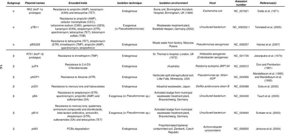
• Table II. Fully sequenced IncP-1 plasmids that represent the full known diversity according to Bahl et al. (2009)...16
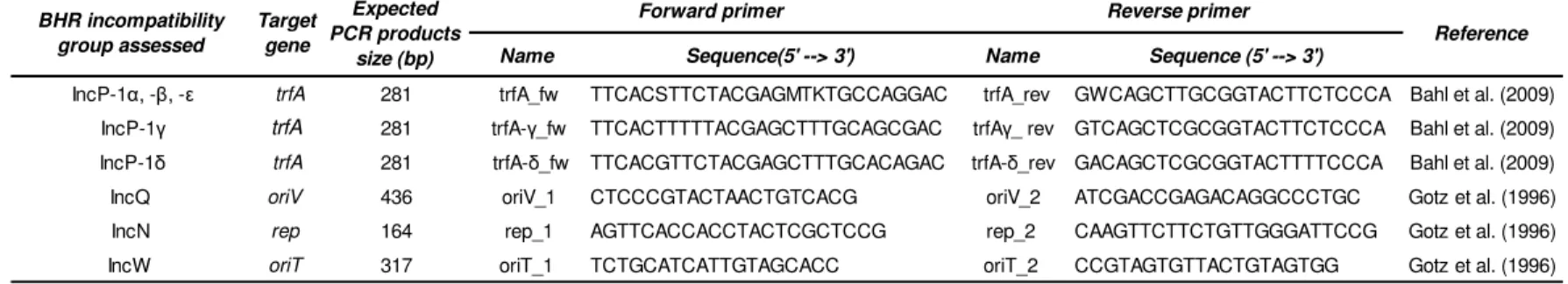
• Table III. Sequence of the primers used for amplification in all polymerase chain reactions...25
• Table IV. Positive controls employed in the PCRs...26
• Table V. PCR conditions followed in each case...25
• Table VI. Compositions of all the solutions used in the dot-blot hybridization...28
• TableVII. Composition and storage temperature of every solution required to perform the plasmid purification...31
• Table VIII. Features of the captured plasmids from the Ria de Aveiro lagoon...36
• Table IX. Features of the captured plasmids from the Ria de Aveiro lagoon and classification into IncP-1 subgroups...40
• Table X. Nucleotide sequences of the seven trfA gene fragments amplified by PCR with the primers trfA...41
• Table XI. Results obtained from the antibiotic resistance assays carried out with the seven captured plasmids harbored by Pseudomonas putida KT2442...50
• Table XII. Results obtained from the antibiotic resistance assays carried out with the four captured plasmids harbored by Escherichia coli Top10...52
• TableXIII. Results obtained from the mercury resistance assay carried out with the seven captured plasmids harbored by Pseudomonas putida KT2442...53
• Table XIV: Comparison between the seven plasmids obtained from the Ria de Aveiro lagoon and the previously described model plasmids, pKJK5 and pB10....59
4
Index of figures
• Fig 1. Schematical structures of IncP-1 and IncQ plasmids...12
• Fig 2. Sampling sites in Ria de Aveiro ...23
• Fig 3. Nucleotide sequence recognition site of endonuclease NotI………..32
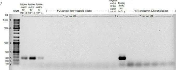
• Figs 4a and 4b. Example of electrophoretical gels showing PCR products obtained
with all employed IncP-1 specific primer sets…...37
• Fig 5a, 5b and 5c. Images obtained from the dot-blot hybridization where the
detection of the products from the PCR screening for IncQ (a), IncN (b) and IncW (c) plasmids performed over endogenous origin bacterial isolates can be seen...38
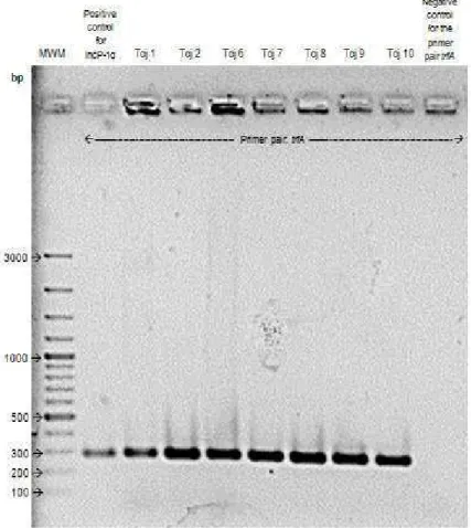
• Fig 6. Image of the electrophoretical gel where the amplification products of the
PCR screening made over seven transconjugants for the IncP-1 α, β or ε BHR plasmids were detected...39
• Fig 7. Score table of the multiple sequence alignment of the amplified trfA gene
fragments from captured plasmids from Ria de Aveiro lagoon (Tcj1, 2, 6-10) performed by ClustalW2...42
• Fig 8. Phylogeny of the amplified trfA gene fragments from captured plasmids from
Ria de Aveiro lagoon (Tcj1, 2, 6-10) in relation to previously described IncP-1 plasmids...43
• Fig 9. Ilustration of the 0,8% agarose gel where every purified plasmid DNA as well
as the genomic DNA extracted from Pseudomonas putida KT2442 (the bacterial host in every cases) are shown...45
• Fig 10. Image of the electrophoretic gel where the restriction fragment profiles of
the plasmids purified from tcj 2,8, 9 and 10 (IncP-1β) and the known pB10 digested with NotI were seen...47
• Fig 11. Image of the electrophoretic gel where the restriction fragment profiles of
the plasmids purified from tcj 1, 6 and 7 (IncP-1ε) and the known pKJK5 digested with NotI were seen...48
5
• Fig 12. Image of the Mueller-Hinton medium plates obtained from the antibiotic
resistance assay made with the plasmids harbored by Pseudomonas putida KT2442...51
• Fig 13. Image of the Mueller-Hinton medium plates obtained from the antibiotic
6
1. Introduction
1.1 Aquatic environments and microbial communities
The specific physical habitat or location of a microorganism is its microenvironment. There, the fluxes and gradients of required oxidants, reductants, and nutrients as well as waste products create a unique niche. However, the microorganisms can create their own microenvironments and niches. When microbial growth happens on surfaces, such as in freshwater and marine environments, biofilms are formed (Prescott et al., 2002b).
The physical frontier between the ocean and the atmosphere is the sea-surface microlayer (SML). This interface between the sea and the air is considered to be the around 1mm-top layer of the ocean (the layer of 10-100 µm-top was employed in this research). If compared with subsurface water (SSW) it is physical-chemically distinct and characteristically enriched with biogenic organic compounds, such as lipids, proteins and polysaccharides. That stratification makes the SML stable enough to exist at typical oceanic wind conditions (Wurl et al., 2009). The main point is that the SML could be made up of gel particles, with similar microbiological and biogeochemical characteristics. Some studies showed an enrichment of transparent exopolymer particles (TEP) in the SML (Wurl et al. 2008). TEPs are normally formed in surface waters from the coagulation of biogenic polysaccharides; chiefly those produced by phytoplankton, and are some of the most ubiquitous gel particles in the marine environment. They are critical in the formation of marine aggregates, acting as the binding matrix or ‘glue’ that holds the aggregate together (Verdugo et al., 2004).
The presence of the surface film and surface tension properties turn the SML into a unique habitat. Bacterial communities that are present in the SML are known as the bacterioneuston. Surface films also take place on all water bodies, marine, estuarine and freshwater, sometimes as visible slicks. The microbial communities established in the SSW are less dense and less structured, and constitute what is called the bacterioplancton (Cunliffe et al., 2009a).
The aggregation of bacterial cells seen in some studies (Franklin et al., 2005; Cunliffe et al., 2009a) most likely takes place in the SML, with bacterial cells attached to
7
the TEP-based gel particles reported by Wurl and Holmes (2008). It has been proposed that this enrichment on attached biofilm-growing cells is the main cause of the distinct properties of bacterioneuston. If, as suggested by Sieburth (1983), the SML is a gelatinous film in which prevails biofilm-growing cells, then biological processes that occur in bacterial communities and that are favored when bacterial cells are in close proximity, such as horizontal gene transfer (HGT) and quorum sensing, could be more frequent. The large specific microbiological diversity supposed to occur in this unique ecological niche supports a much broader molecular and functional diversities. Therefore, its subsequently possible biotechnological value makes its investigation interesting. Nonetheless, studies concerning the molecular microbial ecology of the SML started only recently (Cunliffe et al., 2009a).
1.2 Bacterial plasmids and their importance in shaping the properties of bacterial communities.
Nowadays there is evidence that plasmids and transposable elements can move genetic material between bacterial chromosomes to cause rapid changes in genomes and drastically alter phenotypes (Prescott et al., 2002a). The prokaryotic horizontal gene pool (HGP), defined as the mobile genetic elements (MGEs) and their encoded genes (Thomas, 2000b), plays a vital and essential role in the evolution and adaptation of individual microorganisms and microbial communities by generating genetic variations in bacteria (reviewed in Arber, 2000). Like that, the sequencing of complete bacterial genomes has clearly shown that a large proportion of their genetic diversity have been acquired by HGT and come from distantly related microorganisms. This evidence has been derived from nucleotide sequence comparisons as well as from atypical nucleotide composition (guanine + cytosine content) of large genomic regions or different patterns of codon usage of some genes (Ochman et al., 2000).
Huge efforts have been carried out to catalogue, characterize, and exploit the different aspects of the enormous biodiversity of microorganisms in every natural habitat. In contrast, there is an undeveloped understanding of the abundance, distribution and molecular diversity of MGEs extant in the HGP occurring in most marine, freshwater, and many terrestrial microbial communities (Sobecky, 2002). Probably that is due to the fact
8
that the importance of the HGT in bacterial persistence, diversity and evolution is less widely appreciated (Thomas, 2000b). The first time its significance was accepted match up with the moment when multiple antibiotic resistant pathogens emerged, since MGE played a primary role in the development and dissemination of antibiotic resistance genes and allowed bacterial populations to rapidly adapt to a strong selective pressure. Additionally, plasmid mediated conjugation is probably one of the most frequent mechanisms of HGT (Sota et al., 2008), which explain the particular interest falling on this kind of MGE.
Because of this reshuffling of genes between bacterial cells, populations and communities that makes plasmids remarkably influent in ecological processes, the study of plasmid ecology and transfer together with environmental microbiology and microbial ecology can provide essential information on plasmid evolution. The evolutionary change affects plasmids in the same way as their host bacteria so plasmids have been regarded as units of evolution. In addition, retrospective studies offer a means to infer evolutionary relationships directly from plasmids isolated from geographically similar or remote locations (Sobecky, 2002; Pickup et al., 1996).
Thus, since plasmids represent a large genetic resource of bacterial diversity, research on plasmid distribution, evolutionary relationships, and diversity of plasmids in relation to the natural selection pressures is required in order to comprehend the role they play in the flow of genetic information in natural bacterial communities (Dahlberg et al., 1997; Harada et al., 2006).
Furthermore, the HGT is also manifested in the “plasmids backbones” where recombination within homologous regions of closely related plasmids can be noticed. Since these regions are responsible for replication, maintenance, and plasmid transfer, these recombination events play a key role in increasing or reducing the host range of these plasmids. This could be imperative as much with regard to the species which could obtain new genetic traits as because it sets the genetic backgrounds from which plasmids can purchase new traits (Lipps et al., 2008).
Most of the actual knowledge of HGT is related to plasmids occurring in bacteria of medical and agricultural importance. These plasmids correspond to a limited collection of replicons that surely are not representative of the plasmid populations occurring in every environment.
9
Several plasmid types present in strains isolated from clinical environments can be also found in environmental samples such as manure slurries or marine sediments; however a large part of the plasmids isolated from bacterial strains of natural environments contain replication and incompatibility regions unrelated to those of the known groups of plasmids traditionally found associated to clinical environments. This justifies further evaluation of the prevalence, diversity and evolution of plasmids, especially in nonclinical environments (Thomas, 2000b; Sobecky, 2002; Gstalder et al., 2003).
1.3 The study of plasmids and its biotechnological significance
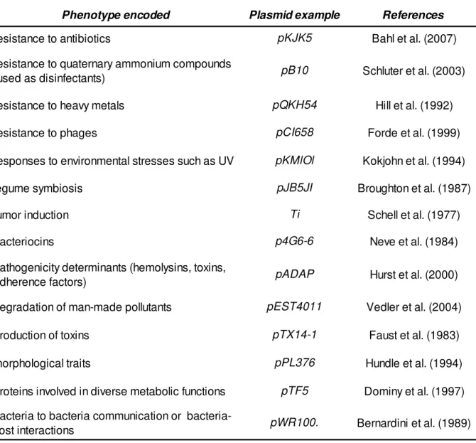
A vast pool of traits usually associated with plasmids rather than chromosomes confer advantages which allow bacterial communities to evolve, adapt to selective pressure and colonize certain habitats. Some examples of those traits are mentioned in the table I (Thomas, 2000b). These kind of habitats, among which a representative case is the SML, due to their extreme characteristics or to the way they bring diverse bacterial species together, may represent a good source of new plasmids or of old plasmid skeletons carrying new recombined segments. Ecological studies that contribute to a deeper knowledge on the distribution of different types of plasmids and their hosts are important in defining which environments are better to exploit, avoiding the inconvenient of blind extensive screenings.
The emergence of large multidrug resistance plasmids due to the strong selective pressure caused by antimicrobial chemotherapy is threatening to reverse development in the treatment of infectious diseases by augmenting human pathogens resistance to antimicrobial agents (Tauch et al., 2003; Tennstedt et al., 2005; Schlüter et al., 2007). Nevertheless, in addition to the clearly imperative significance of increasing the knowledge about resistance genes to clinically relevant antimicrobial drugs, there is also a huge interest in further study other kinds of functions carried by plasmids.
A large number of the xenobiotics that have been introduced to the environment at modern society are recalcitrant. Catabolic plasmids and their host strains have an enormous environmental and ecological significance due to their potential application for environmental biotechnology. Different catabolic pathways may be either chromosomally or plasmid-encoded (Sayler et al., 1990). However, ecologically, plasmid-encoded
10
pathways are beneficial because they provide genetically flexible systems and can be maintained in the population and transferred between bacterial species, without the need of technological support.
In addition to the genes that encode the degradation of man-made organic compounds, those responsible for the metabolism of naturally occurring pollutants are also assumed to be often located on plasmids or other mobile elements (Top et al., 2002).
Besides, since some accessory genes on these plasmids have no homologues and cannot be assigned a function, it is not known yet what phenotypes other than the well-known resistance and degradation functions may be encoded by certain plasmids (Sen et al., 2010). In this context, plasmids seem to be an extraordinary reservoir of molecular determinants of diverse functions and the technological potential of this reservoir deserves to be exploited.
Otherwise, plasmids are powerful vectors of recombinant DNA, allowing its spread in the environment. Its biotechnological importance in this respect lies in its utility as cloning vectors or vehicles of protein expression in different bacterial hosts. A complete knowledge of the distribution, maintenance, recombination, and conjugation of BHR plasmids is consequently essential to evaluate the risk associated with the release of recombinant DNA into the environment (Drönen et al., 1998; Gstalder et al., 2003).
As a conclusion it could be stated that plasmids may have increasingly important biotechnological applications, both respect to the large source of novel traits MGE represent and as useful tools in genetic engineering. Anyway, the first step in researching these potential utilities, as referred before, is further studying plasmid characterization, diversity and evolution.
11
Table I.Examples of traits usually associated with plasmids.
Phenotype encoded Plasmid example References
resistance to antibiotics pKJK5 Bahl et al. (2007)
resistance to quaternary ammonium compounds
(used as disinfectants) pB10 Schluter et al. (2003)
resistance to heavy metals pQKH54 Hill et al. (1992)
resistance to phages pCI658 Forde et al. (1999)
responses to environmental stresses such as UV pKMlOl Kokjohn et al. (1994)
legume symbiosis pJB5JI Broughton et al. (1987)
tumor induction Ti Schell et al. (1977)
bacteriocins p4G6-6 Neve et al. (1984)
pathogenicity determinants (hemolysins, toxins,
adherence factors) pADAP Hurst et al. (2000)
degradation of man-made pollutants pEST4011 Vedler et al. (2004)
production of toxins pTX14-1 Faust et al. (1983)
morphological traits pPL376 Hundle et al. (1994)
proteins involved in diverse metabolic functions pTF5 Dominy et al. (1997)
bacteria to bacteria communication or
bacteria-host interactions pWR100. Bernardini et al. (1989)
1.4 Plasmid biology
1.4.1 Molecular structure of plasmids
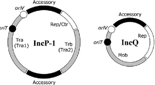
Plasmids can be considered as being made up of two distinct regions: the ‘‘backbone” region, which encodes functions involved in replication, transfer, and
12
Fig 1. Schematical structures of IncP-1 and IncQ plasmids. The white area represents the regions for plasmid replication and control; the grey one, those genes involved in plasmid transfer (or mobilization); black symbolize the accessory genes (Lipps, 2008).
maintenance and control of the plasmid; and the ‘‘accessory” region consisting of genes that may confer specific beneficial traits to the bacterial host (Thomas, 2000a).
In any case, plasmids are not at all static genomes, but instead are plastic genetic mosaics that have evolved over time through the iterative acquisition of various transposons, integrons, gene cassettes, genomic islands and insertion sequences, which are almost always associated with accessory genes, placing them in one or two sites between essential DNA fragments i.e., between oriV (the origin of vegetative replication), and trfA (a gene for plasmid replication) and/or between the two transfer operons that encode mating-pair formation and plasmid transfer trb and tra. Plasmids with almost identical backbones can have entirely different accessory genes (Lipps et al., 2008; Sen et al., 2010; Schlüter et al., 2007). Another essential gene that should be named is oriT, the origin of plasmid transfer. Mob proteins in mobilizable plasmids are necessary to convert the plasmids into the transferable form.
However, plasmids without such functional elements, these accessory elements, have also been described (Kamachi et al., 2006).
In other respects, plasmids are known to have complex recombinational histories. Some survival functions depend on just one genetic locus, but most depend on at least
13
two. The first sorts of modules to emerge would probably have consisted of interdependent functions but, at the same time, independent but mutually beneficial functions probably became clustered, as reviewed in Thomas (2000a).
1.4.2 Incompatibility groups and BHR plasmids.
Plasmid classification has been originally based in a property inherent in them, which is in addition a manifestation of relatedness (since it is the sharing of common elements related to plasmid control of replication and partition). Incompatibility is the inability of two plasmids to be maintained stably in the same cell line (Couturier et al., 1988). Two plasmids which cannot stay in the same cell belong to the same incompatibility group.
Four of these incompatibility groups are considered to be broad-host-range (BHR) plasmids. In spite of the difficulty of determining the host range of a plasmid, as transfer can be tested experimentally only between hosts representing a minute fraction of the bacterial world, Szpirer and co-workers (1999) proposed, in an attempt to define BHR, that plasmids which can transfer and replicate in bacterial species from at least two branches of the Proteobacteria should be viewed as BHR plasmids. Then the conjugative IncP, IncN and IncW, plasmids and the mobilizable IncQ plasmids are regarded as having BHR (Gstalder et al., 2003).
The role of BHR plasmids is remarkable when speaking about interspecies gene exchange since they are most likely the single intermediaries of HGT between distantly related bacterial hosts. IncQ plasmids have the broadest host-range of all of them in gram negative bacteria, followed of those of the group IncP-1 (based in the Pseudomonas classification system). Apart from spreading genes across taxonomically distant species by conjugative transfer, BHR plasmids can partially integrate into the recipient chromosome, or mobilize non-conjugative vectors with a wider host range (Lipps, 2008). They can also retromobilize or retrotransfer i.e., capturing genes or non-self-transmissible plasmids from host in which they cannot replicate (Szpirer et al., 1999). All these reasons make research about BHR plasmids especially interesting.
14
1.5 The group of IncP-1 plasmids
The distinctive characteristic of low copy number plasmids belonging to the incompatibility group IncP-1 is the central control operon coding for at least three global regulators. It gives them flexibility in the replication initiation, multiple stability mechanisms and coordinates regulation of all plasmid backbone functions, providing them with enormous adaptability and stable maintenance in such a wide spectrum of hosts. This ability of replicating and being stably maintained in almost all Gram-negative bacteria as well as being transferred by conjugation to Gram-positive bacteria, yeasts and eukaryotic cell lines justifies the particular interest that fall on IncP-1 plasmids (Adamczyk et al., 2003; Waters, 2001; Harada et al., 2006).
In spite of the high infection transfer rates the IncP-1 plasmids have, horizontal transfer is not enough for them to be maintained as genetic parasites, as they are a burden to their host, so that they need to carry at least occasionally advantageous traits to be maintained in bacterial communities. Consequently, the majority of the IncP-1 plasmids have large regions with acquired genes encoding traits which occasionally might augment the fitness of the bacterial host (Bergstrom et al., 2000).
It appears that the IncP-1 backbone can either carry antibiotic-resistance determinants or degradative operons but to the best of our knowledge, a plasmid that carries both types of genes (antibiotic-resistance and degradative genes) has not yet been identified (Schlüter et al., 2003).
Regardless of the large range of accessory elements among IncP-1 plasmids, they may be phylogenetically grouped into a small number of distinct subgroups based on their backbone sequences. As a result, during the last years, phylogenetic analysis of the 281bp-region of the replication gene trfA, as well as a few other essential backbone genes regions, has allowed the IncP-1 plasmid group to be classified into five main phylogenetic sub-groups: IncP-1α, -β, -γ, -δ and -ε (Sen et al., 2010; Bahl et al., 2009).
The α and β subgroups constitute the two first recognized subgroups and have been extensively studied. The δ and γ subgroups were defined only based on a phylogenetic analysis of their backbone genes. Later, three IncP-1 δ and one IncP-1 γ plasmids were completely sequenced and described (Xia et al., 1998; Vedler et al., 2004; Sen et al., 2010; Hill et al., 1992). The presence of a fifth subgroup, ε, was recently
15
suggested by Bahl (2007) consisting of pEMT3 (from which just a fragment is known) and pKJK5 that has been completely sequenced (Gstalder et al., 2003; Top et al., 1995; Bahl et al., 2007).
In the table II some information about the completly sequenced IncP-1 plasmids that represent the full known diversity within this 281 bp-region of the trfA gene is shown.
1
6
Table II. Completely sequenced IncP-1 plasmids representing the whole phylogeny of this incompatibility group of plasmids according to Bahl et colleagues (2009).
Subgroup Plasmid names Encoded traits Isolation technique Isolation environment Host Acession
number Reference(s) α RK2 (IncP-1α
prototype)
Resistance to ampicilin (AMP), kanamycin
(KAN) and tetracycline (TET) Endogenous
Burns unit, Birmingham Accident,
Hospital, Birmingham, UK (1969) Escherichia coli NC_001621 Datta et al. (1971)
α pTB11
Resistance to ampicilin (AMP), cefaclor monohydrate (CEC), cefuroxime sodium (CMX), gentamicin (GEN),
kanamycin (KAN), streptomycin (STR), spectinomycin, tetracycline (TET), tobramycin
sulfate (TOB)
Exogenous (in Pseudoalteromonas)
Wastewater treatment plant,
Bielefeld-Heepen, Germany (2002) Uncultured bacterium NC_006352.1 Tennstedt et al. (2005)
α pBS228
Resistance to tetracycline (TET), streptomycin (STR), trimethoprim (TMP), ampicilin (AMP),
spectinomycin, streptothricin
Endogenous Waste water from factory, Moscow,
Russia Pseudomonas aeruginosa NC_008357 Haines et al. (2007)
β R751 (IncP-1β
prototype) Resistance to trimethoprim (TMP) Endogenous
St. Thomas’s hospital, London, UK (1972)
Klebsiella aerogenes
(Enterobacter aerogenes) NC_001735 Jobanputra et al. (1974)
β pJP4 Resistance to
2,4-D3-Chlorobenzoate Endogenous (Australia) Ralstonia eutropha JMP134 NC_005912
Don and Pemberton (1981)
β pADP1 Resistance to Atrazine (ATR) Endogenous Herbicide spill site/agricultural soil, Little Falls, Minnesota, USA
Pseudomonas sp. Strain
ADP NC_004956
Mandelbaum et al. (1993) and Mandelbaum et al.
(1995) β pUO1 Resistance to mercury ions and haloacetates Endogenous Industrial wastewater, Japan Delftia acidovorans strain B NC_005088 Sota et al. (2002)
β pB4
Resistance to streptomycin (STR), spectinomycin, ampicillin (AMP) and
sulfonamides (SA)
Exogenous (in Pseudomonas sp.)
Activated sludge from municipal wastewater treatment plant,
Braunschweig, Germany
Uncultured bacterium NC_003430 Tauch et al. (2003)
β pB10
Resistance to mercury ions, quaternary ammonium compounds and disinfectants,
beta-lactam antibiotics, amoxicillin, streptomycin (STR), sulfonamides (SA) and tetracycline (TET)
Exogenous (in Pseudomonas sp.)
Activated sludge from municipal wastewater treatment plant,
Braunschweig, Germany
Uncultured bacterium NC_004840 Schluter et al. (2003)
β pA81 PCBs degradation Endogenous
Polychlorinated biphenyl-contaminted soil, Zamberk, Czech
Republic
Achromobacter
xylosoxidans NC_006830 Jencova et al. (2004)
1
7
Subgroup Plasmid names Encoded traits Isolation technique Isolation environment Host Acession
number Reference(s)
β pB3
Resistance to streptomycin (STR), spectinomycin, ampicillin (AMP), sulfonamides (SA), chloramphenicol (CAP) and
tetracycline (TET)
Exogenous (in Pseudomonas sp.)
Activated sludge from municipal wastewater treatment plant,
Braunschweig, Germany
Uncultured bacterium NC_006388 Heuer et al. (2004)
β pB8 Resistance to streptomycin (STR),
spectinomycin and sulfonamide (SA) Exogenous (in Pseudomonas sp.)
Activated sludge from municipal wastewater treatment plant,
Braunschweig, Germany
Uncultured bacterium NC_007502 Schluter et al. (2005)
β pTP6 Resistance to mercury ions
Exogenous-triparental (in Pseudomonas putida and Escherichia coli K12)
River sediment, river Nura (mercury contaminated), Kazakhstan
(1999–2000)
Uncultured bacterium NC_007680 Smalla et al. (2006)
β pBP136 None Endogenous General hospital, Oita prefecture,
Japan-2002 Bordetella pertussis NC_008459 Kamachi et al. (2006) β pA1 None Endogenous Isolated from ditch sample/soil, Japan (1991) Sphingomonas sp. NC_007353 Harada et al. (2006)
δ pIJB1 2,4-D, malonate degradation Endogenous Garden soil (UK) Burkholderia cepacia 2a NC_013666 Xia et al. (1998)
δ pEST4011 2,4-D malonate degradation Endogenous Agricultural soil sample, Estonia
Achromobacter xylosoxidans subsp. denitrificans EST4002
NC_005793 Vedler et al. (2004)
δ pAKD4 Resistance to mercury ions
Exogenous-bi/triparental (in Pseudomonas putida UWC1/ P putida KT2442,
Escherichia coli CV601, Cupriavidus necator JMP228, and Agrobacterium
tumefaciens UBAPF2)
Agricultural soil in Norway (1998) Uncultured bacterium GQ983559 Sen et al. (2010)
γ pQKH54 Resistance to mercury ions Exogenous (in Escherichia coli ) Epilithic bacteria, River Taff, South
Wales, UK Uncultured bacterium NC_008055 Hill et al. (1992) ε pKJK5 Resistance to trimethoprim (TMP), tetracycline
(TET); slight resistance to spectinomycin
Exogenous (in Escherichia coli
K12 ) Soil/manure, Taastrup, Denmark Uncultured bacterium NC_008272 Bahl et al. (2007)
18
1.5.1 Genetic and phenotypic characterization of IncP-1 plasmids
Plasmid comparison and characterization has often been studied through restriction fragment analyses. There are two important practical aspects to take into account: the first one is the quality of the plasmid DNA, because the analysis may be impaired by partial digest; the second one is the correct selection of the most suitable restriction endonuclease useful to generate appropriate restriction fragment length polymorphism (RFLP) patters, which depends on the plasmid sequence.
Studies concerning the heterologous DNA encoding certain phenotypic traits are of limited value for plasmid classification in its strict sense, since identical transposons can occur on different nonrelated plasmids. Nevertheless, research on these accessory functions is essential to determine the ecological role of plasmids and improve the knowledge about its evolution as a consequence of the responses to environmental stresses. Consequently, the great importance of heterologous DNA makes it be included in the general process for plasmid classification (Tsäpe, 1994; Thomas, 2000b).
1.6 Methodological considerations
Studies in such specific environments as is the case of the SML suppose to play special attention to some difficulties they involve, by establishing singular strategies and methodological approaches as appropriate as possible. The sample collection obtained may vary accordingly to the sampling method and strategy used and that has to be considered. That is the case, for instance, of sampling the SML or the SSW.
1.6.1 Methods of sample harvest
The SSW and even more the SML in the natural environment are submitted to temporal variations at a given place. This temporal and spatial variability of the samples has to be taken into account when analyzing the presence of plasmids in bacteria from environments such as surface waters. The quantity of samples should be a compromise between statistical requirements and practical considerations; the time between sampling, sample processing and analysis should be on the shortest possible way to circumvent shifts in the microbial populations and their activity and the number of parameters to be
19
analyzed should be the highest to increase the impact of spatial and temporal SML heterogeneity on the final results. In addition, natural turbulences may disturb the sampling work. The objective is to attain the closest biological composition of the sample to the original distribution in the SML that can be possible (Agogué et al., 2004; Thomas, 2000b)
The SML sampling techniques used may be therefore the key issue, since different sampling approaches will represent different depths of the SML, thus influencing the samples composition collected. Also, some methods may be biased toward specific cell types, determining this way the results of the research. Consequently, it is essential to carry out a complete comparison of all SML sampling methods utilized, especially when using molecular tools to analyze the microbial ecology of these samples (Cunliffe et al., 2009a).
The most common samplers are the mesh screens (MS), glass plates (GP), hydrophobic (PTFE, polytetrafluoroethylene) and hydrophilic (PC, polycarbonate) membranes (Cunliffe et al., 2009b). Employing these methods bacterial community structures collected in the SML and in SSW can be compared. However, the question of the most suitable method of sampling the SML is far from being answered (Agogué et al., 2004; Cunliffe et al., 2009b).
1.6.2 Methods to carry out the plasmid isolation
The determination of the distribution of plasmids in microbial communities has traditionally been carried out with the initial cultivation of bacterial hosts, with or without the use of selective media types, for the screening and confirmation of plasmids, which is the so called endogenous isolation (Sobecky, 2002).
On the flipside, the small proportion of bacteria accessible to cultivation techniques explains the lack of information on the incidence and abundance of plasmids in the natural environment. In addition, culturable bacteria are known to respond to environmental stress by the formation of viable but nonculturable cells (Roszack et al., 1987).
Conventionally numerous procedures employing a variety of cell lysis and extraction conditions and different commercial extraction kits have been used in the plasmid isolation from culturable bacterial hosts; more recently, methods to isolate plasmids independently from the culturability of their original hosts have become available
20
(Bale et al., 1987). This alternative method, known as exogenous isolation, consists on “capturing” plasmids directly from the bacterial community via mating with a selectable recipient strain. In its two variations, named bi- and tri-parental isolation, it is required that the captured plasmid(s) either be self-transferable or mobilizable and subsequently able to replicate and express (selectable) plasmid-encoded genes in the recipient host (Sobecky, 2002).
Another way of plasmid detection has been established, the screening of environmental DNA. It consists on directly extracting the whole nucleic acids from environmental samples, combined with the amplification of specific plasmid sequences, which is facilitated by the development of DNA probes and PCR primers targeted to conserved plasmid replication and transfer regions (Sobecky, 2002; Thomas, 2000b). However this approach was not used in the present study so it won’t be longer explained. This allows to improve the knowledge about the plasmid distribution in different places, an ecological approach that is, as referred before, essential to focus our research in potentially interesting environments where plasmid isolation strategies should be used.
1.6.3 Phenotypic traits for plasmids isolation.
One of the most toxic heavy metals that can be found in the environment, both due to geological processes and anthropogenic activities, is mercury. Molecular analysis has revealed a huge variety of genes encoding resistance to mercury ions which are reported to occur on a large range of plasmids belonging to various incompatibility groups, contributing that way to bacterial adaptability and reflecting at the same time environmental stresses (Smalla et al., 2006).
Since tetracycline was the first broad spectrum antibiotic with widespread usage in the treatment of diseases, resistance to tetracycline rapidly emerged. Presently, at least 17 different genes conferring tetracycline resistance are known and believed to come from many different ecosystems bacterial communities (Thomas, 2000b).
The known habitual presence of mercury or tetracycline resistance genes in plasmids leads to their use as selective markers to detect or capture plasmids from natural environments by means of the selective isolation of their bacterial hosts.
21
Additionally, the frequent correlation between mercury resistance genes and self-transmissible plasmids, as well as the common genetically link of antibiotic resistance genes to heavy-metal resistance genes, adaptation to mercury pollution may lead the spreading of antibiotic resistance genes throughout the bacterial community (Rasmussen et al., 1998). That adds significance to a deeper study on that kind of plasmids.
22
2. Objectives
Knowing that BHR plasmids may be gene carriers that allow bacterial adaptation to certain special conditions by carrying resistance, degradative or another kind of genes, it is our premise that these genes can have high biotechnological applications and those MGE should be consequently explored.
As referred before, ecological and evolutionary approaches are essential to understand plasmid influence on the flow of genetic information in bacterial communities. Knowledge on the distribution of different types of plasmids and their hosts is also important in defining which environments are more interesting to exploit and have more likely attractive results. The Ria de Aveiro lagoon, due to their definite characteristics and to their special microbial community, may show an important source of new plasmids or of old plasmid skeletons carrying new recombined segments.
Therefore, the current study aims to investigate the presence of BHR plasmids as well as the diversity of IncP-1 plasmid sequences in the plasmids captured from different places of the Ria de Aveiro lagoon and evaluate their phylogenetic relationship together with well characterized IncP-1 plasmids.
Another target of this thesis is a preliminary phenotypic and genetic characterization of the plasmids obtained in order to discuss and justify further research and whole replicons sequencing, both due to potential application in genetic engineering as cloning vectors or as vehicles for protein expression and because of the possible usefulness that the knowledge about some genes contained into it, like, for example, antibiotic or heavy metal resistance genes, may has.
23
Fig 2. Sampling sites in Ria de Aveiro
(Oliveira et al., 2009).
3. Materials and methods
3.1 Sample characterization
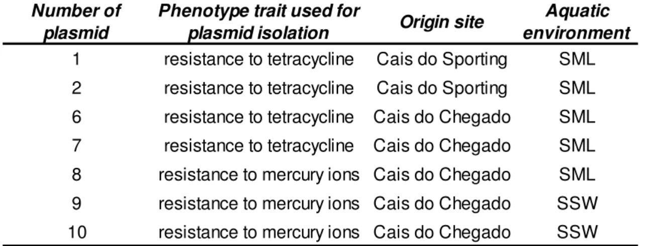
Samples were collected throughout 2008 and 2009 from three contrasting aquatic environments of the Ria de Aveiro lagoon, in the NW coast of Portugal. These sites were different as for salinity, temperature and anthropogenic pressures (run-off from agriculture [1.-Costa Nova-CN], port activities [2.-Cais do Sporting-CS] and industry [3.-Cais do Chegado-CC]). In each sampling site, glass plate method was employed to harvest samples from the SML and a flask submerged at a depth of 40 cm was used to gather samples from the SSW (Ramos, 2009; Oliveira et al., 2009).
On one hand, in order to determine the BHR plasmid distribution by endogenous isolation, the culturable bacterial fraction was analyzed. Right through a previous research investigation (Ramos, 2009) the bacterial strains harvested had been cultured and isolated, resulting in 402 bacterial isolates, 198 from SML and 204 from SSW.
On the other hand, a cultivation-independent approach was carried out by using Pseudomonas putida KT2442 and Escherichia coli CV601 as recipient strains to capture plasmids by conjugation. The 79 transconjugants obtained (through the selective markers ampiciline, tetracycline and mercuric chloride) were confirmed by repetitive extragenic palindromic polymerase chain reaction genomic fingerprints method in a preceding work (Oliveira et al., 2009) in which also the presence of plasmids belonging to the incompatibility groups IncN, IncQ and IncW were tested without any positive result.
3.2 BHR incompatibility group testing
In this work, the PCR amplification of nucleotide sequences of specific replicons of plasmids belonging to the incompatibility groups W, N and Q and to the incP-1 subgroups was performed in order to detect the presence of BHR plasmids in the bacterial isolates
24
representative from SML and SSW. Regarding the transconjugants, PCR amplifications of nucleotide sequences of specific replicons was carried out for the incP-1 incompatibility sub-groups.
All PCR reactions were carried out either on a Bio-Rad iCycler Thermal Cycler (Bio-Rad) or on a Bio-Rad MyCycler Thermal Cycler (Bio-Rad).
The detection of endogenous plasmids by PCR was performed using total DNA extracted from the bacterial isolates with the Silica bead DNA extraction Kit (Fermentas, K0513) as DNA template. Mixtures of the total DNA of each set of 5 bacterial isolates were prepared. After confirming the success of such DNA template dilutions, every mixture was employed as one unique sample for the screening procedure. On the other hand, IncP-1 PCR testing for the 79 transconjugants was carried out. Bacterial suspensions made up by dipping a tiny amount of fresh culture into 20 µl of sterile distilled water were employed, in this case, as DNA template.
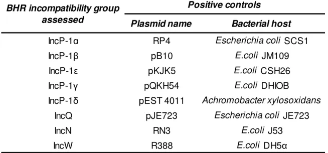
Three different degenerative consensus primer pairs developed by Bahl and co-workers (2009) were used for the amplification of the 281 bp homologous fragments from the gene trfA (which encode the replication initiation protein) from plasmids belonging to different IncP-1 subgroups. Primers, as well as positive controls, are described in tables III and IV, respectively. The reaction mixture consisted in: 1x phusion HF Buffer (Promega); 2,5 mM MgCl2 (Promega); 200 µM dNTP Mix (Bioron); 5% DMSO (Dimethylesulfoxide, Analitical Grade - EuroBio); 0,5 µM each primer; 1 U/µl Taq polymerase (Promega); 1 µl of template DNA in a 25 µl PCR reaction mixture. The PCR cycle conditions are described in table V.
As for IncQ, IncN and IncW plasmids detection, methodology and primers based on these developed by Gotz and co-workers (1996) were used. Every PCR mixture contained 1x phusion HF Buffer (Promega); 3,75 mM MgCl2 (Promega); 200 µM dNTP Mix (Bioron) for IncQ_oriV and 160 µM for IncN_rep and IncW_oriT; 2 % DMSO (Dimethylesulfoxide, Analitical Grade - EuroBio, France); 0,8 µM each primer; 1 U/µl Taq polymerase (Promega); 1µl of template DNA up to 25 µl PCR reaction mixtures. Primers, positive controls and PCR cycle conditions are described in tables III, IV and V, respectively.
2
5
Table III. Sequence of the primers used for the amplification, target genes and expected fragment length of the polymerase chain reaction products correspondent the different incompatibility groups and subgroups tested.
Table V. PCR conditions followed for each gene target.
Name Sequence(5' --> 3') Name Sequence (5' --> 3')
IncP-1α, -β, -ε trfA 281 trfA_fw TTCACSTTCTACGAGMTKTGCCAGGAC trfA_rev GWCAGCTTGCGGTACTTCTCCCA Bahl et al. (2009) IncP-1γ trfA 281 trfA-γ_fw TTCACTTTTTACGAGCTTTGCAGCGAC trfAγ_ rev GTCAGCTCGCGGTACTTCTCCCA Bahl et al. (2009) IncP-1δ trfA 281 trfA-δ_fw TTCACGTTCTACGAGCTTTGCACAGAC trfA-δ_rev GACAGCTCGCGGTACTTTTCCCA Bahl et al. (2009) IncQ oriV 436 oriV_1 CTCCCGTACTAACTGTCACG oriV_2 ATCGACCGAGACAGGCCCTGC Gotz et al. (1996) IncN rep 164 rep_1 AGTTCACCACCTACTCGCTCCG rep_2 CAAGTTCTTCTGTTGGGATTCCG Gotz et al. (1996) IncW oriT 317 oriT_1 TCTGCATCATTGTAGCACC oriT_2 CCGTAGTGTTACTGTAGTGG Gotz et al. (1996)
*primers are ordered to StabVida
Reference BHR incompatibility group assessed Target gene Expected PCR products size (bp)
Forward primer Reverse primer
Temperature Time Temperature Time Temperature Time Temperature Time Temperature Time
trfA (IncP-1α, -β, -ε) 98ºC 30 s. 35 98ºC 20 s. 67ºC 20 s. 72ºC 30 s. 72ºC 5 min
trfA (IncP-1γ) 98ºC 31 s. 35 98ºC 21 s. 67ºC 21 s. 72ºC 31 s. 72ºC 5 min
trfA (IncP-1δ) 98ºC 32 s. 35 98ºC 22 s. 67ºC 22 s. 72ºC 32 s. 72ºC 5 min
oriV (IncQ) 94ºC 5 min 35 94ºC 1 min 57ºC 1 min 72ºC 1 min 72ºC 10 min
rep (IncN) 94ºC 5 min 35 94ºC 1 min 55ºC 1 min 72ºC 1 min 72ºC 10 min
oriT (IncW) 94ºC 5 min 35 94ºC 1 min 51ºC 1 min 72ºC 1 min 72ºC 10 min Final extension Cycles
Initial denaturation Target gene
Denaturation Annealing Extension
26
Plasmid name
Bacterial host
IncP-1α
RP4
Escherichia coli SCS1
IncP-1β
pB10
E.coli JM109
IncP-1ε
pKJK5
E.coli CSH26
IncP-1γ
pQKH54
E.coli DHIOB
IncP-1δ
pEST 4011
Achromobacter xylosoxidans
IncQ
pJE723
Escherichia coli JE723
IncN
RN3
E.coli J53
IncW
R388
E.coli DH5α
Positive controls
BHR incompatibility group
assessed
3.3 PCR amplification products detection: electrophoresis and dot-blot hybridization
The PCR amplification products corresponding to IncP-1 plasmid specific sequences were separated on a 1.5% agarose (Lonza) gel in TAE (Tris-Acetate-EDTA, 5Prime) buffer at 80V during 70 minutes. Either GeneRuler DNA Ladder Mix (Fermentas) or GeneRuler 100bp DNA Ladder Plus, ready-to-use (Fermentas) were loaded in all gels as molecular weight markers. Gels were stained with ethidium bromide and banding patterns were visualized by using a Molecular Imager FXTM system (Bio-Rad Laboratories).
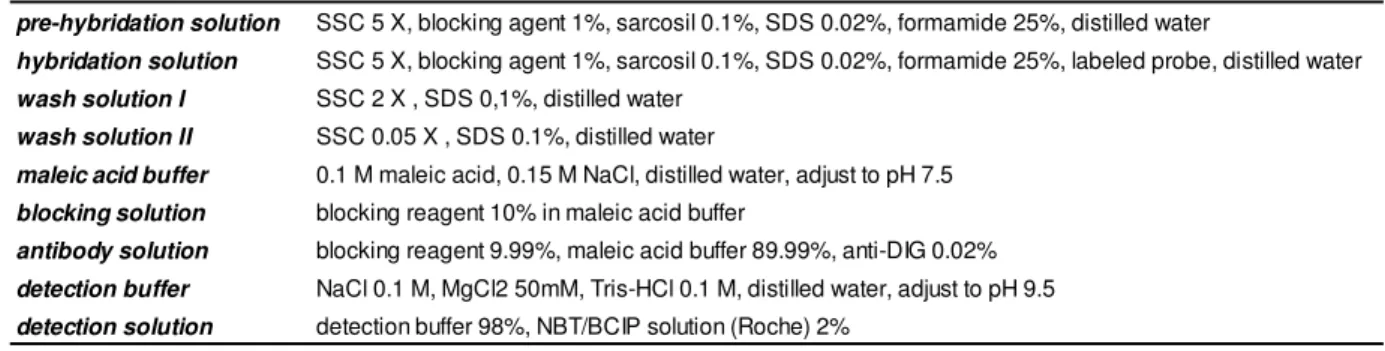
Dot-blot hybridization with digoxigenin-labeled PCR-derived probes from each IncQ, IncN and IncW templates, was employed, by using the DIG Nucleic Acid Detection Kit (Roche), to carry out the detection of the correspondent PCR amplification products. The compositions of all the solutions used in the procedure are described in table VI.
Table IV. Positive controls used in the PCR reactions for every plasmid incompatibility group or subgroup.
27
Detailed protocol:
- The DNA from PCR reactions was denatured by mixing 20 µl of every PCR product with 2 µl of NaOH 0.5 N and incubating at 50ºC for 5 minutes.
- 6 µl of standard sodium citrate (SSC) 20 X were added to every sample to neutralize them. - The DNA transference to a previously soaked in SSC 20X positively charged nylon transfer
membrane (Amersham Hybond –N+, GE Heathcare) was performed under vacuum at 50
mm Hg, by using a VacuGene Pump (VacuGene XL, Pharmacia Biotech) as vacuum
blotting System.
- Dot blotted nucleic acid was UV cross-linked to the nylon membrane during 5 minutes. - The PersonalHyb hybridization oven (Stratagene) with the pre-heated at the same
temperature pre-hybridation solution was employed to carry out the pre-hybridization for 2 hours at 42ºC.
- The probes (made up through the PCR DIG Probe Synthesis Kit, Roche) were heated at 68ºC for 10 minutes to allow the DNA to dissociate.
- The hybridization tubes with the membranes and the hybridization solution (containing each one the correspondent probes) were incubated at 42ºC for 16 hours.
- The membranes were washed several times (twice with wash solution I for 5 minutes at room temperature, with wash solution II for 15 minutes at 42ºC and maleic acid buffer for 5 minutes at 42ºC), blocked (blocking solution for 30 minutes at the room temperature) and incubate with the antibody solution (the conjugate DIG-alkaline phosphatase in dissolved in
blocking solution) for exactly 30 minutes at the room temperature.
- After washing (maleic acid buffer for 15 minutes at the room temperature) and equilibrating (detection buffer for 5 minutes at the room temperature), the membranes were placed on a previously prepared tray in a dark environment (without shaking).
- The chromogenic detection began when the membranes where soaked in the detection
solution. Revealing lasted two hours and the reaction was stopped with abundant distilled
28
pre-hybridation solution SSC 5 X, blocking agent 1%, sarcosil 0.1%, SDS 0.02%, formamide 25%, distilled water
hybridation solution SSC 5 X, blocking agent 1%, sarcosil 0.1%, SDS 0.02%, formamide 25%, labeled probe, distilled water wash solution I SSC 2 X , SDS 0,1%, distilled water
wash solution II SSC 0.05 X , SDS 0.1%, distilled water
maleic acid buffer 0.1 M maleic acid, 0.15 M NaCl, distilled water, adjust to pH 7.5 blocking solution blocking reagent 10% in maleic acid buffer
antibody solution blocking reagent 9.99%, maleic acid buffer 89.99%, anti-DIG 0.02% detection buffer NaCl 0.1 M, MgCl2 50mM, Tris-HCl 0.1 M, distilled water, adjust to pH 9.5 detection solution detection buffer 98%, NBT/BCIP solution (Roche) 2%
3.4 DNA sequencing of PCR products and sequence analysis
The PCR products obtained from the amplification of the nucleotide sequences from specific replicons belonging to the IncP-1incompatibility subgroups were purified with a slightly modified JETQUICK PCR Purification Protocol (Genomed). The DNA elution was performed by applying 20 µl of sterile distilled water at 65ºC.
The purified amplicons were sequenced through the chain termination method (Sanger et al., 1977) by the company StabVida. Then, in order to determine their closest phylogenetic relatives the BLAST software (Altschul et al., 1997) were used to compare the obtained sequences with known sequences. Sequences were aligned with reference taxa within the sequence databases using the CLUSTALW2 program (Larkin et al., 2007).
Finally neighbor-joining trees based on the distance parameter were constructed using the PAUP version4.0b10 program (Swofford, 2003). Bootstrap values from 1000 replicates were also acquired and shown as percentages.
3.5 Plasmid DNA isolation and purification
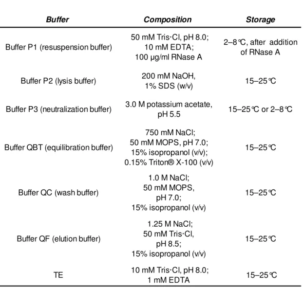
With the purpose of further characterizing the detected BHR plasmids, the Qiagen plasmid Mini Kit was employed to perform the plasmid DNA purification. Extraction was
29
carried out following the manufacturers instructions with adaptations described below. The compositions of the solutions required in this procedure are described in table VII.
Detailed protocol:
- Starter cultures of LB medium (Miller, Merck) containing the appropriate selective markers (30 µg/ml for tetracycline and 25 µg/ml for mercury chloride) were inoculated with single colonies picked from fresh streaked plates of the transconjugants harboring the detected plasmids and incubated overnight at 28°C with shaking of approximately 165 rpm.
- The bacterial cells from culture volumes of 25 ml were harvested by centrifugation (4500 rpm, 7 minutes, 4°C).
- Each pellet was ressuspended with 4 ml of Buffer P1.
- The lysis of the cells was favored by adding 4 ml of Buffer P2, mixing thoroughly and incubating for exactly 5 minutes at room temperature.
- To neutralize, 4 ml of pre-chilled Buffer P3 were added, starting immediately the incubation on ice for at least 15 minutes.
- Centrifugation (13000 rpm, 10 min, 4°C) was carried out twice in order to eliminate unwanted cell material.
- The clear lysate (the supernatant) containing DNA was kept and split into two equal fractions to avoid subsequently over-loading the low capacity column.
- Both fractions of DNA were precipitated by adding 1 volume of room temperature isopropanol.
- Centrifugation at 13000 rpm and 4°C for 30 minutes made unwanted metabolites such as proteins and lipopolysaccharides to be removed. At this time, one of the fractions corresponding to each sample was used to continue the protocol, while plasmid from the second one was kept at 4ºC to be purified afterwards by equilibrating again the same column and following this instructions from here on.
- When no visible liquid could be seen on the DNA pellets, they were redissolved in 100 µl dH2O and 1 ml of Buffer QBT was added.
- The samples were applied to the previously equilibrated with 1 ml Buffer QBT QIAGEN-tip
30
- The QIAGEN-tips were washed with 4 x 1 ml Buffer QC.
- The plasmid DNA was eluted by applying 1 ml Buffer QF (pre-heated at 65ºC to increase the yield).
- The eluted DNA was precipitated by adding 0.7 volumes of room-temperature isopropanol. - A centrifugation at 13.000 rpm and 4°C for 30 minutes was carried out.
- DNA pellet was washed with 1 ml room-temperature 70% ethanol and centrifugated again at 13.000 rpm and 4ºC for 10 minutes.
- The pellets obtained were redissolved in 20 µl of TE buffer.
The success of the plasmid purification was evaluated by loading onto 0.8% agarose (Lonza) gel in TAE (Tris-Acetate-EDTA, 5Prime) buffer at 60V for 200 min. The molecular weight marker used was GeneRuler DNA Ladder Mix (Fermentas). Gels were stained with ethidium bromide and banding patterns were visualized by using a Molecular Imager FXTM system (Bio-Rad).
31
Buffer Composition Storage
Buffer P1 (resuspension buffer)
50 mM Tris·Cl, pH 8.0; 10 mM EDTA; 100 µg/ml RNase A
2–8°C, after addition of RNase A
Buffer P2 (lysis buffer) 200 mM NaOH,
1% SDS (w/v) 15–25°C
Buffer P3 (neutralization buffer) 3.0 M potassium acetate,
pH 5.5 15–25°C or 2–8°C
Buffer QBT (equilibration buffer)
750 mM NaCl; 50 mM MOPS, pH 7.0;
15% isopropanol (v/v); 0.15% Triton® X-100 (v/v)
15–25°C
Buffer QC (wash buffer)
1.0 M NaCl; 50 mM MOPS,
pH 7.0; 15% isopropanol (v/v)
15–25°C
Buffer QF (elution buffer)
1.25 M NaCl; 50 mM Tris·Cl, pH 8.5; 15% isopropanol (v/v) 15–25°C TE 10 mM Tris·Cl, pH 8.0; 1 mM EDTA 15–25°C 3.6 Restriction analysis
A preliminary genetic characterization of the captured plasmids was performed by carrying out DNA digestions with the endonuclease NotI (#ERO591, Fermentas). The enzyme was chosen for the restriction analysis according to the bibliography and the in silico analysis made by means of the NEBcutter software (Vincze et al., 2003). Additionally, it is known that IncP-1 plasmids are characterized by a high guanine + cytosine content. Hence, the use of an endonuclease recognizing DNA regions enriched in
Table VII. Composition and storage temperature of every solution required to perform the plasmid purification.
32
these nucleotides should give back more enlightening restriction patterns which allow to better compare the plasmids with each other. The previously known pKJK5 and pB10 were used as controls.
Firstly the purified plasmid DNA was quantified using a ND-1000 spectrophotometer (Nanodrop). Digestions were carried out by preparing a 20 µl reaction mixture containing 0.05-0.1 mg/µl of plasmid DNA, 0.25U/ µl of NotI, 1 X buffer O (Fermentas) and sterile miliQ water and incubating for 16 hours at 37 ºC. The enzymatic reactions were stopped by freezing at -20ºC.
To analyze the resulting restriction profiles 5 µl of every restriction reaction were loaded onto 0.8% agarose TAE gels, and ran for 200 minutes at 40 V, using the GeneRuler DNA Ladder Mix (Fermentas) as molecular weight marker. Ethidium bromide-stained gels were seen using a Molecular Imager FXTM system (Bio-Rad).
3.7 Electrotransformation of Escherichia coli Top10 with IncP-1 plasmids
With the aim of improving the DNA quality, the purified plasmids known to encode for tetracycline resistance (from the transconjugants 1, 2, 6 and 7) were transferred to previously prepared electrocompetent well known bacterial cells (Escherichia coli Top10, sensible to tetracycline). The MicroPulser™ Electroporation Apparatus (Bio-Rad) was employed to perform a slightly modified electrotransformation procedure based on the Catalog Number 165-2100.
Detailed protocol:
- Both the electrocompetent cells and a sterile 0.1 cm electroporation cuvette (Bio-Rad) were placed on ice.
33
- 40 µl of the cell suspension were mixed with 2 µl of pDNA in a pre-chilled microfuge tube and incubated on ice for 1 minute.
- The mixture of cells and DNA were transferred to the bottom of the cold electroporation cuvette, which were placed in the right position in the chamber slide.
- The Ec1 program was chosen and one fast pulse was made.
- The cuvette was immediately removed from the chamber and 450 µl of SOC medium were added without delay.
- After quickly but gently ressuspending the cells with a Pasteur pipette, the solution was cultured at 37ºC for 1h30min with shaking of 200 rpm approximately.
The success of the procedure was tested by plating in TSA medium (Tryptic Soy Broth, Merck; Rose-Bengal Chloranphenicol Agar, Merck) supplemented with 30 µg/ml of tetracycline and incubating at 37ºC.
3.8 Antibiotic resistance assays
The antibiotic resistance patterns of Pseudomonas putida KT2442 and Escherichia coli Top10 harboring the identified incP-1 plasmids were determined by the agar disk diffusion method on Mueller Hinton media (Merck). Susceptibility testing were performed for resistance against a panel of 9 antibiotics, including representatives of the most common antibiotic resistance traits shown by the previously known IncP-1 plasmids: streptomycin (S10), amoxicillin (AML25), amoxicillin and clavulanic acid (AMC30), tetracycline (TE10), chloramphenicol (C10), trimethroprim-sulfamethoxazole (SXT25), erythromycin (E15), cephalothin (KF30) and aztreonam (ATM30). The antibiotics disks were supplied by the company Oxoid. Escherichia coli 25592 as well as Pseudomonas putida KT2442 (original host) or Escherichia coli Top10 (subsequent host) were used as controls.
Detailed protocol:
- To assure the use of fresh cultures (not more than 24 hours) the antibiotic assay control bacterial strain, as well as all the microorganism to test, were cultivated in LA medium (Lonka) at 30ºC (for Pseudomonas putida) or 37ºC (for Escherichia coli).