IN STI TU T O D E C IÊ N C IA S B IO M ÉD ICA S A B EL SA LA Z A R FA C U LD A D E D E FA R M Á C IA
Daniela Filipa dos Santos Barros.
Engineered platforms with site-specific
immobilized laminin for neural applications
Engineered platforms with site-specific immobilized laminin for neural applications
Daniela Filipa dos Santos Barros
Engineered platforms with site-specific
immobilized laminin for neural applications
Daniela Filipa dos Santos Barros
D
2019D
.IC
B
A
S 2019
D M IN IS T R A T IV A DOUTORAMENTODaniela Filipa dos Santos Barros
Engineered platforms with site-specific immobilized laminin for
neural applications
Tese de Candidatura ao grau de Doutor em
Biotecnologia Molecular e Celular Aplicada às
Ciências da Saúde;
Programa Doutoral da Universidade do Porto
(Instituto de Ciências Biomédicas de Abel
Salazar e Faculdade de Farmácia)
Orientadora – Professora Doutora Ana Paula
Gomes Moreira Pêgo
Categoria – Investigadora Principal, Professora
Associada Convidada
Afiliação – Instituto de Investigação e Inovação
em Saúde (i3S), Universidade do Porto (UP);
Instituto Nacional de Engenharia Biomédica
(INEB), UP; Faculdade de Engenharia (FEUP),
UP; Instituto de Ciências Biomédicas Abel
Salazar (ICBAS), UP.
Co-orientadora – Doutora Isabel Maria Santana
Ramos de Freitas Amaral
Categoria – Investigadora Auxiliar
Afiliação – Instituto de Investigação e Inovação
em Saúde, Universidade do Porto (i3S, UP);
The work presented in this thesis was developed at:
nBTT - nanoBiomaterials for Targeted Therapies Group i3S – Instituto de Investigação e Inovação em Saúde
INEB – Instituto Nacional de Engenharia Biomédica Universidade do Porto, Portugal
García Lab
Parker H. Petit Institute for Bioengineering and Bioscience George W. Woodruff School of Mechanical Engineering
The work presented in this thesis was funded by projects NORTE-01-0145-FEDER-000008 and NORTE-01-0145-FEDER-000012, supported by Norte Portugal Regional Operational Programme (NORTE 2020), under the PORTUGAL 2020 Partnership Agreement, through the European Regional Development Fund (ERDF) and FEDER - Fundo Europeu de Desenvolvimento Regional funds through the COMPETE 2020 - Operacional Programme for Competitiveness and Internationalisation (POCI), Portugal 2020; by Portuguese funds through FCT/MCTES in the framework of the project "Institute for Research and Innovation in Health Sciences" (POCI-01-0145-FEDER-007274) and Santa Casa da Misericórdia de Lisboa through project COMBINE (Prémio Neurociências Melo e Castro-MC-1068-2015). D. Barros was supported by FCT PhD Programs (PD/BD/105953/2014) and Programa Operacional Potencial Humano (POCH), in the scope of the BiotechHealth Program (Doctoral Program on Cellular and Molecular Biotechnology Applied to Health Sciences), Programa FLAD Healthcare 2020 and the project PARES (Prémio Albino Aroso).
Remember what they say There’s no shortcut to a dream It’s all about blood and sweat And life is what you manage in-between
E 5 anos depois, eis que esta longa e desafiante jornada está prestes a terminar! O doutoramento foi sem dúvida uma etapa durante a qual vivi algumas das melhores e também piores experiências da minha vida, e que me fez crescer não só cientificamente, mas também a nível pessoal. Felizmente não fiz este caminho sozinha, tive pessoas espetaculares a apoiar-me e a fazer-me querer ir sempre um bocadinho mais além, e é a todas elas que quero agradecer e também dedicar esta tese.
Às minhas orientadoras Ana Paula e Isabel, obrigada por me terem recebido e me terem dado a oportunidade de explorar uma área que estava totalmente fora da minha zona de conforto...aprendi imenso nestes últimos anos. Uma das coisas mais importantes que aprendi foi que muitas vezes (ou quase sempre) temos de desligar o “descomplicómetro” e olhar para aquilo que vemos como perdido/mau de forma mais positiva. Obrigada pelo vosso entusiasmo, apoio e pelas palavras de incentivo. A vossa orientação e apoio foi fundamental para o meu crescimento científico nestes últimos anos.
To Andrés, thank you for the opportunity to work in your laboratory and for being such an enthusiastic and positive mentor. I had a really great time in your lab. Thank you also, to all the members from García Lab who welcomed me so well, and specially to Woojin and Ricardo, my coffee buddies, for the friendship and support, making my journey at GeorgiaTech, a really happy journey.
Ao Professor Mário Barbosa, pela oportunidade de frequentar o BiotechHealth e pelo esforço em tornar este, um programa doutoral de excelência. À Helena Martins, Catarina Pereira, Daniel Vasconcelos e João Cortez por estarem sempre disponíveis para ajudar e tornar a nossa vida um bocadinho mais fácil. Aos meus colegas do BiotechHealth que fizeram este caminho comigo, foi muito bom contar com o vosso apoio e companheirismo. À Paula Parreira e Cristina Martins, pela ajuda e apoio numa fase crucial desta tese. Obrigada pela vossa persistência e pelas palavras de incentivo...a vossa ajuda foi fundamental para o meu trabalho.
A todas as pessoas dos serviços do i3S, Frederico, Fátima, Maria, Dalila, Ricardo, Eduardo, Tânia, Paula, Hugo e André, muito obrigada por partilharem o vosso conhecimento e me ajudarem nos momentos mais críticos. Um obrigada especial à Joana, que foi essencial na fase inicial do meu doutoramento. A tua paciência e apoio fizeram toda a diferença.
Um muito obrigada a todos os colegas e amigos do INEB/IBMC/i3S...sinto-me uma sortuda pela quantidade enorme de pessoas que tive oportunidade de conhecer e manter na minha vida ao longo destes 5 anos (e não só). Foi muito bom poder contar com o vosso apoio e amizade!!
À Catarina e ao Philippe, os novos “bosses”, por todo o apoio e compreensão nesta fase de transição. A todos os colegas do ICB Lab e do iBiMED por me acolherem tão bem no laboratório e em Aveiro.
À minha família do coração, àqueles que são as “minhas pessoas” Beta, Jorginho, Pedro e Tânia, e ao mais recente elemento, Tiago, obrigada pelo apoio, pelo carinho, pelos serões passados a estupidificar (sempre com o belo do copo de vinho a acompanhar)...obrigada por fazerem parte da minha vida e me deixarem fazer parte da vossa.
À pessoa que durante muitos anos partilhou comigo as angústias, alegrias e que nunca me impediu de sonhar... apesar dos nossos caminhos já não se cruzarem, obrigada Ricardo por nunca teres desistido de mim!
Aos meus pais, não há palavras para lhes agradecer o apoio e amor incondicional. Muito do que sou hoje devo-o a vocês, que me ensinaram a não baixar os braços e a nunca desistir de ir atrás dos meus sonhos. OBRIGADA POR TUDO!!!
À minha irmã, pelo apoio, pelo amor, por mostrar tanto entusiasmo e orgulho pelas minhas conquistas, por nunca me deixar desistir, pelas palavras certas nos momentos mais complicados,...POR TUDO!!! Obrigada ao André pelos conselhos e apoio. E obrigada ao meu Guigas, que é uma das pessoas que eu mais amo neste mundo e que nos seus pequenos gestos me faz sentir tão amada e feliz.
The main outputs of the work conducted during the period of this thesis are listed
below:
Papers in peer-reviewed scientific journals
D. Barros, P. Parreira, J. Furtado, F. Ferreira-da-Silva, E. Conde-Sousa, A.J. Garcia, M.C.L. Martins, I.F. Amaral, A.P. Pêgo, An affinity-based approach to engineer laminin-presenting cell instructive microenvironments, Biomaterials 192 (2019) 601-611
W. M. Han, S. E. Anderson, M. Mohiuddin, D. Barros, S. A. Nakhai, E. Shin, I. F. Amaral, A. P. Pêgo, A. J. García, Y. C. Jang, Synthetic matrix enhances transplanted satellite cell engraftment in dystrophic and aged skeletal muscle with comorbid trauma, Science Advances, 4 (2018).
J. Silva, A. R. Bento, D. Barros, T. L. Laundos, S. R. Sousa, P. Quelhas, M. M. Sousa, A. P. Pêgo, I. F. Amaral, Fibrin functionalization with synthetic adhesive ligands interacting with a6b1 integrin receptor enhances neurite outgrowth of embryonic stem cell-derived neural stem/progenitors, Acta Biomaterialia, 59 (2017) 243-256.
D. Barros, I. F. Amaral, A. P. Pêgo, Biomimetic Synthetic Self-Assembled Hydrogels for Cell Transplantation. Current Topics in Medicinal Chemistry, 15(13) (2015) 1209-26.
Manuscripts in preparation for submission
D. Barros, I. F. Amaral, A. P. Pêgo, Laminin-inspired cell instructive microenvironments for neural tissue engineering. (Literature Review).
D. Barros, A. J. García, I. F. Amaral, A. P. Pêgo, Engineering hydrogels with affinity-bound laminin as 3D neural stem cell culture systems. (Original Research).
Book Chapter
J. Caldeira, F. Sousa, D. Sousa, D. Barros (2018). Extracelular matrix constitution and function for tissue regeneration and repair, in Peptides and Proteins as Biomaterials for
Tissue Regeneration and Repair, M.A. Barbosa and M.C.L. Martins, Editors. Woodhead
Publishing – Elsevier.
Patents
Neural stem cells (NSCs) hold great potential for application in the treatment of central nervous system (CNS) disorders. These multipotent cells reside within a dynamic and complex microenvironment, the NSC niche, where cell-cell interactions and local microenvironmental cues are key to regulate stem cell behavior. Therefore, the development of new platforms allowing a better understanding of niche properties and how they regulate NSC behavior and function is highly desirable. These are expected to provide valuable insight into cell physiology and interactions with the surrounding microenvironment, and ultimately be used for the development of more effective NSC-based regenerative therapies.
Laminin has a key role in the modulation of NSC function and fate within neurogenic niches. Indeed, different studies already explored the immobilization of full-length laminin and its peptide analogues into diverse biological and synthetic matrices, to recapitulate the microenvironment of stem cell niches and/or gain insight into the role of ECM on stem cell behavior. However, the strategies currently explored for laminin immobilization within three-dimensional (3D) matrices do not address a critical aspect influencing laminin bioactivity, which is the control over the exposure of laminin crucial bioactive epitopes.
In this thesis, we propose the design of biologically relevant matrices able to recapitulate the laminin-rich microenvironment of NSC niches. For that, we explored the modification of a degradable synthetic hydrogel with full-length laminin or with a laminin-derived peptide interacting with syndecans - AG73.
One of the specific aims of this thesis was the establishment of an affinity-based approach that takes advantage of the high affinity interaction between laminin and the human N-terminal agrin (hNtA) domain. A recombinant human NtA (rhNtA) domain was produced and successfully conjugated at its N-terminal with a thiol-terminated poly(ethylene glycol) (PEG) – mono-PEGylated rhNtA. Mono-PEGylated rhNtA showed ability to act as an effective natural binding partner for the site-selective immobilization of laminin, while preserving the exposure of laminin key bioactive domains. This translated into an enhanced ability of laminin to polymerize and mediate hNSC adhesion and spreading. Aiming at the establishment of a chemically-defined cell-instructive microenvironment, the proposed affinity-based approach was explored to promote the
properties. The PEG-4MAL macromer was functionalized with the thiol-terminated mono-PEGylated rhNtA domain to mediate the site-selective incorporation of laminin into PEG-4MAL. The developed hydrogels revealed mechanical properties (complex modulus (G*) = 251 Pa) within the range of values preferred for NSC proliferation (100 – 1000 Pa) and neurite branching and extension (200 – 400 Pa). Moreover, the affinity-bound laminin PEG-4MAL hydrogels showed an enhanced ability to mediate hNSC proliferation and neurite outgrowth, when compared to unmodified hydrogels, and most importantly, in contrast to hydrogels containing equal amount of physically entrapped laminin.
In alternative to the incorporation of full-length laminin, a laminin-derived peptide interacting with syndecan receptors (AG73) was explored to confer bioactivity to 3D matrices. The modular nature of the synthetic matrix used, allowed us to explore the effect of AG73 peptide density on cell behavior, independently of other hydrogel biophysical and biochemical properties. The tethering of AG73 peptide to PEG-4MAL hydrogels led to a significant improvement of cell viability after 7 days in culture, independently of the input peptide concentration. In addition, we demonstrated that a higher input AG73 concentration (1 mM) better supports hNSC proliferation and neurite outgrowth, as well as the establishment of a neuronal network.
Overall, the work presented in this thesis contributed to: (i) the development of an affinity-based strategy for site-selective immobilization of laminin, suitable for use as an alternative to conventional immobilization approaches (physical entrapment and non-selective covalent immobilization) in a wide range of applications (e.g. engineered coatings for neuroelectrodes, 2D substrates for cell culture, and biofunctionalization of 3D matrices); (ii) development of engineered matrices for use as defined 3D platforms for the establishment of artificial NSC niches and as temporary mimetic microenvironments to support hNSC transplantation in the context of nervous system regeneration/disorders.
In summary, we strongly believe this work opens new avenues in the design of more efficient hydrogel matrices for application in NSC-based regenerative approaches for the treatment of traumatic CNS disorders or neurodegenerative diseases.
As células estaminais neurais (CENs) apresentam um grande potencial para aplicação no tratamento de doenças do sistema nervoso central (SNC). Estas células multipotentes residem num microambiente dinâmico e complexo, o nicho das CEN, onde as interações célula-célula e os sinais do microambiente local são essenciais para regular o comportamento das células estaminais. Deste modo, o desenvolvimento de novas plataformas que permitam compreender melhor as propriedades do nicho e a forma como estas regulam o comportamento e a função das CENs é altamente vantajoso. É expectável que estas plataformas permitam uma melhor compreensão da fisiologia celular das CENs e das suas interações com o ambiente circundante, e que contribuam para o desenvolvimento de terapias regenerativas baseadas no transplante de CENs mais efetivas.
A laminina tem um papel crucial na modulação da função e diferenciação das CENs nos nichos neurogénicos. De facto, já vários estudos exploraram a imobilização da molécula da laminina e de péptidos derivados da laminina em matrizes biológicas e sintéticas, de forma a recapitular o microambiente do nicho e/ou a compreender melhor o papel da matriz extracelular no comportamento das células estaminais. Contudo, as estratégias exploradas atualmente para a imobilização da laminina em matrizes tridimensionais (3D) não contemplam um aspeto crítico para a bioatividade da laminina, e que é o controlo sobre a exposição dos epítopos bioativos mais importantes.
Nesta tese, propomos o desenvolvimento de matrizes biologicamente relevantes capazes de recapitular o microambiente rico em laminina dos nichos neurogénicos. Para isso, explorámos a modificação de um hidrogel sintético degradável com a molécula da laminina ou com um péptido derivado da laminina capaz de interagir com recetores sindecano – AG73.
Um dos objetivos específicos desta tese foi o desenvolvimento de uma estratégia de imobilização por afinidade, baseada na interação de elevada afinidade entre a laminina e o domínio N-terminal da agrina (NtA). O domínio recombinate humano NtA (rhNtA) foi produzido e conjugado eficazmente no seu N-terminal com um polietilenoglicol (PEG) contendo um grupo tiol numa das suas extremidades – rhNtA mono-PEGilado. O rhNtA mono-PEGilado demonstrou capacidade para atuar como um ligando natural para a imobilização seletiva da laminina, sem comprometer a exposição dos domínios bioativos
diferentes isoformas da laminina (no contexto desta tese avaliámos a laminina-111 derivada de ratinho e a laminina-521 recombinante humana) num hidrogel sintético e degradável. Para o efeito selecionámos um hidrogel de PEG com quatro braços funcionalizados nas extremidades com maleimida (PEG-4MAL) devido à sua natureza modular, biocompatibilidade e propriedades bioquímicas e biofísicas bem caracterizadas. O PEG-4MAL foi inicialmente funcionalizado com o domínio rhNtA mono-PEGilado para lhe permitir mediar a incorporação selectiva da laminina. Os hidrogéis desenvolvidos apresentaram propriedades mecânicas (módulo complexo (G*) = 251 Pa) no gama de valores preferidos para a proliferação das CEN (100 – 1000 Pa) e extensão de neurites (200 – 400 Pa). Os hidrogéis com laminina imobilizada seletivamente revelaram modular de forma mais eficaz a proliferação das CENs e a extensão de neurites, quando comparados com hidrogéis não modificados, e sobretudo em contraste com hidrogéis sem o domínio rhNtA mono-PEGilado, mas contendo a mesma quantidade de laminina fisicamente incorporada.
Alternativamente à imobilização da molécula da laminina, também explorámos um péptido derivado da laminina que interage com os recetores sindecano (AG73), para conferir bioatividade às matrizes 3D. A natureza modular da matriz sintética utilizada, permitiu-nos explorar o efeito da densidade do péptido AG73 no comportamento celular, independentemente de outras propriedades bioquímicas e biofísicas do hidrogel. Os hidrogéis de PEG-4MAL modificados com o péptido levaram a um aumento significativo da viabilidade celular após 7 dias em cultura, independentemente da concentração de péptido. Além disso, a concentração máxima de AG73 testada (1 mM) revelou suportar melhor a proliferação das CENs e a extensão de neurites, tal como favorecer o estabelecimento de uma rede neuronal.
No geral, o trabalho apresentado nesta tese contribuiu para: (i) o desenvolvimento de uma estratégia de afinidade para a imobilização seletiva da laminina, com potencial para ser utilizada em alternativa às estratégias de imobilização convencionais (incorporação física e imobilização covalente não seletiva) em diversas aplicações (p.e. revestimentos para neuro-elétrodos, substratos para cultura celular, e biofuncionalização de matrizes 3D); (ii) desenvolvimento de plataformas 3D definidas com potencial de utilização no estabelecimento de nichos neurogénicos artificiais e como microambientes temporários biomiméticos, para transplante de CENs no contexto da regeneração do sistema nervoso.
baseadas no transplante de CENs para o tratamento de doenças traumáticas ou degenerativas do SNC.
ACKOWLEDGMENTS ... i
LIST OF MAIN OUTPUTS ... iii
ABSTRACT ... v
RESUMO ... vii
TABLE OF CONTENTS ... xi
LIST OF ABBREVIATIONS ... xv
PART 1 CHAPTER I | General Introduction ... 3
CHAPTER II | Aim and Structure of the Thesis ... 23
Aim of the Thesis ... 25
Structure of the Thesis ... 26
CHAPTER III | Laminin-Inspired Cell-Instructive Microenvironments for Neural Tissue Engineering ... 31
Abstract ... 34
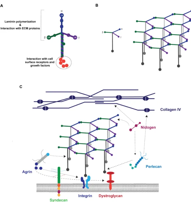
1.Laminin ... 35
1.1. Structure and domain architecture ... 35
1.2. Main Functions ... 36
1.2.1. Laminin polymerization ... 38
1.2.2. Laminin role in the assembly and stability of basement membranes ... 38
1.2.3. Cell adhesion-promoting activity ... 39
2.Laminin in the central nervous system ... 40
5.Engineering laminin-inspired hydrogels: progress and future challenges ... 58
References ... 60
CHAPTER IV | Biomimetic Synthetic Self-Assembled Hydrogels for Cell Transplantation ... 77
Abstract ... 80
Cell Transplantation in the Context of Regenerative Medicine ... 81
Hydrogels as Attractive Vehicles for Cell Transplantation ... 82
Self-Assembled Hydrogels ... 84
Engineered protein-based hydrogels ... 84
Elastin-like polypeptides ... 85
Leucine zipper coiled-coil-based polypeptides ... 86
Amphiphilic block co-polypeptides ... 87
Mixing-induced two-component hydrogel ... 88
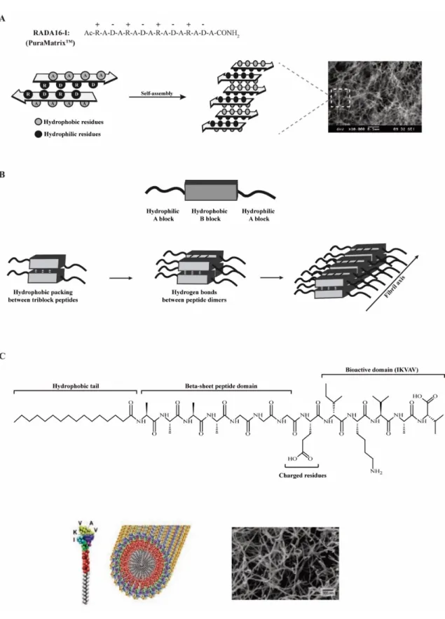
Peptide-based synthetic hydrogels ... 90
Self-assembling peptides ... 90
Self-assembling peptide amphiphiles ... 92
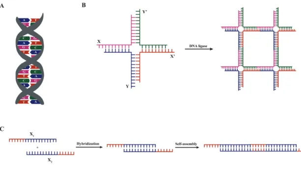
DNA-based hydrogels ... 96
Hybrid hydrogels ... 98
In vivo Biological Performance of Synthetic Self-Assembled Hydrogels ... 101
Translation of Synthetic Self-Assembled Hydrogels into the Clinic: Progress and Future Challenges... 106
Ackowledgments ... 108
References ... 109
PART 2 CHAPTER V | An Affinity-Based Approach to Engineer Laminin Presenting Cell Instructive Microenvironments ... 123
2. Materials and Methods ... 129
3. Results and Discussion ... 133
4. Conclusion ... 146
5. Acknowledgments ... 147
Supplementary Data ... 148
Supplementary Materials and Methods ... 148
Supplementary Figures and Tables ... 152
References ... 161
CHAPTER VI | Engineering Hydrogels with Affinity-Bound Laminin as 3D Neural Stem Cell Culture Systems ... 167
Abstract ... 170
1. Introduction ... 171
2. Materials and Methods ... 174
3. Results and Discussion ... 177
4. Conclusion ... 185
Acknowledgments ... 186
Supplementary Data ... 187
Supplementary Materials and Methods ... 187
Supplementary Figures and Tables ... 188
References ... 190
CHAPTER VII | An Alternative Approach to Engineer Synthetic Hydrogels with Affinity-Bound Laminin ... 195
Supplementary Figures and Tables ... 212
References ... 214
CHAPTER VIII | AG73-Functionalized Synthetic Hydrogels Support Neural Stem Cell Survival, Proliferation and Neurite Outgrowth ... 219
Abstract ... 222
1. Introduction ... 223
2. Materials and Methods ... 226
3. Results and Discussion ... 229
4. Conclusion ... 234
Acknowledgments ... 234
References ... 235
CHAPTER IX | Concluding Remarks and Future Perspectives ... 241
A
A – Avogadro’s constant
Ac-PEG-NHS – Acrylate - Poly(ethylene
glycol) - N-hydroxysuccinimide
ACN – Acetonitrile
ATDPC – Adipose tissue-derived
progenitor cell(s)
B
BDNF – Brain-derived neurotrophic
factor
bFGF – Basic fibroblast growth factor BM – Basement membrane
BMEC – Brain microvascular endothelial
cell(s)
BMP - Bone morphogenic protein BMHP - Bone-marrow homing peptide BMSC - Bone marrow stromal cell(s) BSA - Bovine serum albumin
C
CBD - Collagen-binding domain
CHCA -
alpha-Cyano-4-hydroxycinnamic acid
CLSM - Confocal laser scanning
microscopy
CNS - Central Nervous System CS – Chondroitin sulfate
D
DAPI - 4’,6-diamidino-2-phenylindole DGR - Osteopontin cell-adhesion motif
DRG - Dorsal root ganglia ΔD - Dissipation shift
E
E - Elastic moduli
ECM - Extracellular matrix
EDC - 3-(N,N-dimethylamino)
propyl-N-ethylcarbodiimide
EGF – Epidermal growth factor EG4 - (11-mercaptoundecyl)
tetraethylene glycol
ELISA - Enzyme-linked immunosorbent
assay
ELP - Elastin-like polypeptide ESC - Embryonic stem cell(s)
F
Δf - Frequency shift FBS – Fetal bovine serum FI - Fluorescence intensity
Fmoc - Fluoren-9-ylmethyloxycarbonyl Fmoc-FF -
Fluoren-9-ylmethyloxycarbonyl-diphenylalanine
FOV – Field of view
G
G’ – Storage modulus G’’ – Loss modulus G* - Complex modulus GAG – Glycosaminoglycan
GMEM - Glasgow Minimum Essential
cell(s)
HEPES -
4-(2-hydroxyethyl)-1-piperazineethanesulfonic acid
hiPSC-EC – Human induced pluripotent
stem cell-derived endothelial cell(s)
HRP - Horseradish peroxidase HS - Heparan sulfate
HSPG - Heparan sulfate proteoglycan HUVEC - Human umbilical vein
endothelial cell(s)
I
IPN - Interpenetrating polymer network iPSC - Induced pluripotent stem cell(s)
IPTG - Isopropyl
b-D-1-thiogalactopyranoside
IRRAS - Infrared reflection absorption
spectroscopy
K
ka - Association rate constant
KD - Dissociation constant
kd - Dissociation rate constant
L
LE – Laminin-type epidermal growth
factor like
LG - Laminin globular LN - Laminin N-terminal
LVR – Linear viscoelastic region
M
MC - Methylcellulose
MMP - Matrix metalloproteinase mPEG - mono-PEGylated
msLn-111 - Laminin-111 from mouse
Engelbreth-Holm-Swarm sarcoma
MWCO - Molecular weight cut-off
N
Nap - Naphthalene
NHS - N-hydroxysuccinimide NSC - Neural stem cell NPC – Neural progenitor cell
NSPC – Neural stem progenitor cell NT - Neurotrophin
NtA - N-terminal agrin
O
ON - Overnight
OXMC - Oxidized methylcellulose
P
d - Phase angle
PA - Peptide amphiphile
PBS - Phosphate buffered saline
PCLM - Poly(caprolactone
methacryloyloxyethyl ester)
PCR - Polymerase chain reaction PDL – poly(D-lysine)
PEG - Poly(ethylene glycol)
PEG-4Ac - Four-arm acrylate-end
functionalized poly(ethylene glycol)
PEG-4MAL - Four-arm maleimide-end
functionalized poly(ethylene glycol)
PEG-DA - Poly(ethylene glycol)
diacrylate
PEGDVS - Poly(ethylene glycol) divinyl
sulfone
methacrylamide)
PI – Propidium iodide
PMF - Peptide mass fingerprint
Q
QCM-D - Quartz crystal microbalance
with dissipation monitoring
R
R – Gas constant rhLn-521 - Recombinant human Laminin-521 RIA - Radioimmunoassay RT - Room temperatureS
SAM - Self-assembled monolayer SANPAH - sulfosuccinimidyl
6-(4’-azido-2’-nitrophenylamino)hexanoate
SC – Schwann cell
Sca-1 – Stem cells antigen-1 SCI - Spinal cord injury SD - Standard deviation
SDS-PAGE - Sodium dodecyl sulfate–
polyacrylamide gel electrophoresis
SDF-1a - Stromal cell-derived factor-1a
SELP - Silk-elastin-like polypeptide SEM - Standard error of the mean
SFM - StemProÒ NSC serum-free
medium
SGZ - Subgranular zone
SH-PEG-SGA - thiol-poly(ethylene
SPR - Surface Plasmon Resonance SVZ - Subventricular zone
T
3D - Three-dimensional 2D - Two-dimensional T - Temperature Tt - Transition temperature TBS-T - Tris-buffered saline – 0.1% Tween® 20TCPS - Tissue culture polystyrene TFA - Trifluoroacetic acid
TMB - 3,3’,5,5’ tetramethyl benzidine Trx-His6 - Thioredoxin-poly-His6 (6x
Histidine residues)
U
uPA - urokinase plasminogen activator UV - Ultraviolet
V
VEGF - Vascular endothelial growth
factor
CHAPTER I
_________________________________________________________________________
Central nervous system (CNS) neurological disorders, which may be induced by physical trauma (e.g. spinal cord or traumatic brain injury), or by chronic neural degeneration in the case of neurodegenerative diseases (e.g. Parkinson’s and Alzheimer’s disease and Amyotrophic Lateral Sclerosis)), affect millions worldwide and are commonly characterized by the loss of neurons and glial cells [1, 2]. After an injury/trauma, a physical and chemical barrier to axonal regeneration is created within CNS. This leads to the destruction of neural and vasculature structures; recruitment of inflammatory cells and reactive astrocytes, which favor the formation of a glial scar; and to the release of inhibitory molecules associated with myelin, fibrotic tissue or glial scar that will contribute to the failure of axonal regrowth [3, 4]. Ultimately, the complexity and hostility of the CNS microenvironment established after injury/trauma, limits its ability to repair and regenerate [5].
The treatments currently available for CNS disorders, which include physical therapy, pharmacological intervention and functional electrical stimulation [6, 7], although able to alleviate the symptoms of the disease or injury, do not evidence ability to efficiently promote the regrowth and restoration of damaged central nerve cells, neither the creation of new neurons. Therefore, continuous research to better understand the pathophysiology of CNS disorders and provide new therapeutic strategies is highly needed. In this context, stem cell therapies are being extensively explored, as they allow the targeting of multiple therapeutic mechanisms in a controlled fashion [4, 5, 8]. These therapies rely upon the neuroprotective, trophic and replacement potential of stem cells [4, 9], which may contribute to create a permissive microenvironment for the processes of repair and regeneration to occur. Among the different stem cells being currently investigated in the context of neurologic disorders, neural stem cells (NSCs) constitute one of the most attractive choices [4, 9, 10].
NSCs are multipotent stem cells that differentiate into the main cell phenotypes of the CNS - neurons, oligodendrocytes and astrocytes - and can thus, be used for the replacement of lost neurons or oligodendrocytes [9]. Moreover, through the production and secretion of neurotrophic and immunomodulatory factors, either naturally or through genetic modification, transplanted NSCs have shown to have a neuroprotective effect on endogenous neural cells [9] and are able to promote the regrowth of disrupted axons in vivo [11]. Ultimately, NSC transplantation is expected to support the regeneration of the damaged CNS, through the reestablishment of a relay neuronal circuitry and restoration of the neurological function. NSCs can be isolated from developing or adult CNS, where they reside within distinct and specific microenvironments; differentiated from pluripotent stem cells, including embryonic stem (ES)
Different CNS disorders have already shown promise as targets for NSC transplantation, both in pre-clinical and clinical studies [9, 12, 13]. Nevertheless, and despite the significant advances made in the last few years, towards the clinical implementation of NSC-based therapies [9, 14], these still present limited success. This is mainly related to the rapid clearance and minimal engraftment observed within the host tissue after transplantation. Indeed, different studies showed that less than 5% of transplanted cells remain at the site of injection within days of transplantation [15]. The lack of stable engraftment is thought to be mainly related with the complexity and hostility of the CNS microenvironment established after injury, which adversely impacts stem cell fate and function. In this regard, and to achieve long-term functional integration of transplanted NSCs into the host CNS and better support their survival and differentiation along the neuronal lineage, a better understanding of the surrounding microenvironment, both under physiological and pathological conditions, as well as the underlying mechanisms controlling the NSC fate is highly desirable.
NSCs reside within a dynamic and complex microenvironment, the stem cell niche, where cell-cell interactions and local microenvironmental cues, including those from neighboring cell-cells, humoral factors and extracellular matrix (ECM), are key to regulate stem cell behavior [16-18]. However, the intrinsic regulatory mechanisms allowing NSCs to integrate this complex array of signals remain poorly understood, as the traditional culture systems are unable to recapitulate several important features of these microenvironments [19]. Consequently, in the last few years, emphasis was put on the development of well-defined and tunable three-dimensional (3D) platforms that replicate the physical and chemical elements of the native neurogenic niches. These are expected to provide more accurate insights into in vivo cell physiological function and interactions with the surrounding microenvironment, and ultimately, contribute to the design of more effective NSC-based therapies. Different standard approaches for the 3D culture of cells, including spheroids, porous scaffolds and hydrogels, have been explored in this regard [20, 21]. Among these, hydrogels still constitute the most widespread option, as they are well-defined and tunable matrices that share many key physical properties with native ECM [19]. These include high-water content, good permeability and elasticity, resembling the nature of soft tissue microenvironments [22, 23]. Moreover, the structural, mechanical and chemical properties of hydrogels can be easily tuned to mimic the tissue-specific ECM microenvironment [22, 23]. Natural and synthetic polymer-based hydrogels have been already explored in different studies to mimic critical aspects of the NSC niches [24-27]. Natural polymers constitute an attractive option for the design of biomimetic hydrogels, due to their inherent bioactivity and mechanical properties similar to those of the native ECM. Nevertheless, their use is very often hindered by batch-to-batch variability and limited range of mechanical properties [28, 29]. Synthetic polymers, in turn, have been
receiving more attention for such applications, as they are able to overcome many of the inherent limitations of natural-based hydrogels. These are chemically-defined materials, which can be easily tuned to present the desired mechanical properties [23]. Moreover, although synthetic polymers, usually, may not intrinsically offer biological information (e.g. adhesive cues, growth factors and protease-sensitive sequences), they can be easily engineered to include biological signals [23].
Poly(ethylene glycol) (PEG), the starting material explored in this thesis, is a synthetic hydrophilic polymer widely used in clinic, with low risk of immunogenicity, which has been extensively explored for the development of cell-instructive microenvironments, with application in the framework of regenerative medicine and tissue engineering [30-32]. PEG has linear and branched (multi-arm or star) basic structure that can be easily modified with different functional groups, including acrylate, maleimide, vinyl sulfone, among others [33], to allow hydrogel formation or conjugation with biomolecules. Different crosslinking methods can be explored for PEG hydrogel formation, including free radical polymerization and covalent reaction of PEG macromers with reactive chain ends (e.g. Michael-type addition, click chemistry, enzymatic reaction, etc; for a more comprehensive review see [30]). Michael-type addition, the polymerization approach used in this thesis, is one of the most commonly explored, as it allows the easy and fast incorporation of cysteine-containing peptides within appropriately functionalized PEG macromers, at physiological pH conditions [34, 35]. As previously mentioned, the chemical and mechanical properties of synthetic hydrogels can be easily tuned and optimized to create a favorable cell microenvironment to maximize the survival and function of encapsulated cells and promote interaction with the host microenvironment. Matrix adhesiveness and degradability are two critical factors to take into consideration, when designing cell-instructive microenvironments, as they will be key for the modulation of NSC function (e.g. viability, proliferation, outgrowth and differentiation) and matrix remodeling [27, 36, 37]. Therefore, and despite the bioinert nature of PEG, this polymer can be synthesized to include reactive functional groups enabling the tethering of bioactive cues, such as cell adhesive domains and protease-sensitive sequences. The structural and viscoelastic properties of these hydrogels, can also be tailored by varying the polymer concentration, chain length, chain configuration (e.g. linear, multi-arm, etc.) and cross-linking density [38, 39]. The modulation of these properties will ultimately impact different cellular functions, including NSC survival, proliferation and differentiation [34, 36, 37]. In addition, the fine control over hydrogel mechanical and structural properties will be crucial to direct neural
contributing for basement membrane assembly and stability, and involved on the modulation of NSC behavior (e.g. cell adhesion, viability and neuronal outgrowth and migration) [46-49]. Indeed, different studies have already explored the immobilization of full-length laminin and their peptide analogues, for the design of 3D NSC niche microenvironments (for a more comprehensive review see [50]). The developed 3D matrices have shown ability to create a permissive microenvironment for cell growth and differentiation in vitro, and some of these matrices evidenced promising in vivo biological performance, when explored as vehicles for cell transplantation in the context of neurological disorders.
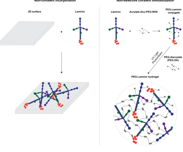
The tethering of bioactive factors, such as proteins and peptides, onto hydrogels requires appropriate synthetic techniques for the preservation of biological function. Nevertheless, the selection of the most appropriate chemistry is not trivial, as it should assure control over peptide/ protein orientation and conformation, which in turn will impact their bioactivity and ability to modulate cellular behavior [51-53]. Specifically, strategies explored, to date, for full-length laminin immobilization, have relied either on its transient non-covalent incorporation or physical entrapment or, alternatively, on its non-selective covalent immobilization by taking advantage of functional groups present in multiple sites of the laminin structure, such as amines and thiols [50] (Fig. 1). Despite widely used, one of the main limitations presented by these strategies is the inability to control the conformation and orientation of bioactive molecules upon immobilization. In this regard, in recent years, immobilization strategies have shifted towards site-specific conjugation, with special focus on biorthogonal chemical reactions (click chemistry), enzymatic ligation and affinity binding, using either unnatural amino acids or engineered site-selective amino acid sequences (for a more comprehensive review see [54]). These strategies are expected to provide a higher retention of bioactivity, by favoring the access to the active sites of immobilized proteins.
Figure 1. Strategies currently explored for laminin immobilization into 2D and 3D cell instructive microenvironments. Non-covalent incorporation approaches are used under the assumption the
adsorbed protein layer forms a molecular network, retaining the overall properties of laminin matrices in vivo. Non-selective covalent immobilization, in turn, takes advantage of functional groups (e.g. amines and thiols) present in multiple sites on the laminin structure. Both strategies lack the ability to control the orientation and conformation of laminin upon immobilization, which may compromise the exposure of key laminin bioactive epitopes.
Affinity-binding has been increasingly explored, as it allows the design of dynamic biomimetic systems, favoring the site-specific and reversible conjugation of proteins and peptides, and, as a result, resembling more closely what happens in vivo. However, its successful implementation is highly dependent on the appropriate selection of the binding pairs. With this in mind, and in alternative to the “artificial” binding systems, such as streptavidin and biotin,
between proteins and peptides, strong non-covalent interactions can be established without the need for protein modification.
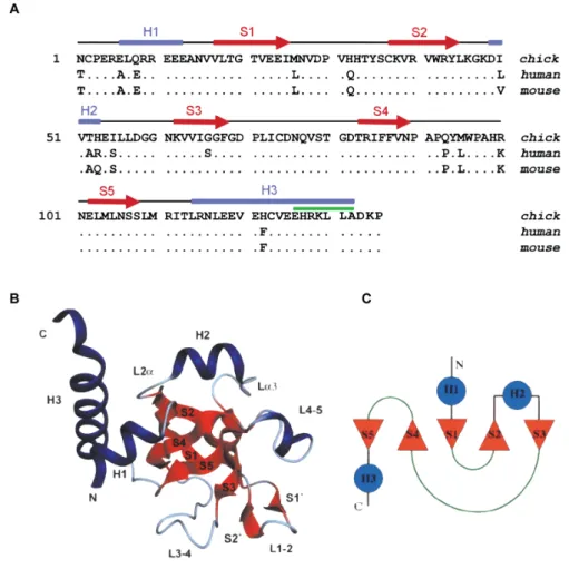
The N-terminal agrin (NtA) domain, which comprises the first 135 amino acids of agrin, mediates a high affinity interaction with laminin (dissociation constant KD @ 5nM) [55], required
for the integration of agrin into synaptic basal lamina and other basement membranes [56]. The amino acid sequence of this domain is highly conserved, with > 90% of the residues being identical, among chick, mouse and human [57] (Fig. 2). The agrin-binding site in laminin was shown to be localized in the central region of the coiled-coil domain of laminin and maps to a sequence of 20 surface-exposed and conserved residues within the g1 chain [58, 59]. This represents more than 50% of the isoforms identified to date [60], with variations in affinity imposed by a and b chains. Interestingly, this interaction requires a coiled-coil conformation of the agrin-binding site [59]. To better understand the interaction of agrin with laminin, the crystal structure of chicken NtA domain, in the presence of a synthetic 20-residue peptide corresponding to the agrin-binding site within the laminin g1 chain, was described [57]. The NtA core domain comprises five b-strands that form two orthogonally packed b-sheets flanked by a-helices at both termini, characterized by a high content of charged amino acids (Fig. 2) [57]. Indeed, charged residues account for > 25% of all amino acid residues that constitutes the NtA sequence. Laminin coiled-coil structures, in turn, were shown to exhibit the typical heptad repeat (abcdefg)n, in which solvent exposed positions are generally occupied by polar
or charged residues while hydrophobic amino acids tend to be oriented towards the core of the superhelix [61, 62]. In line with these evidences, different studies demonstrated that the high affinity interaction between the coiled-coil domain of laminin and the globular NtA domain was mainly of ionic nature and was mediated by the C-terminal helix 3 as the primary binding site and by the highly conserved charged residues at the open face of the b-barrel as an auxiliary site [55].
Figure 2. Structure of the NtA domain. A) Alignment of agrin NtA domain of chick, human and mouse.
The sequences were aligned to the first 135 amino acids of chick agrin, starting after the signal sequence cleavage site. Conserved residues are indicated by dots and the secondary structure elements are indicated above the alignment using correspondent color code in B). B) Ribbon and C) topology diagrams of the NtA structure secondary elements. b-strands (S1-S5) are represented in red; a-helices (H1-H3) are represented in blue; loop regions (L1-2, L3-4 and L4-5) are oriented to the same
surface of the protein in B) and are shown as green lines in C). Adapted from [57], Copyright© 2001,
Springer Nature.
The high affinity interaction between the NtA domain and laminin was already explored for the development of different controlled release systems for therapeutic proteins, such as growth factors [63-66] and neuropeptides [67]. For such purpose, fusion proteins consisting of the
of the growth factors or neuropeptides and hence increase their timely presence at a specific site.
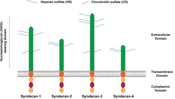
Laminin-derived small adhesive sequences have been increasingly explored, in alternative to the incorporation of the full-length protein, to confer bioactivity to 3D matrices [50]. Most of the works published to date, have explored synthetic adhesive peptides, which mediate cell interaction via integrin receptors [50]. Alternatively, cell binding ligands interacting with transmembrane heparan sulfate proteoglycans (HSPGs), and more specifically syndecans, have gained more attention in the last few years, as attractive alternative to engineer biomimetic matrices, as result of their key role in regulating NSC stemness [68-70]. Syndecans are transmembrane HSPGs, with a key role on the modulation of different biological processes, including neural patterning, angiogenesis, inflammation and wound healing [71]. These cell adhesion receptors are composed by an extracellular domain, comprising glycosaminoglycan (GAG) chains, including heparan sulfate (HS) and chondroitin sulfate (CS) chains, which mediate the interaction with growth factors, ECM proteins, as well as with other receptors (Fig. 3) [71, 72]. Syndecan receptors also present a single transmembrane domain that regulates homo- and hetero-dimerization, and a short cytoplasmic domain, which interact directly with intracellular signaling molecules (Fig. 3) [71, 72]. The extracellular domain of these receptors varies between the four members of the syndecan family identified in mammals (Syndecan-1, -2, -3 and -4), while the transmembrane and cytoplasmic domains,, are highly conserved [71].
Figure 3. Schematic representation of mammalian syndecan receptors. Syndecan receptors are
sulfate (HS) and Chondroitin sulfate (CS) chains, which mediate several cell-cell and cell-matrix interactions. They also have a single transmembrane domain responsible to promote self-association between core proteins, and a cytoplasmic domain that will mediate downstream signaling. Adapted from [73], Copyright® 2014, John Wiley & Sons.
The four members of the syndecan family (Syndecan-1, -2, -3 and -4) present distinct cell- and tissue-specific expression patterns [74]. More specifically, within the NSC niche, syndecans have a key role on the modulation of all stages of stem cell maintenance and neurogenesis (e.g. proliferation, self-renewal, differentiation, migration and maturation), either through independent signaling or by working alongside with other receptors, such as integrins [68-70]. Syndecan-1 is highly expressed during neurogenesis and is key to support NSC proliferation and progenitor cell maintenance [75, 76], while syndecan-4 constitutes a key marker of NSCs differentiation [77]. These evidences are in agreement with previous studies from our lab, showing that human NSCs (hNSCs) derived from the H9 embryonic stem cell line, cultured under basal conditions, express high levels of syndecan-1 (98.6%), whereas syndecan-4 levels were found in much lower amounts (2.62%) [78]. Syndecan-3 is key for the modulation of axon guidance [79, 80] and neurite outgrowth [81, 82], and together with syndecan-4 [83] controls neuronal migration. Lastly, syndecan-2 exerts a remarkable effect in dendritic spine morphogenesis [84].
AG73 (RKRLQVQLSIRT) is a synthetic peptide derived from the laminin globular 4 (LG4) domain of laminin a1 chain, which interacts with syndecan-1 [85, 86] and syndecan-4 [87]. This peptide was reported to promote cell adhesion with membrane ruffling [88-90], and showed great potential to enhance neurite outgrowth of PC12 neuronal cells when tethered onto natural-based hydrogels [91-93]. A previous study from our lab, evidenced that functionalization of fibrin hydrogels with AG73 improves cell outgrowth of hNSCs [78]. Despite the promising results, the intrinsic bioactivity presented by natural-based polymers makes difficult to deconvolute the contribution of the syndecan-binding peptide to the final biological outcome. Synthetic-based polymers, in turn, constitute an attractive alternative in this regard, as they are chemically-defined materials that allow a precise and independent control over the biochemical and biophysical properties of the matrix [23]. Additionally, to the best of our knowledge, to date, no study evaluating the functionalization of synthetic hydrogels with the AG73 peptide and assessing its effect on the modulation of NSC fate and function was conducted.
great potential when trying to improve the efficacy of cell-based therapeutic strategies currently under investigation and development.
References
[1] R.Y. Tam, T. Fuehrmann, N. Mitrousis, M.S. Shoichet, Regenerative therapies for central nervous system diseases: a biomaterials approach, Neuropsychopharmacology 39(1) (2014) 169-88.
[2] L. Binan, A. Ajji, G. De Crescenzo, M. Jolicoeur, Approaches for neural tissue regeneration, Stem Cell Rev 10(1) (2014) 44-59.
[3] G. Yiu, Z. He, Glial inhibition of CNS axon regeneration, Nat Rev Neurosci 7(8) (2006) 617-27.
[4] A.P. Pego, S. Kubinova, D. Cizkova, I. Vanicky, F.M. Mar, M.M. Sousa, E. Sykova, Regenerative medicine for the treatment of spinal cord injury: more than just promises?, J Cell Mol Med 16(11) (2012) 2564-82.
[5] S.A. Goldman, Stem and Progenitor Cell-Based Therapy of the Central Nervous System: Hopes, Hype, and Wishful Thinking, Cell Stem Cell 18(2) (2016) 174-88.
[6] C.H. Ho, R.J. Triolo, A.L. Elias, K.L. Kilgore, A.F. DiMarco, K. Bogie, A.H. Vette, M.L. Audu, R. Kobetic, S.R. Chang, K.M. Chan, S. Dukelow, D.J. Bourbeau, S.W. Brose, K.J. Gustafson, Z.H. Kiss, V.K. Mushahwar, Functional electrical stimulation and spinal cord injury, Phys Med Rehabil Clin N Am 25(3) (2014) 631-54, ix.
[7] I. Vismara, S. Papa, F. Rossi, G. Forloni, P. Veglianese, Current Options for Cell Therapy in Spinal Cord Injury, Trends Mol Med 23(9) (2017) 831-849.
[8] O. Lindvall, Z. Kokaia, Stem cells in human neurodegenerative disorders--time for clinical translation?, J Clin Invest 120(1) (2010) 29-40.
[9] Y. Tang, P. Yu, L. Cheng, Current progress in the derivation and therapeutic application of neural stem cells, Cell Death Dis 8(10) (2017) e3108.
[10] J.A. Steinbeck, L. Studer, Moving stem cells to the clinic: potential and limitations for brain repair, Neuron 86(1) (2015) 187-206.
[11] P. Lu, L.L. Jones, E.Y. Snyder, M.H. Tuszynski, Neural stem cells constitutively secrete neurotrophic factors and promote extensive host axonal growth after spinal cord injury, Exp Neurol 181(2) (2003) 115-29.
[12] K. Reekmans, J. Praet, J. Daans, V. Reumers, P. Pauwels, A. Van der Linden, Z.N. Berneman, P. Ponsaerts, Current challenges for the advancement of neural stem cell biology and transplantation research, Stem Cell Rev 8(1) (2012) 262-78.
[13] D. McLauchlan, N.P. Robertson, Stem cells in the treatment of central nervous system disease, J Neurol 265(4) (2018) 984-986.
[15] M.H. Amer, F. Rose, K.M. Shakesheff, M. Modo, L.J. White, Translational considerations in injectable cell-based therapeutics for neurological applications: concepts, progress and challenges, NPJ Regen Med 2 (2017) 23.
[16] F. Gattazzo, A. Urciuolo, P. Bonaldo, Extracellular matrix: a dynamic microenvironment for stem cell niche, Biochim Biophys Acta 1840(8) (2014) 2506-19.
[17] S.W. Lane, D.A. Williams, F.M. Watt, Modulating the stem cell niche for tissue regeneration, Nat Biotechnol 32(8) (2014) 795-803.
[18] A. Vishwakarma, J. Rouwkema, P.A. Jones, J.M. Karp, The Need to Study, Mimic and Target Stem Cell Niches, in: A. Vishwakarma, J.M. Karp (Eds.), Biology and Engineering of Stem Cell Niches, Elsevier2017.
[19] J.L. Wilson, T.C. McDevitt, Biofunctional Hydrogels for Three-Dimensional Stem Cell Culture, in: A. Vishwakarma, J.M. Karp (Eds.), Biology and Engineering of Stem Cell Niches, Elsevier2017.
[20] F. Ruedinger, A. Lavrentieva, C. Blume, I. Pepelanova, T. Scheper, Hydrogels for 3D mammalian cell culture: a starting guide for laboratory practice, Appl Microbiol Biotechnol 99(2) (2015) 623-36.
[21] Technology Platforms for 3D Cell Culture: A User's Guide, John Wiley & Sons Ltd.2017. [22] M.W. Tibbitt, K.S. Anseth, Hydrogels as Extracellular Matrix Mimics for 3D Cell Culture, Biotechnol. Bioeng. 103(4) (2009) 655-663.
[23] D. Barros, I.F. Amaral, A.P. Pego, Biomimetic synthetic self-assembled hydrogels for cell transplantation, Curr Top Med Chem 15(13) (2015) 1209-26.
[24] K.J. Lampe, S.C. Heilshorn, Building stem cell niches from the molecule up through engineered peptide materials, Neurosci Lett 519(2) (2012) 138-46.
[25] J. Lam, S.T. Carmichael, W.E. Lowry, T. Segura, Hydrogel design of experiments methodology to optimize hydrogel for iPSC-NPC culture, Adv Healthc Mater 4(4) (2015) 534-9.
[26] C. Regalado-Santiago, E. Juarez-Aguilar, J.D. Olivares-Hernandez, E. Tamariz, Mimicking Neural Stem Cell Niche by Biocompatible Substrates, Stem Cells Int 2016 (2016) 1513285.
[27] C.M. Madl, B.L. LeSavage, R.E. Dewi, C.B. Dinh, R.S. Stowers, M. Khariton, K.J. Lampe, D. Nguyen, O. Chaudhuri, A. Enejder, S.C. Heilshorn, Maintenance of neural progenitor cell stemness in 3D hydrogels requires matrix remodelling, Nat Mater 16(12) (2017) 1233-1242. [28] J. Zhu, R.E. Marchant, Design properties of hydrogel tissue-engineering scaffolds, Expert Rev. Med. Devices 8(5) (2011) 607-26.
[29] G.A. Saracino, D. Cigognini, D. Silva, A. Caprini, F. Gelain, Nanomaterials design and tests for neural tissue engineering, Chem. Soc. Rev. 42(1) (2013) 225-62.
[30] J. Zhu, Bioactive modification of poly(ethylene glycol) hydrogels for tissue engineering, Biomaterials 31(17) (2010) 4639-56.
[31] Y.H. Tsou, J. Khoneisser, P.C. Huang, X. Xu, Hydrogel as a bioactive material to regulate stem cell fate, Bioact Mater 1(1) (2016) 39-55.
[32] X. Lu, T.H. Perera, A.B. Aria, L.A.S. Callahan, Polyethylene glycol in spinal cord injury repair: a critical review, J Exp Pharmacol 10 (2018) 37-49.
[33] N.A. Peppas, K.B. Keys, M. Torres-Lugo, A.M. Lowman, Poly(ethylene glycol)-containing hydrogels in drug delivery, J Control Release 62(1-2) (1999) 81-7.
[34] E.A. Phelps, N.O. Enemchukwu, V.F. Fiore, J.C. Sy, N. Murthy, T.A. Sulchek, T.H. Barker, A.J. Garcia, Maleimide cross-linked bioactive PEG hydrogel exhibits improved reaction kinetics and cross-linking for cell encapsulation and in situ delivery, Adv Mater 24(1) (2012) 64-70, 2.
[35] J. Yu, F. Chen, X. Wang, N. Dong, C. Lu, G. Yang, Z. Chen, Synthesis and characterization of MMP degradable and maleimide cross-linked PEG hydrogels for tissue engineering scaffolds, Polymer Degradation and Stability 133 (2016) 312-320.
[36] N.O. Enemchukwu, R. Cruz-Acuna, T. Bongiorno, C.T. Johnson, J.R. Garcia, T. Sulchek, A.J. Garcia, Synthetic matrices reveal contributions of ECM biophysical and biochemical properties to epithelial morphogenesis, J Cell Biol 212(1) (2016) 113-24.
[37] W.M. Han, S.E. Anderson, M. Mohiuddin, D. Barros, S.A. Nakhai, E. Shin, I.F. Amaral, A.P. Pego, A.J. Garcia, Y.C. Jang, Synthetic matrix enhances transplanted satellite cell engraftment in dystrophic and aged skeletal muscle with comorbid trauma, Sci Adv 4(8) (2018) eaar4008.
[38] S. Lin, N. Sangaj, T. Razafiarison, C. Zhang, S. Varghese, Influence of physical properties of biomaterials on cellular behavior, Pharm Res 28(6) (2011) 1422-30.
[39] J. Kim, Y.P. Kong, S.M. Niedzielski, R.K. Singh, A.J. Putnam, A. Shikanov, Characterization of the crosslinking kinetics of multi-arm poly(ethylene glycol) hydrogels formed via Michael-type addition, Soft Matter 12(7) (2016) 2076-85.
[40] L.A. Flanagan, Y.E. Ju, B. Marg, M. Osterfield, P.A. Janmey, Neurite branching on deformable substrates, Neuroreport 13(18) (2002) 2411-5.
[41] K. Saha, A.J. Keung, E.F. Irwin, Y. Li, L. Little, D.V. Schaffer, K.E. Healy, Substrate modulus directs neural stem cell behavior, Biophys. J. 95(9) (2008) 4426-38.
[42] A. Banerjee, M. Arha, S. Choudhary, R.S. Ashton, S.R. Bhatia, D.V. Schaffer, R.S. Kane, The influence of hydrogel modulus on the proliferation and differentiation of encapsulated neural stem cells, Biomaterials 30(27) (2009) 4695-9.
[44] A. Kerever, J. Schnack, D. Vellinga, N. Ichikawa, C. Moon, E. Arikawa-Hirasawa, J.T. Efird, F. Mercier, Novel extracellular matrix structures in the neural stem cell niche capture the neurogenic factor fibroblast growth factor 2 from the extracellular milieu, Stem Cells 25(9) (2007) 2146-57.
[45] I. Kazanis, J.D. Lathia, T.J. Vadakkan, E. Raborn, R. Wan, M.R. Mughal, D.M. Eckley, T. Sasaki, B. Patton, M.P. Mattson, K.K. Hirschi, M.E. Dickinson, C. ffrench-Constant, Quiescence and activation of stem and precursor cell populations in the subependymal zone of the mammalian brain are associated with distinct cellular and extracellular matrix signals, J Neurosci 30(29) (2010) 9771-81.
[46] A. Hyysalo, M. Ristola, M.E. Makinen, S. Hayrynen, M. Nykter, S. Narkilahti, Laminin alpha5 substrates promote survival, network formation and functional development of human pluripotent stem cell-derived neurons in vitro, Stem Cell Res 24 (2017) 118-127.
[47] L. Luckenbill-Edds, Laminin and the mechanism of neuronal outgrowth, Brain Res Brain Res Rev 23(1-2) (1997) 1-27.
[48] S.K. Powell, H.K. Kleinman, Neuronal laminins and their cellular receptors, Int J Biochem Cell Biol 29(3) (1997) 401-14.
[49] S. Plantman, M. Patarroyo, K. Fried, A. Domogatskaya, K. Tryggvason, H. Hammarberg, S. Cullheim, Integrin-laminin interactions controlling neurite outgrowth from adult DRG neurons in vitro, Mol Cell Neurosci 39(1) (2008) 50-62.
[50] D. Barros, I.F. Amaral, A.P. Pego, Laminin inspired cell instructive microenvironments for neural tissue engineering applications, Manuscript in preparation (2019).
[51] B.G. Keselowsky, D.M. Collard, A.J. Garcia, Surface chemistry modulates fibronectin conformation and directs integrin binding and specificity to control cell adhesion, J Biomed Mater Res A 66(2) (2003) 247-59.
[52] J.C. Rodriguez Hernandez, M. Salmeron Sanchez, J.M. Soria, J.L. Gomez Ribelles, M. Monleon Pradas, Substrate chemistry-dependent conformations of single laminin molecules on polymer surfaces are revealed by the phase signal of atomic force microscopy, Biophys J 93(1) (2007) 202-7.
[53] O.M. Ba, M. Hindie, P. Marmey, O. Gallet, K. Anselme, A. Ponche, A.C. Duncan, Protein covalent immobilization via its scarce thiol versus abundant amine groups: Effect on orientation, cell binding domain exposure and conformational lability, Colloids Surf B Biointerfaces 134 (2015) 73-80.
[54] S.A. Fisher, A.E.G. Baker, M.S. Shoichet, Designing Peptide and Protein Modified Hydrogels: Selecting the Optimal Conjugation Strategy, J Am Chem Soc 139(22) (2017) 7416-7427.
[55] J.B. Mascarenhas, M.A. Ruegg, U. Winzen, W. Halfter, J. Engel, J. Stetefeld, Mapping of the laminin-binding site of the N-terminal agrin domain (NtA), EMBO J 22(3) (2003) 529-36.
[56] M.A. Ruegg, J.L. Bixby, Agrin orchestrates synaptic differentiation at the vertebrate neuromuscular junction, Trends Neurosci 21(1) (1998) 22-7.
[57] J. Stetefeld, M. Jenny, T. Schulthess, R. Landwehr, B. Schumacher, S. Frank, M.A. Ruegg, J. Engel, R.A. Kammerer, The laminin-binding domain of agrin is structurally related to N-TIMP-1, Nat Struct Biol 8(8) (2001) 705-9.
[58] A.J. Denzer, T. Schulthess, C. Fauser, B. Schumacher, R.A. Kammerer, J. Engel, M.A. Ruegg, Electron microscopic structure of agrin and mapping of its binding site in laminin-1, EMBO J 17(2) (1998) 335-43.
[59] R.A. Kammerer, T. Schulthess, R. Landwehr, B. Schumacher, A. Lustig, P.D. Yurchenco, M.A. Ruegg, J. Engel, A.J. Denzer, Interaction of agrin with laminin requires a coiled-coil conformation of the agrin-binding site within the laminin gamma1 chain, EMBO J 18(23) (1999) 6762-70.
[60] M. Aumailley, L. Bruckner-Tuderman, W.G. Carter, R. Deutzmann, D. Edgar, P. Ekblom, J. Engel, E. Engvall, E. Hohenester, J.C. Jones, H.K. Kleinman, M.P. Marinkovich, G.R. Martin, U. Mayer, G. Meneguzzi, J.H. Miner, K. Miyazaki, M. Patarroyo, M. Paulsson, V. Quaranta, J.R. Sanes, T. Sasaki, K. Sekiguchi, L.M. Sorokin, J.F. Talts, K. Tryggvason, J. Uitto, I. Virtanen, K. von der Mark, U.M. Wewer, Y. Yamada, P.D. Yurchenco, A simplified laminin nomenclature, Matrix Biol 24(5) (2005) 326-32.
[61] D.A. Parry, R.D. Fraser, J.M. Squire, Fifty years of coiled-coils and alpha-helical bundles: a close relationship between sequence and structure, J Struct Biol 163(3) (2008) 258-69. [62] G. Armony, E. Jacob, T. Moran, Y. Levin, T. Mehlman, Y. Levy, D. Fass, Cross-linking reveals laminin coiled-coil architecture, Proc Natl Acad Sci U S A 113(47) (2016) 13384-13389.
[63] W. Sun, C. Sun, H. Zhao, H. Lin, Q. Han, J. Wang, H. Ma, B. Chen, Z. Xiao, J. Dai, Improvement of sciatic nerve regeneration using laminin-binding human NGF-beta, PLoS One 4(7) (2009) e6180.
[64] Q. Han, B. Li, H. Feng, Z. Xiao, B. Chen, Y. Zhao, J. Huang, J. Dai, The promotion of cerebral ischemia recovery in rats by laminin-binding BDNF, Biomaterials 32(22) (2011) 5077-85.
[65] J. Xie, B. Jin, D.W. Li, B. Shen, N. Gong, T.Z. Zhang, P. Dong, Effect of laminin-binding BDNF on induction of recurrent laryngeal nerve regeneration by miR-222 activation of mTOR signal pathway, Am J Transl Res 7(6) (2015) 1071-80.
[66] B. Wang, J. Yuan, X. Chen, J. Xu, Y. Li, P. Dong, Functional regeneration of the transected recurrent laryngeal nerve using a collagen scaffold loaded with laminin and
laminin-[67] L. Wu, J. Wang, X. Chen, A. Hong, Expression, identification and biological effects of the novel recombination protein, PACAP38-NtA, with high bioactivity, Int J Mol Med 35(2) (2015) 376-82.
[68] M. Ford-Perriss, K. Turner, S. Guimond, A. Apedaile, H.D. Haubeck, J. Turnbull, M. Murphy, Localisation of specific heparan sulfate proteoglycans during the proliferative phase of brain development, Dev Dyn 227(2) (2003) 170-84.
[69] Y. Choi, H. Chung, H. Jung, J.R. Couchman, E.S. Oh, Syndecans as cell surface receptors: Unique structure equates with functional diversity, Matrix Biol 30(2) (2011) 93-9. [70] F.E. Poulain, H.J. Yost, Heparan sulfate proteoglycans: a sugar code for vertebrate development?, Development 142(20) (2015) 3456-67.
[71] N.A. Afratis, D. Nikitovic, H.A. Multhaupt, A.D. Theocharis, J.R. Couchman, N.K. Karamanos, Syndecans - key regulators of cell signaling and biological functions, FEBS J 284(1) (2017) 27-41.
[72] H. Chung, H.A. Multhaupt, E.S. Oh, J.R. Couchman, Minireview: Syndecans and their crucial roles during tissue regeneration, FEBS Lett 590(15) (2016) 2408-17.
[73] J.R. Couchman, S. Gopal, H.C. Lim, S. Norgaard, H.A. Multhaupt, Fell-Muir Lecture: Syndecans: from peripheral coreceptors to mainstream regulators of cell behaviour, Int J Exp Pathol 96(1) (2015) 1-10.
[74] X. Xian, S. Gopal, J.R. Couchman, Syndecans as receptors and organizers of the extracellular matrix, Cell Tissue Res 339(1) (2010) 31-46.
[75] Q. Wang, L. Yang, C. Alexander, S. Temple, The niche factor syndecan-1 regulates the maintenance and proliferation of neural progenitor cells during mammalian cortical development, PLoS One 7(8) (2012) e42883.
[76] L. Morizur, A. Chicheportiche, L.R. Gauthier, M. Daynac, F.D. Boussin, M.A. Mouthon, Distinct Molecular Signatures of Quiescent and Activated Adult Neural Stem Cells Reveal Specific Interactions with Their Microenvironment, Stem Cell Reports (2018).
[77] L.E. Oikari, R.K. Okolicsanyi, A. Qin, C. Yu, L.R. Griffiths, L.M. Haupt, Cell surface heparan sulfate proteoglycans as novel markers of human neural stem cell fate determination, Stem Cell Res 16(1) (2016) 92-104.
[78] A.R. Bento, Improving neurite outgrowth in 3D hydrogel matrices by mimicking cell receptor-ECM interactions occurring in neurogenic niches: an engineering approach to develop more efficient neural stem cell hydrogel carriers, Faculdade de Engenharia, Universidade do Porto, 2018.
[79] A. Kinnunen, T. Kinnunen, M. Kaksonen, R. Nolo, P. Panula, H. Rauvala, N-syndecan and HB-GAM (heparin-binding growth-associated molecule) associate with early axonal tracts in the rat brain, Eur J Neurosci 10(2) (1998) 635-48.
[80] A. Kinnunen, M. Niemi, T. Kinnunen, M. Kaksonen, R. Nolo, H. Rauvala, Heparan sulphate and HB-GAM (heparin-binding growth-associated molecule) in the development of the thalamocortical pathway of rat brain, Eur J Neurosci 11(2) (1999) 491-502.
[81] E. Raulo, M.A. Chernousov, D.J. Carey, R. Nolo, H. Rauvala, Isolation of a neuronal cell surface receptor of heparin binding growth-associated molecule (HB-GAM). Identification as N-syndecan (syndecan-3), J Biol Chem 269(17) (1994) 12999-3004.
[82] T. Kinnunen, E. Raulo, R. Nolo, M. Maccarana, U. Lindahl, H. Rauvala, Neurite outgrowth in brain neurons induced by heparin-binding growth-associated molecule (HB-GAM) depends on the specific interaction of HB-GAM with heparan sulfate at the cell surface, J Biol Chem 271(4) (1996) 2243-8.
[83] H.K. Matthews, L. Marchant, C. Carmona-Fontaine, S. Kuriyama, J. Larrain, M.R. Holt, M. Parsons, R. Mayor, Directional migration of neural crest cells in vivo is regulated by Syndecan-4/Rac1 and non-canonical Wnt signaling/RhoA, Development 135(10) (2008) 1771-80.
[84] I.M. Ethell, Y. Yamaguchi, Cell surface heparan sulfate proteoglycan syndecan-2 induces the maturation of dendritic spines in rat hippocampal neurons, J Cell Biol 144(3) (1999) 575-86.
[85] M.P. Hoffman, M. Nomizu, E. Roque, S. Lee, D.W. Jung, Y. Yamada, H.K. Kleinman, Laminin-1 and laminin-2 G-domain synthetic peptides bind syndecan-1 and are involved in acinar formation of a human submandibular gland cell line, J Biol Chem 273(44) (1998) 28633-41.
[86] L.N. Gama-de-Souza, E. Cyreno-Oliveira, V.M. Freitas, E.S. Melo, V.F. Vilas-Boas, A.S. Moriscot, R.G. Jaeger, Adhesion and protease activity in cell lines from human salivary gland tumors are regulated by the laminin-derived peptide AG73, syndecan-1 and beta1 integrin, Matrix Biol 27(5) (2008) 402-19.
[87] N. Suzuki, N. Ichikawa, S. Kasai, M. Yamada, N. Nishi, H. Morioka, H. Yamashita, Y. Kitagawa, A. Utani, M.P. Hoffman, M. Nomizu, Syndecan binding sites in the laminin alpha1 chain G domain, Biochemistry 42(43) (2003) 12625-33.
[88] M. Nomizu, W.H. Kim, K. Yamamura, A. Utani, S.Y. Song, A. Otaka, P.P. Roller, H.K. Kleinman, Y. Yamada, Identification of cell binding sites in the laminin alpha 1 chain carboxyl-terminal globular domain by systematic screening of synthetic peptides, J Biol Chem 270(35) (1995) 20583-90.
[89] K. Hozumi, N. Suzuki, P.K. Nielsen, M. Nomizu, Y. Yamada, Laminin alpha1 chain LG4 module promotes cell attachment through syndecans and cell spreading through integrin
[90] M. Mochizuki, Y. Kadoya, Y. Wakabayashi, K. Kato, I. Okazaki, M. Yamada, T. Sato, N. Sakairi, N. Nishi, M. Nomizu, Laminin-1 peptide-conjugated chitosan membranes as a novel approach for cell engineering, FASEB J 17(8) (2003) 875-7.
[91] Y. Yamada, K. Hozumi, F. Katagiri, Y. Kikkawa, M. Nomizu, Biological activity of laminin peptide-conjugated alginate and chitosan matrices, Biopolymers 94(6) (2010) 711-20.
[92] Y. Yamada, F. Katagiri, K. Hozumi, Y. Kikkawa, M. Nomizu, Cell behavior on protein matrices containing laminin alpha1 peptide AG73, Biomaterials 32(19) (2011) 4327-35. [93] Y. Yamada, K. Hozumi, A. Aso, A. Hotta, K. Toma, F. Katagiri, Y. Kikkawa, M. Nomizu, Laminin active peptide/agarose matrices as multifunctional biomaterials for tissue engineering, Biomaterials 33(16) (2012) 4118-25.