www.scielo.br/aabc
Role of laminin bioavailability in the
astroglial permissivity for neuritic outgrowth
MARCIENNE TARDY
INSERM U-421, IM3, Medical Faculty and Biochemistry Department CHU Henri Mondor, 94010 Creteil, France
Manuscript received on October 3, 2002; accepted for publication on October 10, 2002; presented byLeny A. Cavalcante
ABSTRACT
The mechanisms involved in the failure of an adult brain to regenerate post-lesion remain poorly understood. The reactive gliosis which occurs after an injury to the CNS and leads to the glial scar has been considered as one of the major impediments to neurite outgrowth and axonal regeneration. A glial scar consists mainly of reactive, hypertrophic astrocytes. These reactive cells acquire new properties, leading to A non-permissive support for neurons. Astrogial reactivity is mainly characteriized by a high overexpression of the major com-ponent of the gliofilaments, the glial fibrillary acidic protein (GFAP). This GFAP overexpression is related to the astroglial morphological response to injury. We hypothesized that modulation of GFAP synthesis, reversing the hypertrophic phenotype, might also reverse the blockage of neuritic outgrowth observed after a lesion. In this article, we review findings of our group, confirming our hypothesis in a model of lesioned neuron-astrocyte cocultures. We demonstrate that permissivity for neuritic outgrowth is related to pheno-typic changes induced in reactive astrocytes transfected by antisense GFAP-mRNA. We also found that this permissivity was related to a neuron-regulated extracellular laminin bioavailability.
Key words: astrogliosis, neuro-glia interactions, neurite outgrowth, neuronal migration, laminin, metallo-proteinases.
INTRODUCTION
Astrocytes are the most numerous cellular compo-nents of the brain. Despite their number and their contribution to brain development and function, the consequences of their dysfunction on the physiology of the nervous system have only been considered re-cently. Astrocytes have been involved in many func-tions including control of brain development and homeostasis (for a review see Tardy 1991), CNS
im-Correspondence to: Dr. Marcienne Tardy Inserm U-421, Medical Faculty 8, rue du Gal Sarrail
94010 CRETEIL (France) Fax: 33+ 149813709 E-mail: tardy@im3.inserm.fr
munity, and in regulation of development, function and efficiency of the synapse (Pfrieger and Barres 1996). Their implication in a defence mechanism against oxydative stress has been reported (Desagher et al. 1996). It has been shown that astrocytes may provide new means of communication in the ner-vous system (Smith 1994, Verkhratsky et al. 1998). Subpopulations of these cells have been described and as many different types of astrocytes may exist as neurons (Kimelberg 1995).
684 MARCIENNE TARDY
These cells are typified by cytoplasmic hypertrophy associated with a profusion of long, thick processes. The most striking ultrastructural finding, however, is the presence of bundles of intermediate filaments which at times appear to fill the entire cytoplasmic compartment (Hozumi et al. 1990). These fila-ments are often heteropolymers, largely consisting of GFAP and, at a lesser degree, of vimentin (Ridet et al. 1997). The glial scar constitutes a physical and chemical barrier which isolates the intact tis-sue, but also contributes to the failure for recovery post-injury (Bush et al. 1999). Reactive astrocytes, however, may also produce trophic factors benefic for axonal regrowth. Whether these elements are beneficial or detrimental for recovery remains enig-matic. In the developing brain, the growth of axons is guided by astrocytes (Silver et al. 1993). In con-trast, in the adult brain, axons do not regrow after axotomy and, in this regard, the inhibitory role of the astroglial scar has been well documented (Reier and Houle 1988). Many phenotypic changes have been observed in astroglial cells during their maturation, among them, a decrease in molecules known to pro-mote axonal growth, like laminin (Rivas et al. 1992), NCAM, heparan sulfate proteoglycan (HSPG) have been reported (McKeon et al. 1995). In the oppo-site, molecules like chondroitin sulfate proteogly-can (CSPG) or Tenascin, known for inhibiting neu-rite outgrowth, increase during astroglial maturation (Faissner and Steindler 1995).
One major change in the astroglial phenotype during maturation as well as during astroglial reac-tivity, is associated with the increase in the major component of their intermediate filament, the glial fibrillary acidic protein (GFAP). This major event, directly associated with morphological changes (Tardy et al. 1993), suggested its involvement in the astroglial functional shift from neurite-promoting (permissive), to neurite-inhibiting (non-permissive) elements.
Mechanically lesioned neurons/astrocytes pri-mary cocultures presenting an astrogliosis around the lesion site, reproduce adequately in vitro, the phenomena characterized in vivo. This model was
used to determine whether a triggered decrease in GFAP could affect neurite outgrowth. Antisense GFAP-mRNA decreased GFAP synthesis and the consecutive phenotypic changes were associated with a functional shift towards neurite-promoting elements. Molecular studies showed a regulation of a major ECM molecule by the astrocytes, in rela-tion to the expression of a member of the family of zinc-dependent metalloproteinases (MMPs) whose activities are involved in tissue remodeling (Yong et al. 1998).
WHAT IS A REACTIVE ASTROCYTE?
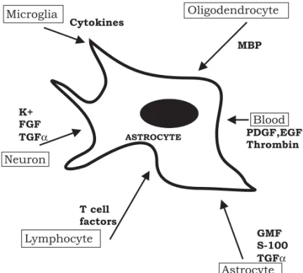
fac-Fig. 1 – Factors triggering the astroglial reactivity in response to an injury in the adult CNS. MBP (myelin basic protein), GMF (glial maturating factor), TGFα (transforming growth factorα), FGF (fibroblast growth factor), EGF (epidermal growth factor), PDGF (platelet-derived growth factor).
tor, interferon-gamma), lesioned neurons (fibrob-last growth factor, potassium, ATP), oligodendro-cytes (myelin basic protein), from blood (throm-bin, epithelial growth factor (EGF), platelet-derived growth factor (PDGF), steroids, insulin), endothe-lial cells (endothelin, ATP), and activated lympho-cytes (Fig. 1). Other factors including the prion protein, fibronectin, putrescine and prostaglandines (Kimelberg and Norenberg 1994) have also been im-plicated.
Reactive astrogliosis is accompanied by an in-duction and an upregulation of many proteins with potential biological effects (Fig. 2). For instance, basic fibroblast growth factor (bFGF) stimulates the production of nerve growth factor (NGF) and pro-tects against excitotoxic injury, but also contributes to the excessive neurite sprouting and dystrophic neurite formation observed inAlzheimer disease (for a review see Norenberg 1994). It becomes evident that reactive gliosis varies depending on the nature of the injury, and the microenvironment of the
in-jury site. Reactive astrocytes are metabolically acti-vated cells. As an example, glutamine synthetase, a metabolic astroglial marker, essential for glutamate and ammonia neutralization, (Tardy 1991), cellu-lar function and brain detoxification, has also been found modified in various pathological conditions associated with astroglial reactivity. Increased in the natural scrapie of the sheep (Lefrançois et al. 1994), it may illustrate an attempt by astrocytes to maintain and control the cerebral homeostasis. Reduced in senile dementia brains of the Alzheimer type, it un-derlines a dysfunction of the astroglial metabolism, might reflect oxidative damage and might have se-vere consequences on the pathological cascade of events (Le Prince et al. 1995).
INHIBITION OF ASTROGLIAL HYPERTROPHY INDUCES FUNCTIONAL CHANGES, BENEFICIAL FOR AXONAL REGROWTH
mechani-686 MARCIENNE TARDY
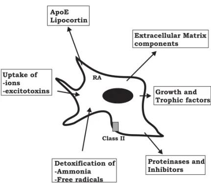
Fig. 2 – Functional properties of reactive astrocytes (RA). Reactive astrocytes produce growth and trophic factors (NGF, FGF, PDGF, CNTF, IGF...), inter-act with remodeling of Extracellular Matrix (glycoproteins, proteinases and in-hibitors), with Inflammation/ Immunity compounds, (class II histocompatibility antigens, apoE, lipocortin) and with detoxification and control of homeostasia (excitotoxins, ions, free radicals).
cal lesion, we chose mechanically lesioned rodent neuron-astrocyte cocultures. Hypertrophic astro-cytes were present, neighboring the lesion and neu-rons were absent from the area of lesion and from its surroundings. These reactive, hypertrophic astro-cytes appeared, therefore, as a non-permissive sub-strate and the coculture model used could be con-sidered valid to study the consequences of astroglial reactivity on axonal growth, in spite of the absence of the three dimensions of a tissue. Transfection of an antisense for GFAP-mRNA in the lesioned cocul-ture, in order to modulate the astrocytic response to the injury, reduced cell hypertrophy and decreased the capacity of the cells to translate GFAP-mRNA (Lefrançois et al. 1997).
After such antisense treatment, neuronal cell bodies and neurites were reobserved over astrocytes of the lesion border and neurites projected into the lesion site. The expression of GAP-43, associated with nerve sprouting (Schreyer and Skene 1991) was
therefore, may be one element involved in the lack of inhibition after antisense treatment.
MANIPULATING THE LAMININ BIOAVAILABILITY INDUCES CHANGES IN PERMISSIVITY
OF NEURITE OUTGROWTH
A dynamic role for the overlaying laminin in neu-ronal migration and neurite outgrowth was con-firmed when permissive cocultures were treated with anti-laminin antibodies (Costa et al. 2002). These antibodies which recognize the astroglial laminin blocked a large part of neurite outgrowth and inhibi-tion of neuronal migrainhibi-tion to the lesion site, whereas an antibody against tenascin C, another component of the extracellular matrix (Faissner and Steindler 1995), did not. This indirect evidence was sup-ported by a more direct one, consisting in the ad-dition of soluble, native laminin to the coculture, which promotes neurite outgrowth and neuronal mi-gration. These results do not exclude a soluble form of laminin released by the astroglial monolayer which may contribute to the permissivity in addition to the overlaying network.
SWITCH TO A PERMISSIVE SYSTEM FOR NEURITE OUTGROWTH IS RELATED TO A RAPID AND
CONSTANT INCREASE IN LAMININ
Laminin level increased rapidly in the permissive antisense-treated cocultures, and remained high. A reverse relationship between GFAP and laminin lev-els was observed in the permissive cocultures. In addition, we found that an enhanced laminin level was under neuronal control and consequent to either secreted factors or to cell interactions. Such signals may regulate both, laminin synthesis and extracel-lular availability, in order to improve neurite out-growth (Costa et al. 2002).
CHANGES IN LAMININ STRUCTURE AND LOCALIZATION MIGHT PARTICIPATE
IN PERMISSIVITY
The switch from a non-permissive to a permissive support for neuronal migration and neurite out-growth is correlated with a differential pattern of laminin expression.
A laminin labeled astroglial monolayer, a dense and regular overlaying network, characterizes the control unlesioned cocultures. Mechanical le-sion modified the labeling distribution in the mono-layer, where it significantly decreases, out of a pop-ulation of large and flat astrocytes around the lesion border which remained labeled with punctate intra-cellular spots (Lefrançois et al. 1997). The laminin labeled network, overlaying the astroglial lesioned monolayer, appeared unstructured and a dense la-beled network was concentrated along the lesion border. Antisense treatment restructured the labeled network, underlining that permissivity involves par-ticular patterns of laminin expression (Costa et al. 2002).
LAMININ BIOAVAILABILITY INVOLVES CHANGES IN PROTEASE/ANTIPROTEASE RATIO
IN PERMISSIVE CONDITIONS
The permissive process, observed in the lesioned an-tisense treated cocultures, might be associated with modulations of the extracellular matrix components by proteinases (Gomez et al. 1997). Among them, matrix metalloproteinases MMP-2 expression, have been involved in this type of process (Campbell and Pagenstecher 1999, Lukes et al. 1999). Associated with our permissive model, a net decrease of MMP-2 expression and activity and an increase in its endoge-nous inhibitor TIMP-2 expression are observed. The ratio between this metalloproteinase and its endoge-nous inhibitor might be directly involved in permis-sive conditions, in the observed laminin stabilization and in cell-matrix interactions.
CONCLUSIONS AND PERSPECTIVES
pre-688 MARCIENNE TARDY
sented as a favorable substrate for neuronal survival and neurite growth, which support our observations (Menet et al. 2000). As a working hypothesis, one may consider changes in the intracellular transport in which gliofilaments are involved or an indirect modulation of genetic transcription.
The observed increase in laminin level, bio-availability and in laminin network structure and dis-tribution, under neuronal control, might result from secreted factors or cell to cell interactions and re-mained to be determined. Such signals may regu-late both laminin synthesis and extracellular laminin availability in order to improve neurite outgrowth. Neuron-glia interactions are believed to play an im-portant role in the regulation of axonal growth and guidance, during both, development and regener-ation. These interactions are mediated by extra-cellular components promoting polymerization or stabilization of elements of the neuronal cytoskele-ton. Laminin appears to be a serious candidate for such an interplay. With respect to cell migration, neural cell may require balanced MMPs activities in order to migrate to the scarred area. New ax-onal growth and synaptic reconnections need to be established in a lesioned brain and their extension through the brain matrix may also require a new MMPs/TIMPs ratio. The decreased ratio observed in our in vitro permissive conditions may actively contribute to the increased laminin steady-state level report here, which, probably in addition with other components of the extracellular domain, assist re-growth permissivity.
ACKNOWLEDGMENTS
This work received its financial support from the In-stitut National de la Santé et de la Recherche Médi-cale (INSERM).
RESUMO
Os mecanismos envolvidos na falha de um encéfalo adulto
a regenerar após uma lesão permanecem escassamente compreendidos. A gliose reativa que ocorre após uma
injúria ao SNC e leva à ‘‘cicatriz’’ glial consiste
prin-cipalmente de astrócitos reativos, hipertróficos. Estas
células reativas adquirem novas propriedades, levando a um suporte não-permissivo para neurônios. A
reativida-de astroglial é caracterizada principalmente por elevada
super-expressão da principal componente dos filamentos
gliais, a proteína acídica fibrilar glial (GFAP). Esta
super-expressão de GFAP está relacionada à resposta morfo-lógica astroglial à injúria. Nós levantamos a hipótese
de que a modulação da síntese de GFAP, revertendo o
fenótipo hipertrófico, poderia também reverter o bloqueio
do crescimento neurítico observado após uma lesão. Neste
artigo, nós revisamos achados de nosso grupo, confir-mando nossa hipótese em um modelo de co-cultura de
neurônios lesados e astrócitos. Nós demonstramos que a
permissividade para crescimento neurítico está
relaciona-da a murelaciona-danças induzirelaciona-das em astrócitos reativos
transfec-tados com o mRNA anti-senso para GFAP. Nós também observamos que esta permissividade estava relacionada
com a bio-disponibilidade de laminina extra-celular.
Palavras-chave: astrogliose, interações neuro-gliais,
crescimento neurítico, migração neuronal, laminina,
metalo-proteinases.
REFERENCES
Bush TG, Puvanachandra N, Horner CH, Polito A, Ostenfeld T, Svendsen CN, Mucke L, Johnson MH and Sofroniew MV.1999. Leukocyte infil-tration, neuronal degeneration and neurite outgrowth after ablation of scar-forming, reactive astrocytes in adult transgenic mice. Neuron 23: 297-308.
Campbell IL and Pagenstecher A.1999. Matrix met-alloproteinases and their inhibitors in the nervous sys-tem: the good, the bad and the enigmatic. Trends Neurosci 22: 285-287.
Costa S, Planchenault T, Charriere-Bertrand C, Mouchel Y, Fages C, Sharon J, Lefrançois T, Barlovatz-Meimon G and Tardy M.2002. As-troglial permissivity for neuritic outgrowth in neuron-astrocyte cocultures depends on regulation of laminin bioavailability. Glia 37: 105-113.
Dell’Albani DF, Kaczmarek L, Messina L, Spamp-inato G, Avolar, Messina A and Giuffrida Stella AM.1990. Glial Fibrillary Acidic Protein mRNA and glutamine synthetase activity after ner-vous system injury. J Neurosci Res 26: 251-257.
Astro-cytes protect neurons from hydrogen peroxide toxic-ity. J Neurosci 16(8): 2553-2562.
Eng LF and Ghirnikar RS.1994. GFAP and astroglio-sis. Brain Pathol 4: 229-237.
Faissner A and Steindler DA. 1995. Boundaries and inhibitory molecules in developing neural tis-sues. Glia 13: 233-254.
Garcia-Abreu J, Cavalcante LA and Moura Neto V.1995. Differential patterns of laminin expression in lateral and medial midbrain glia. Neuroreport 6: 761-764.
Gomez DE, Alonso DF, Yoshiji H and Thorgeirsson UP.1997. Tissue inhibitors of metalloproteinases structure, regulation and biological functions. Eur J Cell Biol 74: 111-122.
Hozumi I, Chiu FC and Norton WT.1990. Biochemi-cal and immunocytochemiBiochemi-cal changes in GFAP after stab wound. Brain Res 524: 64-71.
Junier MP. 2000. What role(s) for TGFalpha in the central nervous system? Prog Neurobiol 62: 443-473.
Kimelberg HK.1995. Receptors on astrocytes – What possible functions? Neurochem Int 26(1): 27-40.
Kimelberg HK and Norenberg MD.1994. Astroglial response to CNS trauma. In Salzman SK and Faden AI(eds). The Neurobiology of central ner-vous system trauma. New York: Oxford University Press: 193-208.
Lefrançois T, Fages C, Peschanski M and Tardy M. 1997. Neuritic outgrowth associated with as-troglial phenotypic changes induced by anti-sense Glial Fibrillary Acidic Protein (GFAP) mRNA in injured neuron-astrocyte cocultures. J Neurosci 17(11): 4121-4128.
Lefrancois T, Fages C, Brugere-Picoux J and Tardy M.1994. Astroglial reactivity in natural scrapie of sheep. Microb Pathogenesis 17: 283-289.
Le Prince G, Delaere P, Fages C, Lefrancois T, Touret M, Salanon M and Tardy M.1995. Glutamine synthetase (GS) expression is reduced in senile dementia of the Alzheimer type. Neurochem Res 20: 859-862.
Lukes A, Mun-Bryce S, Lukes M and Rosenberg GA.1999. Extracellular matrix degradation by met-alloproteinases and central nervous system diseases. Mol Neurobiol 19: 267-284.
McKeon RJ, Höke A and Silver J.1995. Injury-induced proteoglycans inhibit the potential for laminin-mediated axon growth on astrocytic scars. Exp Neurol 136: 32-43.
Menet V, Gimenez Y, Ribotta M, Saudillon F and Privat A.2000. GFAP null astrocytes are favorable substrate for neuronal survival and neurite growth. Glia 313: 267-272.
Norenberg MD. 1994. Astrocyte responses to CNS injury. J Neuropath Exp Neur 53: 213-220.
Pfrieger FW and Barres BA.1996. New views on synapse-glia interactions. Curr Opin Neurobiol 6: 615-621.
Rabchevsky AG, Weinitz JM, Coulpier M, Fages C, Tinel M and Junier MP.1988. A role for transform-ing growth factor alpha as an inducer of astrogliosis. J Neurosci 18: 10541-10552.
Reier PJ and Houle JD. 1988. The glial scar: its bearing, on axonal elongation and transplantation ap-proaches to CNS repair. In Advances in neurology: functional recovery in neurological diseases Waks-man SG(ed.) New York: Raven, 87-138.
Ridet JL, Malhotra SK, Privat A and Gage FH.1997. Reactive astrocytes: cellular and molecular cues to biological function. Trends Neurosci 12: 570-577.
Rivas RJ, Burmeister DW and Goldberg DJ.1992. Rapid effects of laminin on the growth cone. Neuron 8: 107-115.
Schreyer DJ and Skene JH.1991. Fate of gap-43 in ascending spinal axons of DRG neurons after pe-ripheral nerve injury: delayed accumulation and cor-relation with regenerative potential. J Neurosci 11: 3738-3751.
Silver J, Edwards MA and Levitt P.1993. Immuno-cytochemical demonstration on early appearing as-troglial structures that form boundaries and pathways along axons tracts in the fetal brain. J Comp Neurol 328: 415-436.
Smith SJ.1994. Neuromodulatory astrocytes. Curr Biol 4: 807-810.
Suarez I, Bodega G and Fernandez B.2002. Glu-tamine synthetase in brain: effect of ammonia. Neu-rochem Int 4: 123-142.
690 MARCIENNE TARDY
Tardy M, Le Prince G, Babajko S, Riol H, Fages C and Rolland B.1993. GFAP gene expression in normal and reactive astrocytes. Biology and Pathol-ogy of Astrocyte-neuron Interactions. Fedoroff et al.(eds). New York: Plenum, p. 153-161.
Verkhratsky A, Orkrand RK and Kettenmann H. 1998. Glial calcium: homeostasis and signaling function. Physiol Rev 78: 99-142.