Roberto Alexandre
dos Santos Dias
Fosforilação da PPA na colocalização da PPA/G
αoe
ativação da STAT3
APP phosphorylation on APP/G
αocolocalization and
STAT3 activation
Roberto Alexandre
dos Santos Dias
Fosforilação da PPA na colocalização da PPA/G
αoe
ativação da STAT3
APP phosphorylation on APP/G
αocolocalization and
STAT3 activation
Dissertação apresentada à Universidade de Aveiro para cumprimento dos requisitos necessários à obtenção do grau de Mestre em Biomedicina Molecular, realizada sob a orientação científica da Professora Doutora Sandra Vieira, Professora Auxiliar Convidada da Secção Autónoma de Ciências da Saúde da Universidade de Aveiro.
Este trabalho contou com o apoio do Centro de Biologia Celular (CBC) da Universidade de Aveiro, e é financiado por fundos FEDER através do Programa Operacional Factores de Competitividade – COMPETE e por Fundos nacionais da FCT – Fundação para a Ciência e a Tecnologia no âmbito dos projectos PTDC/QUI-BIQ/101317/2008, PTDC/SAL-NMC/111980/2009 e PEst-OE/SAU/UI0482/2011.
Dedicada aos meus pais por todo o apoio que me deram ao longo deste percurso
o júri
presidente Professora Doutora Odete Abreu Beirão da Cruz e Silva
Prof. Auxiliar com Agregação, Secção Autónoma de Ciências da Saúde, Universidade de Aveiro
Professora Doutora Sandra Isabel Moreira Pinto Vieira
Prof. Auxiliar Convidada, Secção Autónoma de Ciências da Saúde, Universidade de Aveiro
Doutora Maria Gomez Lazaro
agradecimentos À minha orientadora, Sandra Vieira, por toda a dedicação e ajuda que me deu não só na elaboração deste trabalho mas durante todo o meu percurso como estudante universitário.
À professora Odete da Cruz e Silva, pela oportunidade de realizar este trabalho no laboratório de Neurociências do Centro de Biologia Celular.
À minha família, pelo seu incentivo e apoio contínuo que sempre me deram. A todos os meus colegas do CBC, em especial à Regina Cerqueira, pela grande ajuda que me deu e sem a qual não teria sido possível concluir este trabalho, e à Joana Rocha, por ter sido como uma irmã mais velha, sempre pronta a ajudar e a partilhar momentos de boa disposição.
A todos os meus amigos de Ciências Biomédicas, em especial à Maria João, à Joana Tavares, ao Bruno Pimparel, ao Rui João e ao Igor, pelos momentos de lazer e descontração que foram fundamentais para vencer o cansaço.
Aos meus amigos, em especial ao Hugo, João, Joni, Ana Isabel, Denise e Patrícia, por me fazerem lembrar que a vida não é só trabalho.
À FCT pelo financiamento dos projetos PTDC/QUI-BIQ/101317/2008 e PTDC/SAL-NMC/111980/2009.
palavras-chave Proteína precursora de amilóide de Alzheimer (PPA); fosforilação da PPA na S655; sinalização da STAT3; proteína Gαo; colocalização, neuritogénese;
células SH-SY5Y
resumo A proteína Gαo é uma das subunidades alfa da família das proteínas G
heterotriméricas, proteínas envolvidas na transdução de sinais a partir recetores membranares. Gαo é principalmente expressa nos neurónios,
encontrando-se localizada ao longo da membrana plasmática, incluindo nos cones de crescimento dos neurónios, onde também se encontra a Proteína Precursora de Amilóide de Alzheimer (PPA), uma proteína membranar com um papel central na doença de Alzheimer. Algumas funções semelhantes já foram atribuídas às duas proteínas, incluindo papéis na diferenciação e migração celular. A ligação da PPA à Gαo já foi descrita, o que pode indicar que a PPA modula as suas funções através da ligação e ativação da Gαo. Também já foi
demonstrado que uma das vias pela qual a Gαo leva à neuritogénese é a
ativação do transdutor de sinal e ativador da transcrição 3 (STAT3). A Gαo
liga-se ao domínio C-terminal da PPA, nos aminoácidos His657-Lis676, que pertencem a uma região hidrofóbica imediatamente a jusante do sorting motif
653
YTSI656. Isto leva a crer que a ligação entre a PPA e a Gαo pode ser mediada
pela fosforilação da PPA no resíduo Serina 655 (S655). Assim, a fosforilação da PPA poderia influenciar a ativação da Gαo e consequente sinalização via
STAT3.
Neste trabalho, nós estudámos os efeitos da fosforilação da PPA e ativação da Gαo na interação funcional da PPA/Gαo. Isto foi analisado por cotransfeção de
células de neuroblastoma humano não diferenciadas (SH-SY5Y) com PPA-GFP selvagem ou fosfomutante (PPA com a S655 constutivamente fosforilada ou desfosforilada) e com a Gαo selvagem ou Gαo constutivamente ativa. As
células foram sujeitas a ensaios de imunofluorescência e vários parâmetros foram analisados, tais como a colocalização subcelular da PPA e da Gαo, o
número de prolongamentos celulares e o seu comprimento, e a intensidade da STAT3 no núcleo. Toda a análise quantitativa feita nas microfotografias foi realizada no software Fiji (ImageJ). Ensaios de Western Blot também foram realizados para avaliar a influência da co-expressão da PPA e Gαo em
proteínas do citoesqueleto.
Os nossos resultados mostram que a Gαo e a PPA parecem colocalizar
principalmente no aparelho de Golgi e nos prolongamentos celulares. Ambas as proteínas parecem cooperar entre si e induzir alterações neuritogénicas, com a Gαo a levar principalmente a uma formação inicial dos prolongamentos e
a PPA a atuar na extensão dos mesmos, num modo dependente da fosforilação da S655. Além disso, observou-se que a PPA diminui a ativação da STAT3 induzida pela Gαo, devido provavelmente a um efeito de
retro-inibição. Estes resultados provam que estas proteínas interagem funcionalmente e o seu potencial valor em aplicações neuritogénicas terapêuticas será estudado em maior detalhe.
keywords Alzheimerʼs Amyloid Precursor Protein (APP); APP phosphorylation at S655; STAT3 signalling; Gαo protein; colocalization; neuritogenesis; SH-SY5Y cells
abstract The Gαo protein is an alpha subunit of the heterotrimeric G proteins, a protein
family vastly involved in signal transduction from membranar receptors. Gαo is
enriched in the plasma membrane of neurons, including neurons growth cone, together with the Alzheimerʼs amyloid precursor protein (APP), a transmembranar protein with a central role in Alzheimer's Disease. A few similar functions have been attributed to both proteins, such as roles in cell differentiation and migration. APP binding to Gαo has been reported, which
could indicate that APP modulates its functions by binding and activating Gαo.
Further, one of the pathways by which Gαo leads to neurite outgrowth is by
activation of the signal transducer and activator of transcription 3 (STAT3). Gαo
binds to the APP C-terminal domain, namely at amino acids His657-Lys676, which belong to a hydrophobic pocket localized immediately downstream the APP 653YTSI656 basolateral sorting motif. Therefore, APP binding to Gαo may be
potentially mediated by APP phosphorylation at the Serine 655 residue (S655), and thus APP phosphorylation could influence Gαo activation and STAT3
signalling.
In this work we have studied the effects of APP phosphorylation and Gαo
activation in Gαo/APP functional interactions. This was analysed by
cotransfecting human non-differentiated neuroblastoma (SH-SY5Y) cells with either Wt or phosphomutants of APP-GFP (APP with either a constitutively phosphorylated or dephosphorylated S655) and Gαo or a constitutively active
Gαo cDNA. Cells were subjected to immnofluorescence assays, and several
parameters analysed, such as the subcellular colocalization of Gαo and APP,
the number of cellular projections and their length, and nuclear STAT3 intensity. Every quantitative analysis done upon microphotographs was performed using the Fiji (ImageJ) software. Western blot assays were also performed to evaluate the influence of APP and Gαo co-expression in
cytoskeleton-related proteins.
Our results show that Gαo and APP appear to colocalize mainly in the Golgi
apparatus and in cellular projections. Both proteins appear to cooperate with each other and induce neuritogenic changes, with Gαo mainly driving an initial
formation of cellular projections and APP influencing their elongation, in an APP S655 phosphorylation-dependent manner. Moreover, APP was observed to decrease Gαo-induced STAT3 activation, probably by a retro-inhibition effect
upon earlier STAT3 activation. These results prove that these proteins functionally interact, and their potential value in neuritogenic therapeutic applications will be further studied.
Index
Abbreviations ... 3
1. Introduction ... 5
1.1. G protein-coupled receptors ... 5
1.2. Guanine Nucleotide-Binding Proteins ... 6
1.2.1 G proteins families ... 7
1.2.2. Modulators of G protein activity ... 9
1.3. The Other Guanine Nucleotide-Binding Protein ... 10
1.3.1. Go genetics ... 11
1.3.2. Modulators of Go activity ... 11
1.3.3. Go expression pattern ... 12
1.3.4. Go functions ... 13
1.4. The Alzheimer's Amyloid Precursor Protein ... 15
1.4.1. APP processing ... 17
1.4.2. APP trafficking ... 18
1.4.3. APP phosphorylation ... 19
1.5. Go binding to APP ... 20
1.5.1. Go:APP and Alzheimer's Disease ... 21
1.5.2. APP and Gαo in neuritogenesis ... 21
2. Aims ... 23
3. Methods ... 25
3.1. Wild type (Wt) and S655 Phosphomutants APP-GFP cDNAs ... 25
3.2. Wt and Constitutively Active Gαo cDNAs ... 25
3.3. Gαo and constitutively active Gαo cDNAs amplification and purification ... 25
3.3.1. Bacterial transformation ... 25
3.3.2. MegaPrep DNA Purification ... 26
3.3.3. Ethanol precipitation of plasmid DNA ... 26
3.4. Culture and maintenance of the SH-SY5Y cell line ... 27
3.5. Transfection of the SH-SY5Y cell line with APP-GFP and Gαo cDNAs ... 27
3.5.1. JetPRIME® transfection ... 27
3.5.2 TurboFectTM transfection ... 28
3.5.3 CombiMagTM transfection ... 28
3.7. Antibodies ... 29
3.8. Western Blot assay ... 31
3.9. Ponceau Red staining of protein bands ... 32
3.10. Immunocytochemistry assay ... 32
3.11. Image analysis ... 33
3.12. Data analysis ... 33
4. Results ... 35
4.1. Optimization of the transfection method and antibodies dilutions ... 35
4.2. Effects of APP S655 phosphorylation and Gαo activation in APP/Gαo subcellular colocalization ... 36
4.3. Gαo/APP-induced alterations in cellular morphology ... 42
4.4. Analysis of cytoskeleton-related protein profiles ... 44
4.5. Effects of APP in Gαo-induced STAT3 activation ... 46
4.5.1. Nuclear STAT3 changes in response to Gαo overexpression ... 46
4.5.2. APP phosphorylation influences Gαo-induced STAT3 activation ... 49
5. Discussion ... 55
6. Conclusion ... 63
References ... 65
Abbreviations
AD Alzheimer’s disease
ADP Adenosine diphosphate
AGS Activator of protein signaling AICD APP intracellular C-terminal domain APP Alzheimer's Amyloid Precursor Protein
Aβ Amyloid β-peptide
BACE1 β-site APP-cleaving enzyme
BSA Bovine serum albumin
Ca2+ Calcium
cAMP Cyclic adenosine monophosphate CBR1 Cannabinoid 1 receptor
Cdk5 Cyclin-dependent kinase 5
cDNA Complementary deoxyribonucleic acid CNS Central nervous system
CTF C-terminal fragment
DAPI 4',6-diamidino-2-phenylindole
ECL Enhanced chemiluminescence
ER Endoplasmic reticulum
FAD Familial Alzheimer’s disease GAIP Gα-interating protein
GAP GTPase-activating protein
GAP43 Growth cone-associated protein 43 Gαo CA Constitutively Active Gαo
GDP Guanosine diphosphate
GEF Guanine nucleotide exchange factor GFP Green fluorescent protein
G protein Guanine Nucleotide-Binding Protein GPCR G protein-coupled receptor
GRIN1 G protein-regulated inducer of neurite outgrowth 1 GSK-3β Glycogen synthase kinase 3β
GTP Guanosine triphosphate
JAK Janus Kinase
K+ Potassium
LB Luria Bertani growth medium
MAPK Mitogen-activated protein kinase
O/N Overnight
PKC Protein kinase C
PM Plasma membrane
PreN Pre-neurite
P-STAT3 STAT3 phosphorylated
PTX Pertussis toxin
RGS Regulators of G protein signaling
RT Room temperature
SA APP Constitutively Dephosphorylated S655 APP mutant
sAPP Secreted APP
SDS Sodium dodecylsulfate
SE APP Constitutively Phosphorylated S655 APP mutant SEM Standard error of the mean
Src Sarcoma-related tyrosine kinase
STAT Signal transducer and activator of transcription STAT3 Signal transducer and activator of transcription 3
TGN Trans-Golgi network
TEMED Tetramethylethylenediamine
WB Western Blot
1. Introduction
1.1. G protein-coupled receptors
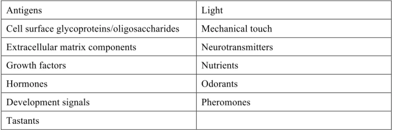
The ability of cells to interact with each other or with the environment surrounding them is essential for organisms’ survival. From simple bacteria to advanced multicellular eukaryotes, cells are gifted with mechanisms that enable them to receive a signal from the extracellular environmentand transform it in an intracellular response, a process called signal transduction (Table 1) (Lodish, 2003; Lehninger et al., 2005).
Table 1 - Some of the signals to which cells respond. Adapted from Lehninger et al., 2005
Antigens Light
Cell surface glycoproteins/oligosaccharides Mechanical touch Extracellular matrix components Neurotransmitters
Growth factors Nutrients
Hormones Odorants
Development signals Pheromones
Tastants
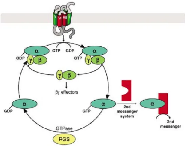
One of the main mechanisms by which cells perform signal transduction is based on the activation of guanine nucleotide-binding proteins (G proteins) by G protein-coupled receptors (GPCRs). The human genome has over 800 genes that encode GPCRs, making them the largest family of transmembrane receptors. Several hormones, neurotransmitters and sensory stimuli act upon cells through GPCRs (Offermanns, 2003; Milligan and Kostenis, 2006). Each GPCR contains seven transmembrane helical regions, an extracellular N-terminus and an intracellular C-terminus (Fig. 1). Upon the binding of a ligand to a GPCR, the receptor suffers a conformational change that enables it to interact and activate a heterotrimeric G protein (Kroeze et al., 2003; Lodish, 2003).
1.2. Guanine Nucleotide-Binding Proteins
Heterotrimeric G proteins consist of three subunits, α, β and γ. The Gα subunit
has a GTPase activity and alternates between an active state when bound to GTP and an inactive state when bound to GDP. The β and γ subunits form a single complex normally designated Gβγ subunit (Lodish, 2003; Offermanns, 2003). While initially it
was thought that only Gα interacted with the effector proteins, it is now known that both
the Gα and Gβγ are responsible for transducing the signal to their respective effector
proteins (Offermanns, 2003; Milligan and Kostenis, 2006).
In an inactivated state, the Gα subunit is bound to a GDP molecule and forms a
complex with the Gβγ subunit. As aforementioned, after the binding of a ligand to a
GPCR, a conformational change occurs that allows it to bind to the heterotrimeric G protein, which results in the release of GDP and binding of GTP to Gα. This reaction
results in the dissociation of Gα from Gβγ, allowing both subunits to interact and
modulate the activity of several effector proteins. G proteins remain active only for short periods of time due to the intrinsic GTPase activity of Gα, which hydrolyzes GTP
to GDP, leading to the reassociation of Gα with Gβγ, returning the G protein to its
inactive form (Fig. 2) (Kroeze et al., 2003; Offermanns, 2003; Milligan and Kostenis, 2006).
Figure 1 - Representation of a G protein-coupled receptor. Reproduced
1.2.1 G proteins families
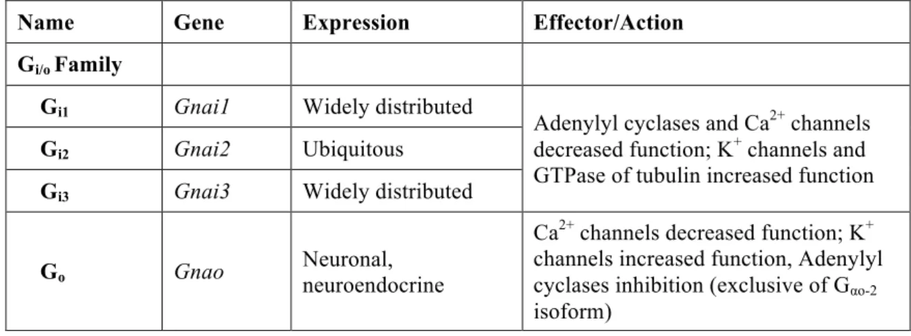
Several mammalian genes that codify Gα subunits have already been identified,
16 of which are present in humans (Gomperts et al., 2009). Since the main properties of G proteins are related to their α subunits, these have been used to divide G proteins in four subfamilies according to their functional and structural homologies: Gi/o, Gs, Gq/11
and G12/13 (Table 2) (Offermanns, 2003; Jiang and Bajpayee, 2009).
The main members of the Gi/o family are Gi proteins. These type of G proteins
have an inhibitory effect on adenylyl cyclase activity (hence G"i"), preventing it to
catalyze the formation of the second messenger cAMP and thus affecting processes within the cell (Offermanns, 2003; Lehninger et al., 2005; Jiang and Bajpayee, 2009). Gt (transducin) and Ggust (gustducin) proteins are also members of the Gi/o family and are
involved mainly in visual and taste functions, respectively (Offermanns, 2003; Milligan and Kostenis, 2006). The Go protein, which also belongs to this class, will be discussed
further ahead.
The Gs family members have the primary function of activate adenylyl cyclases
("stimulatory"), leading to the production of cAMP. Gs and Golf are the main members,
with Gs being ubiquitously expressed and Golf (olfactory) being mainly expressed in
olfactory sensory neurons and in the central nervous system (Simonds, 1999; Offermanns, 2003).
The Gq/11 proteins are widely expressed in humans and act as mediators in the
regulation of phospholipase C β-isoforms. These proteins, unlike Gi/o, are insensitive to
the action of pertussis toxin (PTX), a protein that catalyzes ADP-ribosylation of the Gαi
and Gαo subunits, rendering them incapable of performing their normal functions
(Offermanns, 2003; Milligan and Kostenis, 2006; Locht et al., 2011).
The G12 and G13 are also widely expressed in human cells, although the
receptors by which they are activated and the effectors with which they interact are not yet fully understood. Studies with constitutively active forms of both G proteins have led to the implication of these proteins in cell proliferation, morphology and cadherin-mediated signalling (Riobo and Manning, 2005; Milligan and Kostenis, 2006).
There are also different β and γ subunits (5β and 12γ have already been identified). Even though the βγ subunits have important functions in transducing signals, the different combinations between both subunits do not appear to affect their action, nor do the combination between different α and βγ subunits, although this has not been tested well enough. Since there are a few significant differences between some of the subunits, there is reason to believe that they may have different functions. Also, not all possible combinations are present in every cells (Offermanns, 2003; Milligan and Kostenis, 2006).
Table 2 - Gα subunits genes, expression patterns and known effectors. Adapted from Offermanns,
2003; Milligan and Kostenis, 2006.
Name Gene Expression Effector/Action Gi/o Family
Gi1 Gnai1 Widely distributed
Adenylyl cyclases and Ca2+ channels
decreased function; K+ channels and
GTPase of tubulin increased function
Gi2 Gnai2 Ubiquitous
Gi3 Gnai3 Widely distributed
Go Gnao Neuronal, neuroendocrine
Ca2+ channels decreased function; K+
channels increased function, Adenylyl cyclases inhibition (exclusive of Gαo-2
Gz Gnaz Neuronal, platelets Ca
2+ channels decreased function; K+
channels increased function
Ggust Gnag Taste cells, brush cells Not known
Gt1 Gnat1 Retinal rods, taste cells
cGMP-PDE increased function
Gt2 Gnat2 Retinal cones
Gs Family
Gs Gnas Ubiquitous Adenylyl cyclases, Src tyrosine kinases, GTPase of tubulin increased;
Golf Gnal Olfactory neurons, brain Adenylyl cyclases increased
Gq/11 Family
Gq Gnaq Ubiquitous Phospholipase Cβ isoforms and K
+
channels increased function
G11 Gna11 Almost ubiquitous p63-RhoGEF increased function
G14 Gna14 Kidney, lung, spleen
Not known
G16 Gna15 Hematopoietic cells
G12/13 Family
G12 Gna12 Ubiquitous Phospholipase D and Cε increased
function; E-cadherin-mediated cell adhesion increased
G13 Gna13 Ubiquitous
1.2.2. Modulators of G protein activity
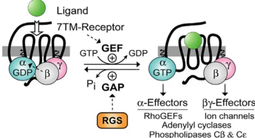
The activation/deactivation cycle of G proteins can be regulated by several proteins that can be divided into two groups: Regulators of G-protein Signalling (RGS) and guanine nucleotide exchange factors (GEFs) (Fig. 3).
RGS have over 30 intracellular proteins already discovered. These can modulate G protein activity mainly by increasing their rate of hydrolysis of GTP by the Gα
subunit and thus inactivating G proteins more quickly. Because of this function, RGS proteins are also called GTPase-activating proteins (GAPs), however this designation is used more often to describe the proteins that have this same function but interact with small monomeric G proteins, like Ras (Gomperts et al., 2009). Besides GAP activity, RGS proteins can physically block the interaction between Gα and effector proteins, and
form of G proteins after GTP hydrolysis, thus preventing Gβγ subunits of interacting
with their effector proteins (De Vries et al., 2000; Roman and Traynor, 2011).
GEFs are able to activate G proteins by increasing the rate of exchange of GDP by GTP. GPCRs are the most well known proteins that act as GEFs of heterotrimeric G proteins (De Vries et al., 2000; Jiang and Bajpayee, 2009).
1.3. The Other Guanine Nucleotide-Binding Protein
The Go protein is the most expressed G protein in the central nervous system,
where it accounts for about 1% of total membrane protein. It was accidentally discovered in 1984 when researchers were trying to isolate Gi protein from bovine brain
and detected an extra Gα subunit with a molecular weight of 39 kDa, naming it the
"other" G protein, to differentiate from Gi and Gs (Sternweis and Robishaw, 1984; Jiang
and Bajpayee, 2009). Since then, it has been intensively studied and several GPCRs, among other proteins, had already been discovered to interact with Go; however, its
main functions are not yet fully understood. Being highly expressed in brain tissue, Go
as been implicated in neuronal development, migration, and such diseases as Alzheimer's and Parkinson's (Jiang et al., 1998; Jiang and Bajpayee, 2009).
Figure 3 - Modulation of G proteins action by Regulators of G-protein signalling (RGS) and guanine nucleotide exchange factors (GEFs). Reproduced from(Siderovski and Willard, 2005)
1.3.1. Go genetics
Go protein has already been identified in several species, such as rat, mouse,
bovine, Drosophila and human, among others. Comparison between DNA sequences of the different Go genes showed that this protein is highly conserved across species,
which suggests that Go signalling is extremely important for organisms to receive,
integrate and execute extracellular signals (Murtagh et al., 1991; Jiang and Bajpayee, 2009).
The human Gαo gene comprises over 100 kb and contains 11 exons, being
localized in chromosome 16 (Tsukamoto et al., 1991). Analysis of bovine and mouse brains have led to the discovery of two isoforms of Gαo, Gαo-1 and Gαo-2 (also called G αo-A and Gαo-B), which were later found in human cells. Both isoforms are identical in the
first two thirds of the amino acid sequence, differing only in 20 amino acids in the latter third of the protein. Study of the human gene found that the exon 8 and 9 are duplicated, and that Gαo-1 contains exon 7-A and 8-A while Gαo-2 contains exon 7-B and 8-B. This
indicates that both isoforms are a product of alternative splicing, and since exon 7 and 8 are thought to be necessary to receptor and effector binding, these isoforms may have different functions in the human brain (Lang, 1989; Hsu et al., 1990; Tsukamoto et al., 1991; Jiang and Bajpayee, 2009).
1.3.2. Modulators of Go activity
Being a member of the Gi/o family, Go suffers ADP-ribosylation by PTX in its
carboxyl terminal cysteine residue of the α subunit, inactivating it (Jiang and Bajpayee, 2009; Locht et al., 2011). A few RGS proteins have been identified that accelerate the process of Go inactivation, being one of those proteins the Gα-interacting protein
(GAIP). This protein has been found to specifically interact with Go, only losing that
specificity when its N-terminal was deleted (Diverse-Pierluissi et al., 1999; Jiang and Bajpayee, 2009).
Several GPCRs that are able to exert their action trough Go proteins have already
been identified, including opioid, α2-adrenergic, M2 muscarinic and somatostatin
receptors (Jiang et al., 1998). All of these act as GEFs, causing Go to dissociate from
Go, such as the growth cone-associated protein with a molecular weight of 43 kDa
(GAP43), the activator of G protein signalling (AGS), the Presenilin I enzyme and the Alzheimer's Amyloid Precursor Protein (APP) (Jiang and Bajpayee, 2009). GAP43, a protein expressed in developing neurons, is thought to act upon Go in a manner similar
to GPCRs. However its action is not affected by the presence of PTX or the βγ subunit, which leads to the believe that is mechanism of action is somehow different of other GEFs (Strittmatter et al., 1991; Jiang and Bajpayee, 2009). AGS studies in vitro showed that this protein enhanced GTPγS binding to both Gi and Go (Cismowski et al., 2000). A
physical interaction between Presenilin I and Go has been described, which led to the
possibility of this protein performing its functions through Go. Besides that, the
interaction of the two proteins could also be implicated in Alzheimer's disease (Smine et al., 1998; Jiang and Bajpayee, 2009). The interaction between Go and APP will be
discussed in more detail further ahead.
Other factors have also been identified that interact and possibly modulate Go
activity. However, their exact mechanism and effect are not fully comprehended. Such is the case of GoLoco motif, a 19 amino acid sequence originally found in the RGS12 protein that binds to Gαo, though is not known whether it acts as a GEF or rather
decreases Go activity by stabilizing it in the GDP-bound form (Siderovski et al., 1999;
Jiang and Bajpayee, 2009).
1.3.3. Go expression pattern
Go protein is expressed mainly in the central nervous system (CNS), however, its
distribution along the brain does not appear to be equal. Immunohistochemical studies in rat brains showed that Go is highly present in the cerebral cortex, cerebellum,
hypothalamus, hippocampus and substantia nigra, being located mainly in the cytoplasmatic face of the plasma membrane, including cell-to-cell contacts (Worley et al., 1986; Gabrion et al., 1989). Besides CNS, Go has also been located in the heart
tissue, pituitary gland and pancreatic isles (Wolf et al., 1998; Jiang and Bajpayee, 2009).
Go expression was also studied in different stages of neuronal differentiation. In
undifferentiated neuroblastoma x glioma hybrid cells, Go levels were very low but after
Go2 isoform is expressed in undifferentiated cells, whereas both Go1 and Go2 were
expressed in differentiated cells, a factor that contributes to the increase in Go protein
levels (Mullaney and Milligan, 1989; Brabet et al., 1991).
1.3.4. Go functions
The Go protein seems to have several functions mediated through a large number
of effector molecules. Most of those functions involve the modulation of ion channels, kinases activity and vesicular transporters (Jiang and Bajpayee, 2009).
Calcium channels: Several neurotransmitters and hormones that inhibit
voltage-gated Ca2+, such as noradrenaline and somatostatine, have been known to act through GPCRs. Initial studies using PTX on neurons led to the discover that the inhibitory effect of the neurotransmitter over the calcium channels was lost when the toxin was present, which suggested that PTX-sensitive G proteins, such as Gi and Go, were involved (Holz et al., 1986; Hille, 1994; Jiang and Bajpayee, 2009). Further studies using antibodies specific for the Gαo subunit showed that the inhibitory effect of
noradrenaline was diminished when the antibodies were applied (Caulfield et al., 1994).
Potassium channels: Like in the calcium channels, there are neurotransmitters
that interact with K+ channels through PTX-sensitive G proteins. Experiments using
membranes of hippocampal pyramidal cells allowed to identify four K+ channels that were activated by purified Go proteins and a recombinant Gαo subunit (VanDongen et
al., 1988; Jiang and Bajpayee, 2009).
The activation of K+ channels by Go was also studied in the heart muscle and
peripheral neurons. However the results showed that it was the βγ subunit of the heterotrimeric complex rather than the α subunit that interacted and activated the K+ channels (Huang et al., 1997; Lei et al., 2000).
Besides potassium and calcium channels, Go also appears to be involved in the
regulation of sodium channels, however the receptors involved in this pathway have not yet been identified (Jiang and Bajpayee, 2009).
MAPK pathway: several studies indicate that Go is involved in the activation of
the mitogen-activated protein kinase (MAPK) pathway (Jiang and Bajpayee, 2009). For example, one of those studies demonstrated that a constitutively active Gαo was able to
activate B-Raf in a Protein Kinase C (PKC)-dependent manner and consequently stimulate the MAPK pathway (Antonelli et al., 2000).
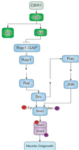
STAT3 pathway: The signal transducer and activator of transcription 3 (STAT3) is a known proto-oncogenic protein that is involved in the development of several cell lines, from hematopoietic cells to neurons. It was recently found that STAT3 can be activated by receptors coupled to Go (He et al., 2005; Hankey,
2009). One of those receptors is the cannabinoid receptor (CBR1), which, when activated, leads to the stimulation of the STAT3 pathway. This mechanism appears to involve the direct interaction between Go
and regulators of small monomeric G proteins, such as Rap1 and its GAP protein, that are present in the STAT3 pathway. This results in the activation of Scr tyrosine kinase, culminating in the phosphorylation of STAT3 (Fig. 4). This Go-signalling was
observed in Neuro-2A cells, which led to neurite outgrowth (He et al., 2005).
Vesicular transporters: Go proteins are present in both secretory granules and
small synaptic vesicles in bovine and rat brain. The exact mechanism in which Go is
involved in these structures is still not well known, however a few possible functions have been proposed. One of those is the control of the uptake of neurotransmitters, since activation of Go2 inhibits the uptake of noradrenaline in PC12 cells (Ahnert-Hilger et al., Figure 4 - STAT3 Pathway leading to neurite outgrowth. Reproduced from(He et al., 2005)
1994; Ahnert-Hilger et al., 1998). It was also demonstrated that Go interacts with small
G proteins (Rho) in a mechanism necessary for the reorganization of neurons cytoskeleton in the exocytosis process (Jiang and Bajpayee, 2009).
Go protein seems to have significant importance in the brain due to its abundance
in this tissue. Studies in mice lacking Go revealed that the absence of the protein led to
severe motor control deterioration, hyperactive behaviour and hyperalgesia (Jiang et al., 1998). The molecular role of Go in the brain has been studied and its interaction,
directly or indirectly, with other brain proteins have led to the discovery of a few specific roles. One of those signalling pathways has already been described above, the STAT3. Another one is the Growth Associated Protein 43 (GAP-43), a protein highly expressed in the growth cones of developing and regenerating neurons, which indicates that it may be involved in neurite growth (Strittmatter et al., 1991; Strittmatter et al., 1994). Like stated above, GAP-43 is a known GEF for Go protein, leading to the believe
that GAP-43 is able to modulate neurite growth and motility through Go activation.
Strittmatter et al have already produced some results that indicate just that. In their studies, GAP-43 was shown to increase neurite outgrowth by stimulating Go in
neuroblastoma cells (Strittmatter et al., 1994). Another study using constitutively activated Gαo (a mutated Gαo lacking its GTPase activity) showed that increase in
neurite outgrowth could be caused by inhibition of PKC and modulation of intracellular calcium release (Xie et al., 1995).
Another important role of Go in the brain appears to be its involvement in
Alzheimer's Disease, an hypothesis that arrived with the discovery that Go interacts with
the Alzheimer’s Amyloid Precursor Protein (Nishimoto et al., 1993).
1.4. The Alzheimer's Amyloid Precursor Protein
Alzheimer's Amyloid Precursor Protein (APP) is a transmembrane protein mainly present in the Golgi and plasma membrane of neuronal cells. Its specific functions are still not very clear, but it is thought to be involved in cell adhesion signalling, neurite outgrowth and synaptic contact, being also the precursor protein of β-amyloid (Aβ) (Small, 1998; Thinakaran and Koo, 2008). The Aβ peptide is the main
component of amyloid plaques (also called senile plaques), one of the main features of Alzheimer's Disease (AD), and it has been associated to most of the pathological changes that occur during the progress of AD, such as induction of inflammatory reactions, neuronal dysfunction, synapse loss and cell death (Hardy and Higgins, 1992; Sommer, 2002; Henriques et al., 2010). Its production depends on the processing of APP, a process that will be explained further ahead.
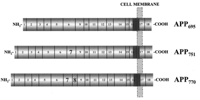
APP arises from the expression of a gene located in the chromosome 21. There are several isoforms resulting from alternative splicing, with APP695 being the most common in the brain (the number represents the amino acids content of the protein) (Fig. 5). APP has only one transmembranar domain, with most of the protein being located in the extracellular space and just a small C-terminal domain being located in the intracellular space (Small, 1998; Ling et al., 2003). After its expression, APP suffers several post-translational modifications that are essential for its function, such as glycosylation and phosphorylation.
Figure 5 - Different isoforms of APP. Reproduced from Edgar F. da Cruz e Silva and Odete A. B. da
1.4.1. APP processing
APP can be cleaved by several secretases, giving rise to some peptides with important physiological and pathological function. Those secretases are mainly divided in α, β and γ secretases and depending on which of these cut APP, Aβ may or may not be generated (Figure 6). γ-secretase cuts the APP transmembrane domain on several sites. Its action gives rise to AICD (APP intracellular domain), a peptide that has been implied in a few signalling pathways. AICD can bind to Fe65, which results in the formation of a complex responsible for the activation of the transcription of several genes, including p53, GSK-3β, Kai1, among others. However, the precise mechanism by which AICD binding to Fe65 leads to gene transcription is not well known (Ling et al., 2003; Thinakaran and Koo, 2008; Chow et al., 2010).
The action of the other two secretases defines the processing pathway of APP. In the non-amyloidogenic pathway (Fig. 6 - left), α-secretase cuts APP in the Aβ region, generating secreted APPα (sAPPα) and a C-terminal fragment (α-CTF), the latter being then cleaved by the γ-secretase, originating the p3 fragment and AICD. In this pathway, no Aβ is generated (hence non- amyloidogenic), which is why this process is normally dubbed neuroprotective. Further, the formation of sAPPα also contributes to the neuroprotective function of the non-amyloidogenic pathway. The precise mechanisms
Figure 6 - Non-amyloidogenic (left) and amyloidogenic pathway (right). Reproduced from
in which sAPPα is involved are not fully understood, but studies indicate that it promotes neurite outgrowth, synaptogenesis and cell adhesion, and protects neurons against oxygen deprivation and excitotoxicity (Ling et al., 2003; Gakhar-Koppole et al., 2008; Chow et al., 2010). No function as yet been assigned to the non pathogenic p3 fragment (Ling et al., 2003).
On the other hand, the cut of APP by the β-secretase is termed amyloidogenic pathway (Fig. 6 - right). The β-secretase cuts APP in its β-cleavage site, generating sAPPβ and C99 (or β-CTF). The C99 is then cleaved by γ-secretase, giving rise to Aβ (hence amyloidogenic) and AICD. The main β-secretase involved in this pathway is BACE1 (β-site APP-cleaving enzyme 1). The sAPPβ has still not a clear function (Ling et al., 2003; Cole and Vassar, 2008; Chow et al., 2010).
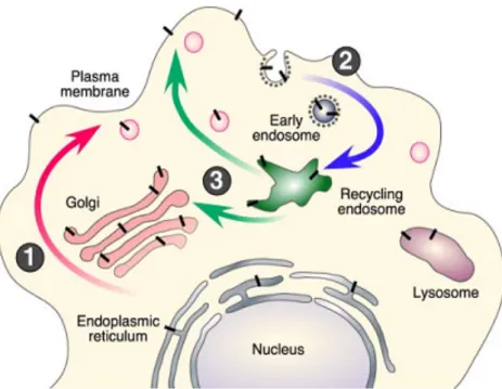
1.4.2. APP trafficking
The first steps in APP post-translational modification are its N- and O-glycosylation, followed by its phosphorylation and tyrosine sulfation. These occur during its transport from the endoplasmic reticulum to the plasma membrane (Figure 7, step 1). However, only a small portion of the expressed APP is localized to the plasma membrane. Most of it remains in the Golgi apparatus or in the Trans-Golgi network (TGN) (Thinakaran and Koo, 2008; Zheng and Koo, 2011).
In the plasma membrane, APP can suffer non-amyloidogenic processing, due to the presence of the α-secretase in the membrane. On the other hand, shortly after its integration in the membrane, APP is internalized by endocitosis (Figure 7, step 2) and can either be degraded in lysosomes or recycled back to the membrane (Fig. 7, step 3). During this transport, APP comes in contact with BACE1 in several compartments, such as the endosome and the TGN. This promotes the amyloidogenic processing of APP and consequently Aβ generation. It has been shown that several proteins, like Fe65, interact with the APP C-terminal and modulate its intracellular trafficking, interfering with the production of Aβ (Small and Gandy, 2006; Thinakaran and Koo, 2008).
Figure 7 - APP trafficking. Reproduced from(Thinakaran and Koo, 2008)
1.4.3. APP phosphorylation
The processing, trafficking and function of APP is regulated in many ways, with phosphorylation appearing to have an important part in all of these processes. A few phosphorylation sites have been identified in both the intracellular and extracellular domain of APP. The serines 198 and 206 are the amino acids phosphorylated in the APP ectodomain, but no relevant function have been so far attributed to these phosphorylations (Walter et al., 1997; da Cruz e Silva and da Cruz e Silva, 2003).
On the other hand, some of the phosphorylation sites discovered in the intracellular domain of APP have a significant impact in its life. These include the Thr654, Ser655, Thr668, Tyr682 and Tyr687. The Thr668 is one of the best studied
phosphorylatable amino acids of APP, being phosphorylated by GSK-3β, Cdk5 and JNK, although its role is still controversial. Thr668 phosphorylation has been linked to both increases and decreases in Aβ formation and in the regulation of the binding of AICD to Fe65 followed by translocation of the complex to the nucleus, or not having any effect in this process (Suzuki and Nakaya, 2008; Schettini et al., 2010).
Phosphorylation of Serine 655 (S655), present in the YTSI sorting sequence, potentially by protein kinase C, has been described as an important event in APP processing, trafficking and its interaction with other proteins. A study using APP phosphomutants mimicking a constitutively phosphorylated and a constitutively
dephosphorylated S655 (S655E and S655A, respectively) showed that S655 phosphorylation increased APP trafficking through the Golgi to the plasma membrane, and an increase in sAPPα production. The two events are most likely related since the continuous trafficking of APP renders it more available to α-secretase cleavage (Vieira et al., 2009). Another study showed that S655 phosphorylation leads to an increase in APP recycling through a retromer mediated process. It also decreases APP targeting to lysosomes, thus expanding its half-life (Vieira et al., 2010).
Interestingly, the binding of APP to Gαo (discussed in more detail further ahead)
occurs immediately downstream of the sorting motif 653YTSI656, which can indicate that APP S655 phosphorylation may influence the interaction between both proteins.
1.5. G
obinding to APP
APP was found to selectively bind to Gαo, but not Giα, through APP His657-Lys676
domain, and described to activate Gαo (Nishimoto et al., 1993). Further studies
involving phospholipid vesicles containing both APP695 and Go identified APP as a
GPCR-like protein, with a possible natural ligand not yet identified but who's action was mimicked by 22C11, an antibody directed against the extracellular domain of APP. When the antibody bound to APP, Go activation was greatly enhanced due to an
increase in the GDP/GTP exchange rate. The ability of APP695 to activate Gi in the
presence of 22C11 was also tested, with negative results, indicating that APP binds specifically to Go protein (Okamoto et al., 1995).
A more recent study shed a new light in the mechanism by which APP activates Go. This study also evaluated the action of the 22C11 in the interaction of APP to Go,
however the results showed a decrease in the GTPaseactivity of Go, a phenomenon not
detected in the earlier study. The authors attributed this difference of results to the fact that the study conducted by Okamoto et al, was done using phospholipid vesicles, which probably lack a few cofactors needed to the decrease in the Go GTPase activity. Since
this decrease would make Go active for longer periods of time, this could be an
alternative mechanism by which APP increases the activity of Go proteins. However,
1.5.1. Go:APP and Alzheimer's Disease
Mutations in the APP gene have already been identified in cases of genetic-linked Familial Alzheimer's Disease (FAD). One class of specific mutations is the V642, where the valine at the 642 position is mutated into isoleucine (V642I), phenylalanine (V642F) or glycine (V642G) (Hardy, 1992; Karlinsky et al., 1992). All these mutations were shown to cause apoptosis in COS-NK1 cells in a Go-mediated
mechanism, since knockout of Go, but not Gi, led to the suppression of apoptosis (Fig.
8). Go involvement was also tested by transfecting COS cells with V642-APPs mutants
lacking the His657-Lys676 sequence. In these cells, apoptosis was also greatly diminished in comparison with V642-APPs mutants, giving stronger evidences of the mediating function of Go in APP-induced apoptosis (Yamatsuji et al., 1996). To clarify which of
the Go subunits was responsible for the activation of the apoptosis cascade, a series of
tests were made in NK1 cells involving constitutively activated Gαo (Q205L) and
βARK1 C-terminus, a binding site to Gβγ subunits that inhibits its functions. The results
showed that constitutively activated Gαo induced little apoptosis while βARK1
C-terminus inhibited NK1 apoptosis, leading to the conclusion that Gβγ subunit, and not
Gα, was responsible for triggering apoptosis (Giambarella et al., 1997).
1.5.2. APP and Gαo in neuritogenesis
Since some of the functions of APP have also been attributed to the Go protein, it
was hypothesized that APP could mediate its functions through Go, acting most likely as
a Gαo GEF (Nishimoto et al., 1993; Okamoto et al., 1995; Okamoto et al., 1996).
One of those functions is neuritogenesis. As stated above, Gαo induction of
neurite outgrowth has already been described via STAT3 pathway. A different pathway
Figure 8 - Mediation of V642-APP-induced apoptosis by Go protein. Adapted from Yamatsuji et
by which Gαo could lead to neurite growth is by activation of the G Protein-Regulated
Inducer of Neurite Outgrowth 1 (GRIN1) (Nakata and Kozasa, 2005), however in this work we will focus on the STAT3 pathway.
APP role on neuritogenesis has also been described. Initial studies showed that APP expression increased neurite length and branching in PC12 cells, possibly by mediating the Nerve Growth Factor action (Milward et al., 1992). Another study analyzed the effect of down-expression of APP on neuronal primary cultures. The results indicated that the blockage of APP expression leads to a decrease in axon and dendritic growth (Allinquant et al., 1995).
More recent studies indicate that while APP clearly influences neuritogenesis, it may have an inhibitory effect rather than stimulatory. Also, sAPPα seems to be fundamental to regulate APP function. In one of those studies, loss of APP expression in primary neuronal culture culminated in increased neurite elongation, with this same result occurring when sAPPα was applied to the cells expressing APP. Both these effects were blocked with the use of antibodies directed against Integrin β1. The hypothesis defended was that full-length membranar APP is able to interact with Integrin β1 leading to the inhibition of neurite outgrowth, while sAPPα blocks this interaction and thus allows neurite elongation (Young-Pearse et al., 2008).
Previous work developed in our lab was dedicated to understanding the role of APP in neuronal differentiation (Rocha, 2011). The study was conducted in SH-SY5Y cells and showed that while long-term overexpression of APP could have a negative effect on neuritogenesis, short-term expression (24h) leads to an increase in neurite outgrowth. Also, with the use of phosphomutants APP-GFP cDNAs, the positive neuritogenic action of APP was found to be enhanced by APP S655 phosphorylation.
2. Aims
This work focused on characterizing the interaction between the Alzheimer's Amyloid Precursor Protein and Gαo protein, with its main aims being:
• To evaluate the effects of APP S655 phosphorylation and Gαo constitutive
activation in APP-Gαo colocalization in SH-SY5Y cells.
• To study the effect of APP-Gαo co-expression in the induction of cellular
morphological changes, and how APP phosphorylation and Gαo activation
regulate that effect(s).
• To analyse the impact of Gαo and APP co-expression in cytoskeleton-related
proteins.
• To evaluate the effect of APP phosphorylation in Gαo-induced STAT3
3. Methods
3.1. Wild type (Wt) and S655 Phosphomutants APP-GFP cDNAs
APP cDNAs, S655 point mutated and fused to GFP (Green Fluorescent Protein) were already available at the lab. Briefly, human APP isoform 695 cDNA was used as a template to generate S655 cDNA point mutations, namely Serine 655 to Alanine (S655A) or to Glutamate (S655E), using site-directed mutagenesis. Due to their structure, these amino acids mimic a constitutively phosphorylated (Glutamate) and dephosphorylated (Alanine) S655. These mutants, together with parental Wt cDNA, were subcloned into the pEGFP-N1 vector (Clontech), a plasmid that encodes GFP. The result was a fusion gene of APP with GFP in its N-terminal (Vieira et al., 2009).3.2. Wt and Constitutively Active G
αocDNAs
The human Gαo cDNAs were acquired from the Missouri S&T cDNA Resource
Center. The Gαo subunit open reading frame (isoform Gαo-1) had been amplified from
human whole brain cDNA (Clontech) by PCR and subcloned into pcDNA3.1+ vector (Invitrogen) between Kpn I (5') and Xba I (3') restriction sites. The Constitutively Active Gαo (Gαo CA) had been engineered by substituting Glutamine 205 by a Lysine
(Q205L), using the Quickchange mutagenisis kit (Stratagene). This substitution significantly reduces the GTPase activity of Gαo, rendering it constitutively active. Both
Gαo inserts have a size of 1070 bp.
3.3. G
αoand constitutively active G
αocDNAs amplification and
purification
3.3.1. Bacterial transformation
E. Coli XL1-Blue competent cells were thawed on ice and 0,25 µg (5 µL) of Gαo
and Gαo constitutively active cDNAs were each added to a 100 µl aliquot of competent
cells. The cells were incubated on ice for 30 min. After this, they were subjected to an heat shock by incubation in a 42 ºC bath for 90 sec and then rapidly transferred to ice
for 2 min. 900 µl of SOC medium was added to the cells and a 45 min incubation at 37 ºC followed. Finally, the cells were centrifuged at 14000 rpm for 30 sec, the supernatant was discarded and the pellet was resuspended in the remaining volume (100 µL), plated onto LB/ampicillin agar plates and incubated at 37 ºC during 16-18 h.
3.3.2. MegaPrep DNA Purification
One colony of transformed bacteria with each Gαo cDNA was isolated and added
to 3 mL of LB medium and incubated during 16-18 h at 37 ºC, with agitation. After bacterial growth, 1 mL of cells was transferred to 1 L of LB medium and again incubated overnight (O/N) at 37ºC with agitation. Cells were then centrifuged at 1500 x g, for 20 min (Beckman Avanti J-25 I - Beckman Coulter), the supernatant discarded and 30 mL of Cell Resuspension Solution was added. After resuspension, 30 mL of Cell Lysis Solution was added and mixed. After the solution became clear and viscous, 30 mL of Neutralization Solution was added, mixed and centrifuged at 20000 x g for 15 min. The supernatant was filtered using filter paper and the remaining volume was measured. 0,5 volume of isopropanol was added and then centrifuged at 14000 x g for 15 min. The supernatant was discarded and the pellet was resuspended in 4 mL of sterile water. 20 mL of Wizard® Megapreps DNA Purification Resin was added to the
solution and mixed. The mix was then transferred to a Megacolumn, inserted in a vacuum manifold port, and vacuum was applied. The column was then washed 2 times with Column Wash Solution, also by applying vacuum. 5 mL of ethanol 80% was added to the column and vacuum was applied. The column was then transferred to a 50 mL screw cap tube and centrifuged at 1300 x g for 5 min (Eppendorf Centrifuge 5810R). The liquid was discarded and the column was again placed in the vacuum manifold and dried by applying vacuum for 5 min. The column was then removed, placed in a new 50 mL screw cap tube, and 3 mL of pre-heated (65-70 ºC) nuclease-free water was added to the column. After 1 min, the column was centrifuged at 1300 x g for 5 min, the liquid transferred to microcentrifuge tubes (400 µL per tube) and stored at -20 ºC. This procedure had been previously done in laboratory for the APP-GFPs cDNAs
3.3.3. Ethanol precipitation of plasmid DNA
microcentrifuge tube containing the DNA solution. 2,5 volume of ethanol 100% was further added, mixed and incubated O/N at -20 ºC. After, the tubes were centrifuged (14000 x g, 15 min, 0 ºC), the supernatant discarded and 750 µL of ethanol 70% (half of the tube capacity) was added and mixed. The solutions were incubated during 5 min, at -20 ºC, and then centrifuged (14000 x g, 5 min, 0 ºC). The supernatant was discarded and the pellet was dried. After all traces of ethanol disappeared, the DNA in each tube was resuspended in 200 µL of sterile water and stored at -20ºC.
3.4. Culture and maintenance of the SH-SY5Y cell line
The SH-SY5Y human neuroblastoma cells are derived from the SK-N-SH cell line, originally established from a bone marrow biopsy of a neuroblastoma patient. The SH-SY5Y cells were maintained in a 10% FBS MEM:F12 (1:1) growth medium, in a 5% CO2 humidified incubator at 37 ºC. Cells were divided when they reached 70-80%
confluence, and transfected with the appropriated cDNAs as follows.
3.5. Transfection of the SH-SY5Y cell line with APP-GFP and G
αocDNAs
In order to be able to study the effects of APP phosphorylation and Gαo activity
in cells, Wt and CA Gαo cDNAs, along with Wt, S655A and S655E APP-GFP cDNAs,
were transfected into SH-SY5Y cells. Three different transfection reagents were first tested in order to optimize SH-SY5Y cells transfection: jetPRIME®
(Polyplus-transfection), TurboFectTM (Fermentas Life Sciences) and CombiMagTM (OZ Biosciences). For each of the reagents used, cells were seeded in 6-well plates 24 h before transfection, so that cells could be around 80 to 90% at the time of transfection. Also, the growth medium was changed immediately before the transfection procedure.
3.5.1. JetPRIME® transfection
The jetPRIME® transfection reagent is a molecule based on a non-liposomal formulation produced by Polyplus-transfectionTM. For each well, 1 µg of cDNA was diluted in 100 µL of jetPRIME® buffer. Then, 2 µL jetPRIME® reagent was added,
vortexed and the mix was incubated for 10 min at room temperature (RT). After this, the mixture was added to each well and mixed by gently agitating the plate. The cells were then incubated at 37 ºC in a 5% CO2 incubator. After 4 h, the cell medium was
changed, and 24 h after transfection cells were collected in 250 µL 1% SDS for Western Blotting (WB).
3.5.2 TurboFectTM transfection
The TurboFectTM transfection reagent is a cationic polymer that forms a positively charged complex with DNA that is easily endocytosed by eukaryotic cells. Briefly, for each well, 1 µg of cDNA was diluted in 100 µL of serum-free growth medium. After gently vortexing it, 2 µL of TurboFectTM reagent was added to the DNA, mixed and incubated during 15 min at RT. It was then added to each well and mixed by gently agitating the plate. The cells were incubated at 37 ºC in a 5% CO2 incubator.
After 6 h, the cell medium was changed, and at 24 h of total transfection time, cells were collected in 250 µL 1% SDS for WB.
3.5.3 CombiMagTM transfection
CombiMagTM transfection reagent is a solution containing magnetic particles that is used in combination with any other transfection reagent to improve its efficiency. In this case it was used in combination with TurboFectTM. It uses the principle of
MagnetofectionTM, in which magnetic particles associate with vectors and then come
into contact with cells by applying a magnetic force, thus enhancing the transfection efficiency (Scherer et al., 2002). First, for each well, 1 µL of CombiMagTM reagent was transferred to a microtube. Then, the TurboFectTM mixture was prepared by mixing 1 µL of the reagent to 1 µg of DNA in a microtube containing 100 µL of serum-free growth medium. Without incubating, the mixture was transferred to the microtube containing the CombiMagTM and mixed by pipetting vigorously. It was then added to the cells mixed by gentle agitation of the plate. Finally, the 6-well plate was placed upon a magnetic plate (OZ Biosciences) and incubated for 15 min. Cells were further incubated at 37 ºC in a 5% CO2 incubator and the cell medium was changed after 6 h of
3.6. Cell collection and quantification of protein content
Upon being collected in 250 µL 1% SDS, cells were immediately boiled at 90 ºC for 10 min, followed by a 20 sec sonication period, and stored at -20 ºC.
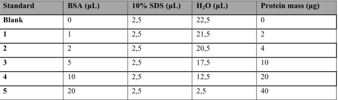
Quantification of the protein content of the cell lysates was made using the Pierce's bicinchoninic acid (BCA) protein assay kit (Thermo Scientific). All the procedure was performed using a 96-well plate. The standard samples needed for quantification were prepared using increasing known amounts of bovine serum albumin (BSA), as depicted in Table 3. The remaining samples were prepared by mixing 5 µL of the cell lysate to 20 µL 1% SDS.
Table 3 - Standards used in the BCA protein assay method. BSA, Bovine serum albumin solution (2 mg/ml).
Standard BSA (µL) 10% SDS (µL) H2O (µL) Protein mass (µg)
Blank 0 2,5 22,5 0 1 1 2,5 21,5 2 2 2 2,5 20,5 4 3 5 2,5 17,5 10 4 10 2,5 12,5 20 5 20 2,5 2,5 40
The Working Reagent (W.R.) was prepared by mixing the BCA reagent A with the BCA reagent B in the proportion of 50:1, and 200 µL were added to each well. The plate was incubated during 30 min at 37 ºC, after which the absorbance at 562 nm was measured using a microplate reader (Infinite M200, Tecan).
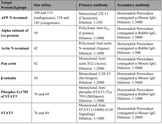
3.7. Antibodies
There were several antibodies used in western blot assays. The primary antibodies were the 22C11 (Chemicon), directed against APP N-terminus, recognizing full-length APP, and thus enabling the evaluation of the transfection levels of the APP-GFP cDNAs; anti-Gαo antibody (Upstate), for detecting Wt and CA Gαo transfection
levels; both anti-actin N-terminal (Sigma) and anti-actin 2G2 (Acris antibodies) antibodies, and anti-β-tubulin (Invitrogen) antibody, for analysing the effects of Gαo and
but with no satisfactory results (see chapter 4.1). To evaluate the effect of Gαo and APP
in STAT3 signalling two antibodies were used: the monoclonal anti-phospho-STAT3 (Millipore), directed against the phosphorylated Tyr705 residue, and the monoclonal anti-STAT3 (Cell Signalling Technology) antibodies, with both of them recognizing the alpha and beta STAT3 isoforms.
All the secondary antibodies, either recognizing mouse or rabbit antibodies, were goat antibodies labelled with horseradish peroxidase, purchased from Amersham Pharmacia. In Table 4 are represented the antibodies used, their respective dilutions and secondary antibody used.
Table 4 - Antibodies used in the Western blots, respective target proteins and their sizes, and specific dilutions used.
Target
Protein/Epitope Size (kDa) Primary antibody Secondary antibody APP N-terminal 109 and 115 (endogenous); 139 and 145 (exogenous) Monoclonal 22C11 (Chemicon); Dilution: 1:250 Horseradish Peroxidase conjugated α-Mouse IgG; Dilution: 1:5000
Alpha subunit of Go protein 39
Polyclonal Anti-Gαo
(Upstate); Dilution: 1:5000
Horseradish Peroxidase conjugated α-Rabbit IgG; Dilution: 1:5000 Actin N-terminal 42 Polyclonal Anti-actin N-terminal (Sigma); Dilution: 1:1000 Horseradish Peroxidase conjugated α-Rabbit IgG Dilution: 1:500 Pan actin 42 Monoclonal Anti-actin 2G2 (Acris); Dilution: 1:2000 Horseradish Peroxidase conjugated α-Mouse IgG; Dilution: 1:5000 β-tubulin 50 Monoclonal 2-28-33 (Invitrogen) Dilution: 1:2000 Horseradish Peroxidase conjugated α-Mouse IgG Dilution: 1:5000 Phospho-Tyr705 of STAT3 76 and 84 Monoclonal Anti-phospho-STAT3 (Tyr 705) (Millipore) Dilution: 1:3000 Horseradish Peroxidase conjugated α-Rabbit IgG Dilution: 1:1000 STAT3 76 and 84 Monoclonal Anti-STAT3 (124H6) (Cell Signaling) Dilution: 1:1000 Horseradish Peroxidase conjugated α-Mouse IgG Dilution: 1:2000
For the immunocytochemistry procedures, the rabbit polyclonal anti-Gαo and
anti-APP 22C11 antibodies were used, together with the mouse monoclonal anti-STAT3 (Cell Signaling). This antibody was used to analyze the influence of Gαo activation and
APP phosphorylation in STAT3 signalling. Like in the WB, another anti-Gαo antibody
4.1). The list of the antibodies and their respective dilution and secondary antibody are present in Table 5.
Table 5 - Antibodies used in immunocytochemistry, respective target proteins specific dilutions used.
Target
Protein/Epitope Primary antibody Secondary Antibody
APP N-terminal Monoclonal 22C11 (Chemicon)
Dilution: 1:50
Alexa Fluor® 488 Goat Anti-Mouse IgG (Life
Technologies) Dilution: 1:300
Alpha subunit of Go protein Polyclonal Anti-Gαo (Upstate)
Dilution: 1:250
Alexa Fluor® 594, 488 and 350 Goat Anti-Rabbit IgG (Life Technologies) Dilution: 1:300
STAT3
Monoclonal Anti-STAT3 (124H6) (Cell Signaling) Dilution: 1:450
Texas Red®-X and Alexa Fluor® 350 goat anti-mouse IgG (Life Technologies) Dilution: 1:300
3.8. Western Blot assay
Mass-normalized cell lysates were subjected to electrophoresis on a 5-20% gradient sodium dodecylsulfate (SDS) polyacrylamide gel and then transferred to a nitrocellulose membrane. After transferring the proteins, the membranes were first soaped in 1X TBS for 10 min, and then the blockage of unspecific antibody binding sites was ensured by incubating the membrane for 1-2 h with 5% non-fat dry milk in 1X TBS-T solution. Incubation with the primary antibody was performed according to the manufacturer indications, or according to previous optimization experiments for the antibodies in use (the incubation time ranged from 2 h to O/N incubation). After 3 washes with 1X TBS-T, the membranes were incubated for 2 h with the secondary antibody. All antibodies were diluted according to the manufacturer indications (dilutions used present in table 4), normally either in 3% non-fat dry milk in 1X TBS-T solution or in 3% BSA in 1X TBS-T solution. Membranes were additionally washed 3X with TBS-T and submitted to signal development using either a home made enhanced chemiluminescence (ECL) or LuminataTM Crescendo (Millipore) reagents. Both of the reagents work as a substrate to horseradish peroxidase with which the secondary antibodies are labelled, producing a chemiluminescent signal. In a dark room, the membranes were incubated for 1 min at RT with ECL or 5 min with LuminataTM
Crescendo, and then an autoradiography film was placed on top of the membrane and placed inside a film cassette. After the exposure (with time varying depending on the protein being detected), the film was developed in developing solution (Sigma Aldrich), washed in water and fixed in fixing solution (Sigma Aldrich). The membrane was further washed with 1x TBS-T and deionised water before drying. The autoradiograms were scanned in a GS-800 calibrated imaging densitometer (Bio-Rad) and protein bands quantified using the Quantity One densitometry software (Bio-Rad).
3.9. Ponceau Red staining of protein bands
Ponceau Red staining is normally applied to assess successful electrotransfer of proteins to the membrane. However it can also be used as a loading control as an alternative to actin or β-tubulin, when these proteins vary with the experimental conditions. This type of staining has been described as a fast, inexpensive, and nontoxic method and it is fully reversible in a few minutes (Romero-Calvo et al., 2010).
The nitrocellulose membranes were incubated in Ponceau S solution (Sigma Aldrich) for seven min, followed by a wash with deionised water for making the protein bands clearly visible. The membrane was then scanned in a GS-800 calibrated imaging densitometer (Bio-rad). The membranes were then washed extensively with 1X TBS-T and water for completely removing the staining and thus be further used in WB assays.
3.10. Immunocytochemistry assay
Cells grown on coverslips were fixed with a 4% paraformaldehyde PBS solution for 30 min, washed 3 times with PBS and permeabilized with a 0,2% TRITON PBS solution (10 min). After another 3 washes, the cells were covered with PBS 1X/3%BSA blockage solution for 30 min. The cells were then incubated with the primary antibodies diluted in PBS 1X/3%BSA (the respective dilutions are present in Table 5) for 2 h at RT. The antibodies were removed by washing 3 times with PBS and the specific secondary antibodies were incubated for 2 h at RT (see Table 5). After 3 washes with PBS and one with deionised water, the cells were mounted with VECTASHIELD®
mounting medium, with or without DAPI (Vector Laboratories). Fluorescence microscopy was carried out using a LSM 510 Meta confocal microscope (Zeiss).
3.11. Image analysis
Every image analysis was performed using the ImageJ Fiji software. For STAT3 nuclear intensity analysis, the nuclei were selected using the LiveWire plugin and STAT3 intensity was measured. To try to optimize the comparison between different conditions without the quantifications being affected by some fluorescence/antibody labelling issues (variation in noise, signal intensity), the signal intensity of STAT3 in the nuclei of transfected cells was divided by the signal intensity in the nuclei of non-transfected cells (taken as control). Also, in every image, three measures of the background were performed and subtracted to the nuclei measure.
For colocalization analysis, the images were first processed using the DeconvolutionLab plugin for better removal of background noise and thus improving colocalization analysis (Landmann and Marbet, 2004). The colocalization analysis was conducted using the Coloc2 plugin. This plugin uses several pixel intensity spatial correlation methods to determine colocalization between two different colour channels, such as the Manders, the Costes, the Pearson or the Li methods. In the work here described, results were obtained using the Manders' method, since it is normally the most used when comparing two objects of different amounts (in this case two proteins who have different expression levels) (Manders et al., 1993). The results are presented as the percentage of colocalization of one protein with the other.
3.12. Data analysis
Data is expressed as mean ± SEM (standard error of the mean) of the different experiments. Statistical significance analysis was conducted by one way analysis of variance (ANOVA) followed by the Turkey test.